3972
Improved Specificity of Functional Mapping of Thalamocortical Connections at Ultra High Field
Mark J Lowe1, Anna Crawford1, and Stephen E Jones1
1Imaging Institute, Cleveland Clinic, Cleveland, OH, United States
1Imaging Institute, Cleveland Clinic, Cleveland, OH, United States
Synopsis
We compare whole brain functional connectivity to the Vim thalamic nucleus at 3T and 7T, and demonstrate that the higher spatial resolution and higher contrast-to-noise ratio at 7T results in maps that are more specific to the function of that thalamic region and the connectivity maps from the 3T data are likely lacking in specificity due to partial volume of signal from neighboring thalamic regions.
Introduction
A principal advantage of performing fMRI at ultra high field is the ability to use the much higher contrast-to-noise ratio to perform high spatial resolution studies. This advantage is particularly useful in regions of the brain where functional relationships vary over small spatial scales, such as the thalamus. The different thalamic nuclei can be as small as 1mm in spatial extent and separated by as little as a few millimeters (fig 1). It has been shown that 7T fMRI can provide better fine-scale descriptions of functional networks in the brain(1). Using a concatenated dataset of resting state data in cohorts of healthy control subjects, we present a comparison of resting state fMRI connectivity of an important thalamic nucleus to the entire brain using data acquired in a typical 3T resting state scan protocol to that of a high spatial resolution 7T resting state protocol.Methods
In order to improve sensitivity to detecting functional networks, while preserving spatial specificity, we adopted a strategy of using nonlinear warping to a common space following by concatenation of resting state data in cohorts of subjects.Data acquisition: Nineteen healthy control subjects were scanned in a resting state fMRI protocol in a Siemens Tim Trio 3T MRI scanner. Another eighteen healthy control subjects were scanned in a resting state protocol on a Siemens Magnetom 7T scanner.
Scan protocols:
3 tesla: One hundred thirty two repetitions of 31-4mm thick axial slices acquired with GE-EPI: TE/TR=29ms/2800 ms, 128x128 matrix, 2mm x 2mm in-plane resolution. The subject is instructed to rest with eyes closed and refrain from any voluntary motion.
7 tesla: One hundred thirty two repetitions of 81-1.5mm thick axial slices acquired using simultaneous multi-slice (SMS) GE-EPI: TE/TR=19ms/2800 ms, 128x128 matrix, MB=3, Grappa=2, 1.2mm x 1.2mm in plane resolution. The subject is instructed to rest with eyes closed and refrain from any voluntary motion.
Data Analysis
Standard processing pipeline was used for each field strength data: Physiologic noise removal, retrospective motion correction, temporal filtering (lowpass < 0.1Hz). No spatial filtering was applied to either data set.
Concatenation:
Individual T1w images were aligned to the EPI scan using linear registration. Then, the T1w images were transformed to the MNI template using a non-linear transformation with ANTS. Each time point of the EPI was then registered to MNI space using the transformation matrix from the T1w registration. Once aligned to MNI, the data was detrended using a third order polynomial. Finally, all of the studies were concatenated together for each field strength using AFNI’s 3dTcat.
Functional Connectivity Analysis:
Using Instacorr from the AFNI toolbox, we calculated whole brain correlation to a voxel determined anatomically to be in the ventral intermediate nucleus (Vim) of the thalamus. This is an important motor-related region of the brain, often used for example as a target for deep brain stimulation to relieve Parkinson’s disease-related symptoms. The same brain voxel was selected in both 3T and 7T. The primary output from this brain region is to cortical motor and premotor regions(2).
Results
Figure 2 shows the relative voxel size superposed on a 200um isotropic resolution MRI (obtained on an ex-vivo brain in 13 hours). The T2* weighted image is zoomed to focus on the Vim region of the thalamus. It is clear that, while the 7T seed voxel can be localized to the Vim, the size of the 3T voxel results in partial voluming of the surrounding thalamic regions. The whole brain connectivity maps are shown in Figure 3. It is clear that the 7T connectivity map is much more specific to the cortical motor regions known to have anatomic connections to this brain region(2). The 3T thalamic connectivity map shows very widespread cortical connections, as well as bilateral subcortical connections.Discussion
We show here that the high spatial resolution of 7T rsfMRI can be used to identify cortical networks connected to thalamic nuclei with much more specificity than lower field strength rsfMRI. The observed widespread cortical connections observed in the 3T concatenated dataset is likely the result of signal partial volume from tissue surrounding the thalamic nucleus of interest.The higher specificity of mapping of thalamocortical connections has implications for a number of applications, such as presurgical planning for deep brain stimulation or high-intensity focused ultrasound treatment. The use of non-linear registration combined with large concatenated rsfMRI datasets proved to be a useful tool to enhance sensitivity while preserving spatial specificity.
Acknowledgements
This work was supported by Siemens Healthineers, Inc. The authors gratefully acknowledge the help of Tobias Kober of Siemens Healthineers for use of WIP944 (MP2RAGE) and Thomas Benner for the use of WIP 770B (MB-EPI).References
1. A, TV, Jamison, K, Glasser, MF, Smith, SM, Coalson, T, Moeller, S, Auerbach, EJ, Ugurbil, K, Yacoub, E: Tradeoffs in pushing the spatial resolution of fMRI for the 7T Human Connectome Project. Neuroimage, 154, 23-32, (2017).
2. Thalamic Organization, in NeuroScience. Edited by Swenson R, Dartmouth Medical School, 2006
Figures

Figure 1: Freesurfer Thalamic nucleus parcellation of the
MRI underlay used in Figure 3. The red bar indicates 10mm of tissue,
illustrating the spatial scale of the thalamic nuclei.
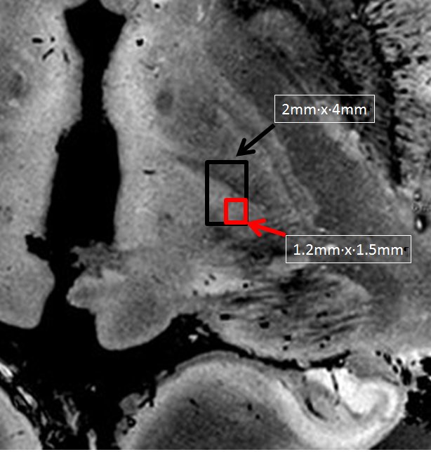
Figure 2: Superposition in black (3T) and red (7T) of the
voxel size used for each field strength on an axial slice of a 200 um isotropic
resolution T2* image obtained of an ex-vivo brain (zoomed to the region of the
Vim). The red box is localized to the Vim of the thalamus while the 3T voxels
encompasses other neighboring regions of the thalamus, in this example the VO
nucleus, which has projections to other cortical and subcortical brain regions.
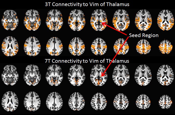
Figure 3: Resting state functional connectivity of a seed
placed in the Vim of the thalamus for both 3T (upper) and 7T (lower). The threshold for the false color overlay is
p<10-10 single voxel, uncorrected.