3967
Alteration resting state networks functional connectivity associated with Hypothyroidism using Graph theory1NMR Research Center, INMAS, Delhi, India, 2Thyroid research Center, INMAS, Delhi, India, 3Cognitive Neuroscience Center, NIMHANS, Bangalore, India
Synopsis
The aim of this study is to explore the alteration RSNs connectivity and its association with underlying neurobehavioral symptoms in hypothyroid as compared to controls and consequences of therapeutic intervention using graph theory-based network analysis. Our finding revealed significantly reduced clustering coefficient, small world connectivity and local network efficiency in hypothyroid as compared to controls in sub-regions FPN, DMN, cingulo-opercular network, SMN and got reversed back in euthyroid patients in the network sub-regions of frontoparietal network, cingulo-opercular network, SMN after thyroxin therapy. These findings of reduced clustering coefficient suggest abated activities of motor, attention, working memory and cognition in hypothyroid.
Introduction:
Resting-state brain networks (RSNs) is a set of regions that show coherent neural activity in the resting state. Thyroid hormones (THs) play an important role in the formation of a normal neurological network and neural differentiation during brain development and adult brain1. To understand the neurocognitive impairment and consequences of therapeutic intervention in hypothyroid patients, we applied an advanced mathematical model based graph theoretical network analysis on the resting-state fMRI data. In graph-theoretical network analysis, the brain is modeled as a complex network, collection of “nodes” representing neural elements (brain regions) and “edges” representing functional connections among the nodes. The clustering coefficient and path length are used to characterize functional brain networks, clustering coefficient quantifies the local connectivity and path length quantifies the global connectivity. A “small world network” is described as a network having a high clustering coefficient and low inter-nodal path length. The objective of this study is to explore the alteration in RSNs connectivity and its association with underlying neurobehavioral symptoms in hypothyroid population as compared to healthy controls and consequences of therapeutic intervention using graph theory-based network analysis.Materials and Methods:
Twenty-five hypothyroid patients (20 females, 5 males), 26 age-gender matched healthy controls (21 females, 5 males), Twenty-five euthyroid patients (20 females, 5 males) were recalled for follow-up after thyroxin therapy (range=6-16 months, mean ± SD=13.28±6.20 months). Drug-naive hypothyroid patients with elevated TSH (> 10 µIU/ml) and suppressed T4, T3 levels were recruited by the group of doctors at the thyroid research center, INMAS. The experimental procedure was approved by the institutional ethics committee, written and informed consent was obtained from all the participants.Image Acquisition: Study was carried on 3T MRI (Siemens), 20 channel matrix head and neck coil. T1-image 160 sagittal slices, matrix size=256*256, slice thickness = 0.9 mm, FOV = 240 mm, TR = 1900 ms, TE = 2.49ms. 205 functional brain volumes with 30 interleaved 5mm thick slices without any inter-slice gap (TE = 30 ms, TR = 2000 ms, matrix size=64*64, FOV = 240 mm, flip angle = 90°, voxel size = 3.75 * 3.75 *5 mm3).
Resting-state functional connectivity data analysis: The data were pre-processed using SPM12 (http://www.fil.ion.ucl.ac.uk/spm/software/spm12/) with the following steps: slice-time correction, realignment, motion correction, co-registration and transforming the functional images to the MNI standard space and normalization. Structural image segmented into gray matter, white matter, and CSF and regress out of the signals related to white matter and CSF. rsfMRI data filtered with a 0.01-0.09Hz band-pass filter and parcellated into 160 sphere nodes (radius= 5 mm) brain regions using Dosenbach atlas2 through MarsBaR toolbox® (http://marsbar.sourceforge.net).
Results:
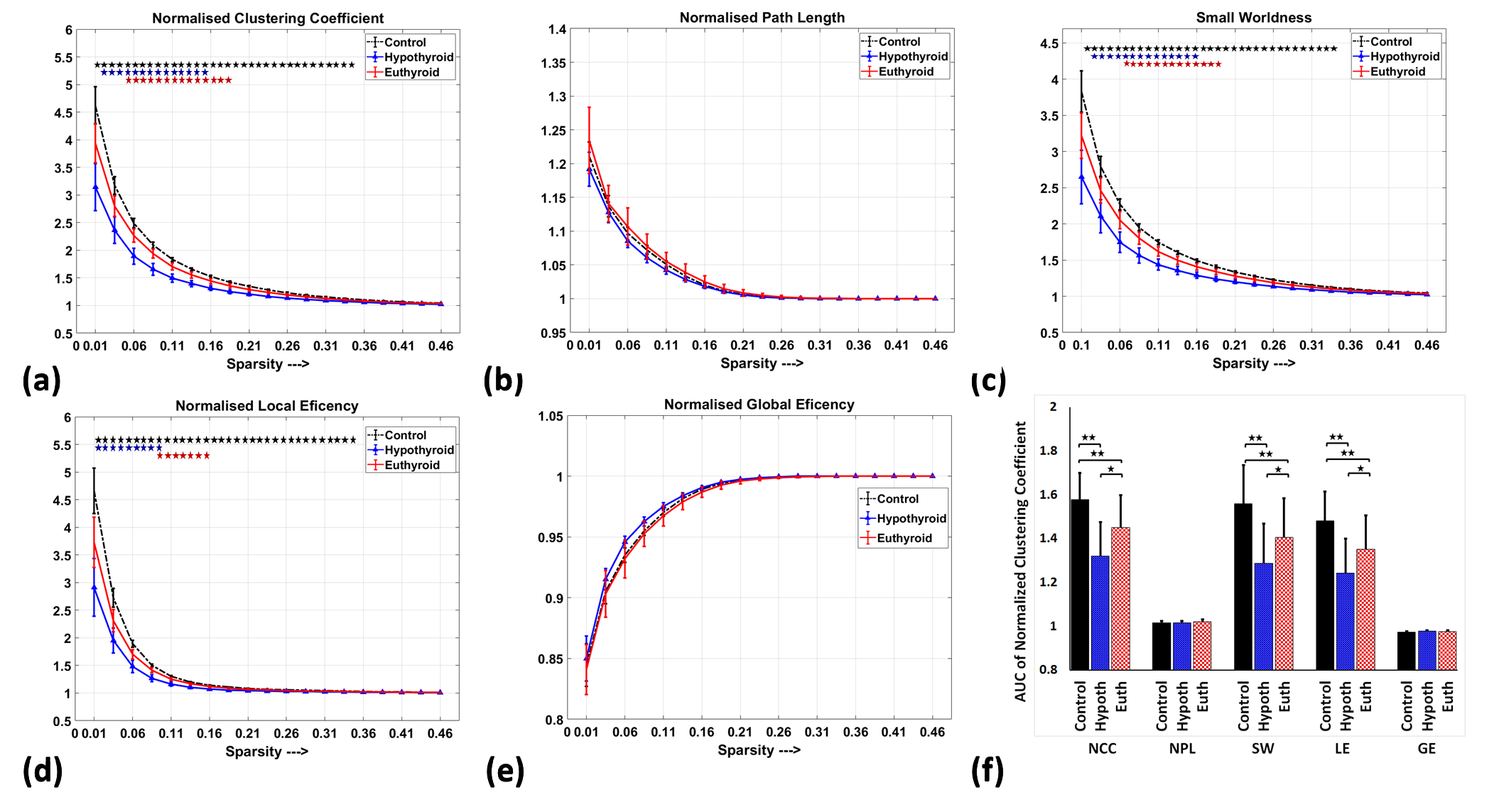
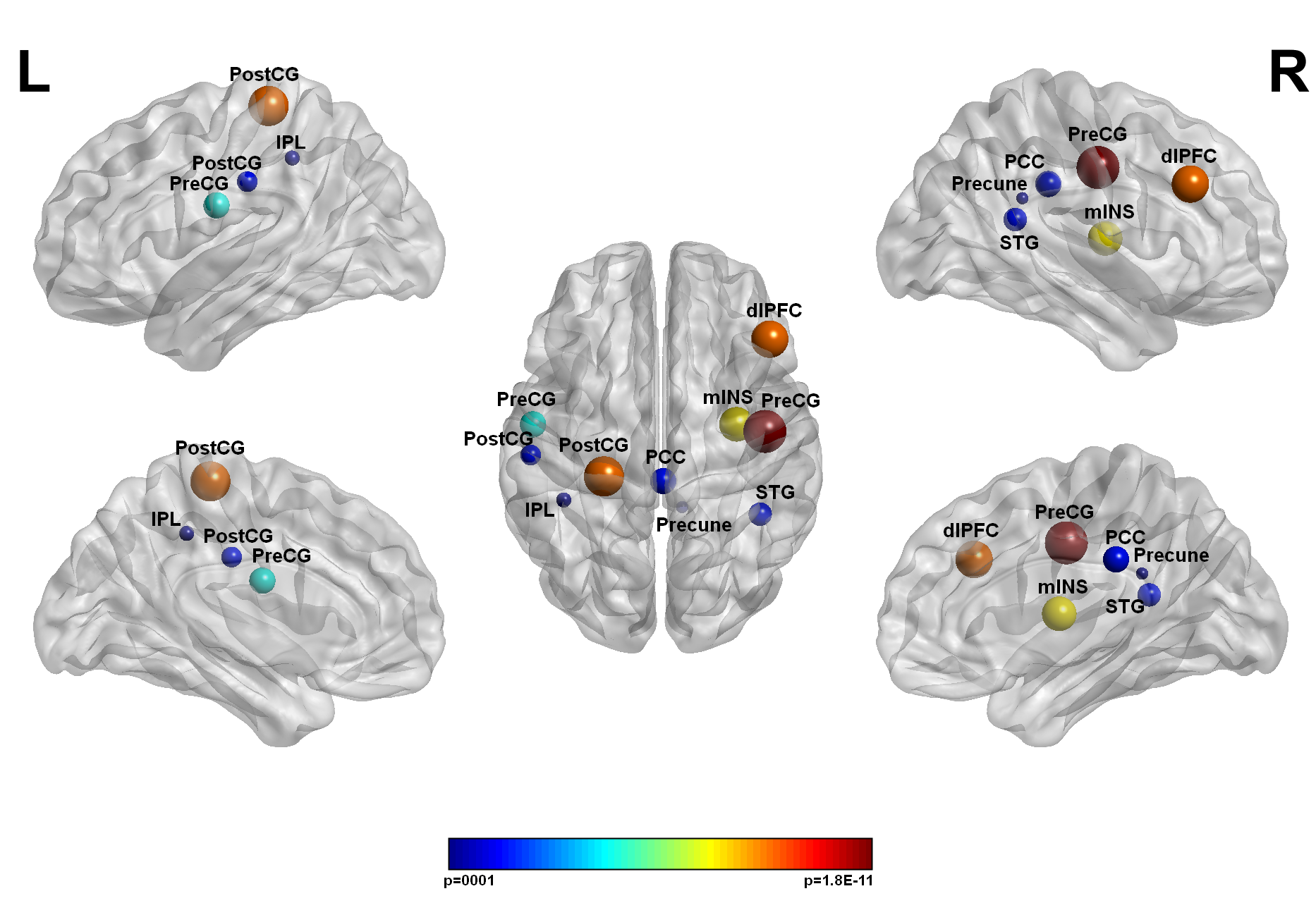
Our finding revealed significantly reduced clustering coefficient, small world connectivity and local network efficiency in hypothyroid subjects as compared to controls in sub-regions of frontoparietal network, default mode network, cingulo-opercular network and sensorimotor network and got reversed back in euthyroid patients in the network sub-regions of frontoparietal network (right dorsolateral prefrontal cortex), cingulo-opercular network (right middle insula) and SMN (right precentral gyrus) after thyroxin therapy (Figure1, Figure2). The clustering coefficient of left middle insula positive correlated with delay recall memory; post cingulate cortex negatively correlated with TSH; post cingulate cortex positively correlated with T4; right DLPFC positively correlate with working memory; left inferior parietal lobule (IPL) positively correlated with the recognition of objects memory.Discussion:
This is the first longitudinal study in patients with thyroid dysfunction that report alterations in graph-theoretical measures (network clustering coefficients, path length, small-worldness, local efficiency, and global efficiency) of resting-state functional connectivity and also report outcomes of the therapeutic intervention. Our finding demonstrates decreased local interconnectivity in the sub-regions of the cingulo-opercular network and frontoparietal network. Both networks play an important role in several cognitive domains and increase local interconnectivity within-network region rise cognitive performance3. Our correlation results demonstrate the impairment in working memory and executive function in hypothyroid patients. Decreased local interconnectivity in DMN correlated with TSH and T4. Decreased local interconnectivity in DMN might be modulating mind, cognition, and mood in hypothyroid patients. Decreased local interconnectivity in SMN might underlie disruption of involuntary movement, motor function alteration and psychomotor slowness in hypothyroid patients4. Our findings are in line with an earlier task-based functional study in thyroid dysfunction5,6. Abnormalities in brain local functional connectivity as obtained in our study might underlie the impairment in delay recall memory, working memory, motor function, mood and executive functions in hypothyroid patients. After thyroxin therapy, the local interconnectivity in the regions associated with the CON (right middle insula cortex), frontoparietal network (DLPFC) and SMN (right precentral gyrus) in euthyroid hypothyroid patients. These findings suggest that cognitive function improves after thyroxin therapy.Conclusion:
The overall study concluded that cognitive functions such as memory, motor functions, executive functions, psychomotor functions, and attention are impaired in hypothyroidism and improve after suitable therapeutic intervention. In addition correlation analysis between clinical and behavioral measures with functional connectivity, changes provide interesting insights into our understanding of the action of thyroid hormone on the functioning of the adult human brain that may aid in the better clinical management of these patients.Acknowledgements
We acknowledge that this research was supported by the 'Defense Research and Development Organization' (DRDO), Ministry of Defense, Government of India.References
1. Bernal J, Guadaño-Ferraz A, Morte B. Perspectives in the study of thyroid hormone action on brain development and function. Thyroid. 2003 Nov 1; 13 (11):1005-12.
2. Dosenbach NU, Nardos B et al. Prediction of individual brain maturity using fMRI. Science. 2010 Sep 10; 329(5997):1358-61.
3. Dosenbach NU, Fair DA et al. Distinct brain networks for adaptive and stable task control in humans. Proceedings of the National Academy of Sciences. 2007 Jun 26; 104 (26):11073-8.
4. Khushu S, Kumaran SS et al. Cortical activation during finger tapping in thyroid dysfunction: a functional magnetic resonance imaging study. Journal of biosciences. 2006 Dec 1;31(5):543-50
5. Yin JJ, Liao LM et al. Spatial working memory impairment in subclinical hypothyroidism: an FMRI study. Neuroendocrinology. 2013;97(3):260-70.
6. He XS, Ma N et al. Functional magnetic resource imaging assessment of altered brain function in hypothyroidism during working memory processing. European journal of endocrinology. 2011 Jun 1; 164 (6):951-9.
Figures