3945
Probing human somatosensory cortex using ultra-high field (7T) fMRI and 3D-printed fractal textures
Alexander M Puckett1, Zoey Isherwood2, Ashley York1, Catherine Viengkham3, and Branka Spehar3
1University of Queensland, Brisbane, Australia, 2University of Wollongong, Wollongong, Australia, 3University of New South Wales, Sydney, Australia
1University of Queensland, Brisbane, Australia, 2University of Wollongong, Wollongong, Australia, 3University of New South Wales, Sydney, Australia
Synopsis
Recent advances in fMRI, particularly accelerated image acquisition and ultra-high field (7T), have enabled high-resolution measurements and thus have provided the capability to non-invasively study the organization and function of smaller cortical areas in humans. One such area is primary somatosensory cortex, S1, in which the neuronal processing of tactile information largely occurs. Here we used 7T fMRI to (1) locate and map the fingertip representations in human S1 and then (2) to examine the activity at these locations in response to touching 3D-printed fractal textures. Robust and overlapping activation was seen across both conditions throughout primary somatosensory cortex.
INTRODUCTION
Our understanding of human somatosensory cortex is limited compared to other, more well-studied sensory cortices (e.g., visual and auditory regions). One reason for this is that primary somatosensory cortex (S1) is a relatively small region, and hence more difficult to non-invasively access in awake and behaving humans. Although there have been PET studies of S1 processing1 dating back to the 1980’s as well as conventional resolution fMRI studies2 in the subsequent couple decades, it wasn’t until the advent of high-resolution (sub-millimeter) fMRI that it became possible to map the finer details of human S1 – such as the somatotopic representations of individual body parts (e.g., fingertips)3. Another factor limiting somatosensory research has been the difficulty associated with constructing well-controlled and appropriate stimuli. Natural objects vary across many dimensions making it difficult to compare responses to different materials, and simple man-made stimuli such as tactile gratings often lack ecological validity. Recent advances in 3D-printing offers a way forward by permitting the generation and modification of a wide variety of textures and objects - all using MR-safe materials.Natural forms, often characterized by irregularity and roughness, have a unique complexity that exhibit self-similarity across different spatial scales or levels of magnification4. This fractal-like scaling characteristic is ubiquitous in many physical and biological domains, and recent research has indicated that the cortex – at least in response to visual stimuli – appear to be tuned for the particular fractal structure found in nature5. Despite both the high ecological validity of fractal structures and the prominent role that the scale-specific processing plays in various sensory modalities, the fractal-scaling framework is largely absent in both research and theory of perceptual processing and experience. Here we aimed to investigate the possible utility of 3D-printed fractal textures for probing processes in human somatosensory cortex using 7T fMRI.
METHODS
Data were acquired on a MAGNETOM 7T whole-body research scanner (Siemens Healthcare) with a 32-channel head coil. Whole-brain anatomical images were collected using an MP2RAGE sequence with an isotropic voxel size of 0.5mm. All functional data were collected using a 3D-EPI sequence6 with an isotropic voxel size of 0.8mm and an effective volume TR of 2s. Models of the 3D tactile stimuli4 were generated following the three steps illustrated in Figure 1. First, a series of 2D, 1/f noise images with varying amplitude spectra slopes were created (top row). Next, to create 3D solids (middle row), the tonal values of each pixel in the image was converted to a height value in three-dimensional space. These models were then 3D printed onto one face of a 10 x 10 x 1cm hard, MR-safe, synthetic block (bottom row). Note that the scale invariance of natural scenes can be captured by a geometric scaling parameter known as the fractal dimension (D). The D values of the textures used in our MRI-experiment were: 1.25, 1.50, 1.75, 2.00, 2.25, and 2.50.Each subject (n=3) underwent two scan sessions: (1) somatotopic digit mapping and (2) the fractal touch experiment. For digit mapping: the 4 fingertips (index, middle, ring, and little) on the right hand were stimulated sequentially using a phase-encoded design7. To this end, a single fingertip was stimulated using a piezoelectric stimulator (Figure 2A) for 8s before moving to the next. Stimulation began at the first finger (index) and ended with the fourth finger (little). Stimulation then returned to the first finger to begin another cycle of stimulation. The frequency of vibrotactile stimulation changed every 2s, and three different frequencies were used (5, 20, and 100Hz). For the fractal touch experiment, the 6 different textured stimuli were affixed around the edge of a disk (Figure 2B). During each run, the participant was prompted by visual cues to stroke one of the textured stimuli using all four fingers in an ON/OFF manner. The participant then rotated the disk clockwise or counter-clockwise and repeated until all 6 textured stimuli had been used. Four runs were collected, two clockwise and two counter-clockwise – presented in alternating order. The order of the stimuli was randomized across participants.
RESULTS
The 3D-printed fractal textures were found to be suitable and effective for use in the MR-environment. Simple stroking of the blocks elicited robust signals throughout primary somatosensory cortex for each participant and these overlapped with the individual digit representations defined using the data from the somatotopic mapping condition (Figure 3). Despite the robust activation elicited by stroking the textured stimuli, we found no evidence at the individual level that the BOLD activity in S1 was sensitive to the fractal dimension of the stimuli. Instead, we found that all stimulus textures elicited similar levels of activation (Figure 4).DISCUSSION
This study shows that 3D-printed fractal textures can be used to elicit strong and wide-spread activity across the digit representations in primary somatosensory cortex. The strength of activation was found to be similar across all textures suggesting that S1, or at least the BOLD signal therein, is not sensitive to the fractal dimension within the range tested. Although we demonstrate its utility for generating fractal textures, 3D-printing is flexible and well-suited for producing a range of other interesting textures and objects for use in the MR-environment.Acknowledgements
We thank Aiman Al-Najjar, Nicole Atcheson, Saskia Bollmann, and Markus Barth for help with data collection, and the authors acknowledge the facilities of the National Imaging Facility at the Centre for Advanced Imaging, University of Queensland. This work was supported by the Australian Research Council (DE180100433) as well as the National Health and Medical Research Council (APP 1088419).References
- Greenberg J H, et al. Metabolic mapping of functional activity in human subjects with the [18F] fluorodeoxyglucose technique. Science 212, 678-680 (1981)
- Gelnar P A, et al. Fingertip representation in the human somatosensory cortex: an fMRI study. Neuroimage 7, 261-283 (1998).
- Sanchez-Panchuelo R M, et al. Mapping human somatosensory cortex in individual subjects with 7T functional MRI. J. Neurophysiol 103, 2544-2556 (2010).
- Viengkham C, et al. Fractal-scaling properties as aesthetic primitives in vision and touch. Axiomathes (2019).
- Isherwood Z J, et al. The tuning of human visual cortex to variations in the 1/fa amplitude spectra and fractal properties of synthetic noise images. Neuroimage (2017).
- Poser B A, et al. CAIPIRINHA-accelerated 3D EPI for temporal and/or spatial resolution EPI acquisitions. Proc. Of the ISMRM (2014).
- Puckett A M, et al. Measuring the effects of attention to individual fingertips in somatosensory cortex using ultra-high field (7T) fMRI. Neuroimage (2017).
Figures

Figure 1. Generating the tactile
stimuli. Images of grayscale 1/f noise images with
varying amplitude spectra slopes (top row), computer-generated 3D solids
(middle row), and the final 3D printed tactile surfaces (bottom row). Adapted
from Viengkham et al., 2019.
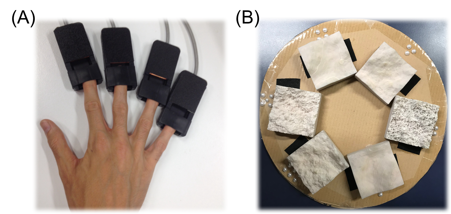
Figure 2. Set-up of
stimuli for MRI experiment. (A) A set of four
piezoelectric vibrotactile stimuli were used to map the digit representations
in S1. (B) A set of 6 fractal textures were affixed around the edge of a disk,
which was then placed on participant’s lap during the fMRI scan.
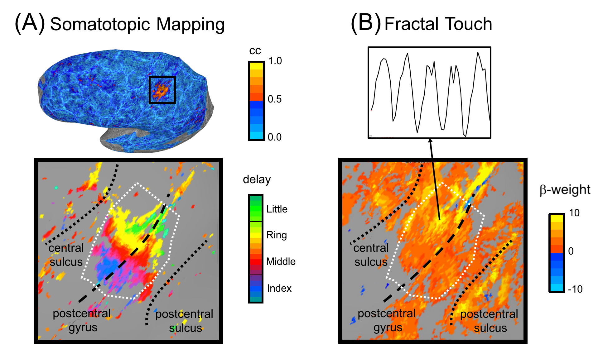
Figure 3. Somatotopic fingertip mapping and the fractal touch
experiment elicit overlapping activity. (A) Somatotopic
mapping data projected onto an inflated cortical surface model. Top shows an un-thresholded activation map.
Bottom shows the fingertip map within a zoomed in area around the post-central
gyrus. White dashed line illustrates the S1 ROI boundary. (B) Bottom shows the activation
map in and near S1 during the fractal touch experiment, thresholded at a
FDR-corrected q < 0.001. An example of a single voxel time-course is shown
above.
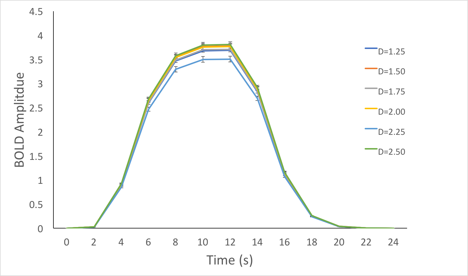
Figure 4. Similar levels
of activity were elicited in S1 regardless of fractal dimension. Mean response estimated for each of the 6 different fractal textures
(defined by their fractal dimension, D). Error bars represent SEM across the S1
voxels.