3943
Chemically-Induced Hypothermia Altered Hypothalamic Functional Connectivity with Limbic System in the Monkey Brain1Yerkes Imaging Center, Yerkes National Primate Research Center, Emory University, Atlanta, GA, United States, 2Department of Anesthesiology, Emory University School of Medicine, Atlanta, GA, United States, 32Department of Anesthesiology, Emory University School of Medicine, Atlanta, GA, United States, 4Center for Visual and Neurocognitive Rehabilitation, , Atlanta Veterans Affairs Medical Center, Decatur, GA, United States, 5Division of Neuropharmacology and Neurologic Diseases, Yerkes National Primate Research Center, Emory University, Atlanta, GA, United States
Synopsis
Rhesus monkeys were used to investigate whether ABS-201 induces similar hypothermia effect in large animals as seen in rodents and how this drug influences the hypothalamus centered limbic functional network. Functional connectivity (FC) between anterior hypothalamus and limbic system was examined by using resting state fMRI (rsfMRI). It is found that ABS-201 caused significant hypothermia effects in monkeys and significantly decreased hypothalamic FC with limbic regions as well. The findings suggest ABS-201 may have profound effects on the neurobehavior of subjects mediated by the hypothermia and antipsychotic effect.
Introduction
Prior rodent studies suggest that ABS-201, one of the NTR1 agonists, can induce mild-to-moderate hypothermia in a dose-dependent manner and has the potential for neuroprotection in stroke injury [1, 2]. The compound acts presumably on the hypothalamus and can induce regulated hypothermia minutes after administration without triggering shivering [2]. Given that the hypothalamus is known as an important area involved in temperature regulation [3, 4], we hypothesized that functional connectivity (FC) between the hypothalamus and other brain regions would be influenced after administration of ABS-201. In this study, rhesus monkeys, as a highly translational model of stroke, were used to investigate whether ABS-201 can produce hypothermia effect in large animals and how the hypothalamus centered limbic functional network is affected.Methods
Five rhesus monkeys (3 females and 2 males, 11-18 years old, 8-10kg) were anesthetized with 1% isoflurane and scanned on a Siemens 3T TIM Trio scanner with an 8-channel Tx/Rx volume coil. Physiological parameters (such as O2 saturation, blood pressure, heart rate, respiration rate, body temperature, and end-tidal CO2) were monitored continuously and maintained in normal ranges during each scanning session. Resting state functional MRI (rsfMRI) data were acquired using the single-shot EPI sequence with TR/TE=2060ms/25ms, FOV = 96 mm × 96 mm, spatial resolution= 1.5×1.5×1.5mm3, 32 contiguous slices, 232 volumes (acquisition time = 8 minutes). The rsfMRI data were collected at baseline (97.5˚F) and then repeated at the end of session (>2 hours after administration). The solution of 20 ml saline with/without ABS-201 (50mg/kg) was infused into monkey over 20 minutes after the baseline scan was completed. T1 weighted images and field map images were acquired also. The rsfMRI data were preprocessed for image distortion correction using the FSL software. Temporal filtering (0.009 Hz ~0.0237 Hz) [5] , spatial smoothing (FWHM = 2.5mm) and normalizing individual brains to a template brain [6] were performed with AFNI. The averaged time courses of rsfMRI signal in anterior hypothalamus (HypothA, Fig.2A) was used for seed-based correlation analysis. Z transformation was applied to the individual correlation maps to show normalized correlation. The averaged z values of functional connectivity (FC) between hypothalamus and posterior cingulate cortex (PCC), anterior cingulated cortex (ACC), amygdala (Amy), hippocampus (hippo), posterior hypothalamus (hypothP) and orbitofrontal cortex (OFC) were examined. Multivariate analysis of variance (MANOVA) in SPSS 26.0was conducted to compare FC results at the baseline (before saline or ABS-201 infusion) or the end time point (After, the final resfMRI scan after saline or ABS-201 infusion) across the saline and drug groups; LSD (Least significant difference) Post Hoc test were conducted to perform pair-wise comparison. Spearman correlation was carried out to measure the correlation between temperature and FC in PCC and other brain regions. P-values less than 0.05 were considered statistically significant.Results
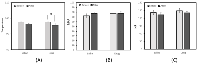
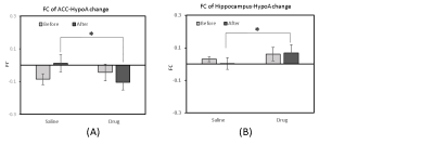
The body temperature of monkeys showed significant decrease after drug infusion 1- 3 hours compared to baseline (Before vs After, Fig.1A). No significant changes in mean arterial pressure (MAP) and heart rate (HR) were observed after drug infusion (Fig.1 B and C). Significantly decreased hypothalamic FC with limbic regions (including ACC (Fig. 2B, Fig. 3A) and Hippo (Fig.2C, Fig.3B)) were seen in monkeys after drug administration. Significant correlation between FC in HypothA- PCC and temperature change is seen in saline group but not drug group. In addition, the correlation between FC in HypothA- Amy and HypothA- OFC and temperature changes indicated obvious difference between saline and drug groups though significant statistic result were not detected probably due to the small sample size.Discussion and conclusion
ABS-201 showed hypothermia and neuroprotective effects and improved functional recovery after stroke and traumatic brain injury as reported in previous rodent studies [1, 2]. The present results suggest that ABS-201 caused similar hypothermia effect in adult rhesus monkeys although the effect is milder than that seen in rodents [1, 2]. Alteration in functional connections was observed between the hypothalamus and limbic areas (including ACC and Hippo which usually regulate emotion expression functions) [7]. The disappeared correlation of FC in PCC-HypothA, increased correlations between FC in HypothA-Amy and HypothA-OFC and temperature change further indicated the relationship between hypothalamus and limbic areas (Amy, OFC) might be affected by ABS-201. Since limbic system has intimate relationship with psychiatric disorders and ABS-201 has been found significantly improved post-stroke depression/anxiety-like symptom in mice [8], the altered FC between hypothalamus and limbic areas suggests that ABS-201 may have profound effects on hypothermia and antipsychotic effect in monkeys as well. Further investigation in stroke models is needed to elucidate the underlying neuroprotection mechanism of hypothermia and antipsychotic effect induced by ABS-201.Acknowledgements
The project was funded by NIH/STTR 2R44NS073378-05 (Yu) and P51-OD011132.References
1. Lee, J.H., et al., Therapeutic effects of pharmacologically induced hypothermia against traumatic brain injury in mice. J Neurotrauma, 2014. 31(16): p. 1417-30.
2. Lee, J.H., et al., Improved Therapeutic Benefits by Combining Physical Cooling With Pharmacological Hypothermia After Severe Stroke in Rats. Stroke, 2016. 47(7): p. 1907-13.
3. Dampney, R.A., Brain control of body temperature: Central command vs feedback. Temperature (Austin), 2016. 3(3): p. 362-363.
4. Morrison, S.F., Central control of body temperature. F1000Res, 2016. 5.
5. Li, C.X. and X. Zhang, Effects of Long-Duration Administration of 1% Isoflurane on Resting Cerebral Blood Flow and Default Mode Network in Macaque Monkeys. Brain Connect, 2017. 7(2): p. 98-105.
6. Rohlfing, T., et al., The INIA19 template and NeuroMaps atlas for primate brain image parcellation and spatial normalization. Frontiers in Neuroinformatics, 2012(6).
7. Rajmohan, V. and E. Mohandas, The limbic system. Indian J Psychiatry, 2007. 49(2): p. 132-9.
8. Weiwei Zhong, Y.Y., Xiaohuan Gu, Samuel In-young Kim, Ryan Chin, Modupe Loye, Thomas A Dix, Ling Wei, Shan Ping Yu, Neuropsychological Deficits Chronically Developed after Focal Ischemic Stroke and Beneficial Effects of Pharmacological Hypothermia in the Mouse. Aging and disease, 2020. 11(1).
Figures


