3917
Assessing drug cue-induced brain response in heroin dependents treated by methadone maintenance and protracted abstinence measures one year1radiology department, Tangdu Hospital, the Fourth Military Medical University, Xi'an, China
Synopsis
Our research aims to compare PA with MMT to reveal which abstinence way is better to recover the brain function in heroin-dependent individuals. Twenty-four heroin-dependent male males with about 12 months of PA, 21 heroin-dependent male patients stabilized on MMT for about 12 months and 20 demographically HC completed an event-related fMRI task including heroin-related and neutral cues. In the last part of this study, we proved PA is closer to HC group. This study showed PA is more beneficial for the heroin-dependent patients to lower the salience value of drug related cues, in turn to reduce relapse risks.
Introduction
Addiction, the most severe form of substance use disorder, is a chronic brain disorder molded by strong biosocial factors that has devastating consequences to individuals and to society[1].Heroin addiction in particular has become an increasingly serious problem for China in recent years[2].Methadone maintenance treatment (MMT) and protracted abstinence (PA) are common methods of therapy in heroin addiction as both suppress the craving for drug use. However, the difference in patterns of brain function between two groups is unknown. We combined fMRI and drug cue-induced task to explore PA and MMT for heroin-dependent and compare the differences between subjective craving and brain response to heroin-related cues in heroin-dependent males, and to evaluate the curative effect of these two methods in treatment.Methods
24 heroin-dependent males with about 12 months of PA, 21 heroin-dependent male patients stabilized on MMT for about 12 months and 20 demographically matched healthy controls (HC) completed an event-related fMRI task including heroin-related and neutral cues.There were two types of image cues, including 24 heroin related and 24 neutral image cues. The former included images of heroin injection, paraphernalia and preparation, and the latter included household objects or chores. Differences among the three groups were analyzed with respect to heroin cue induced brain responses. The fMRI data were acquired by a 3.0T MRI, preprocessed and analyzed by DPABI 4.3 and SPM12 software package.Results
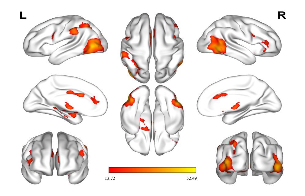
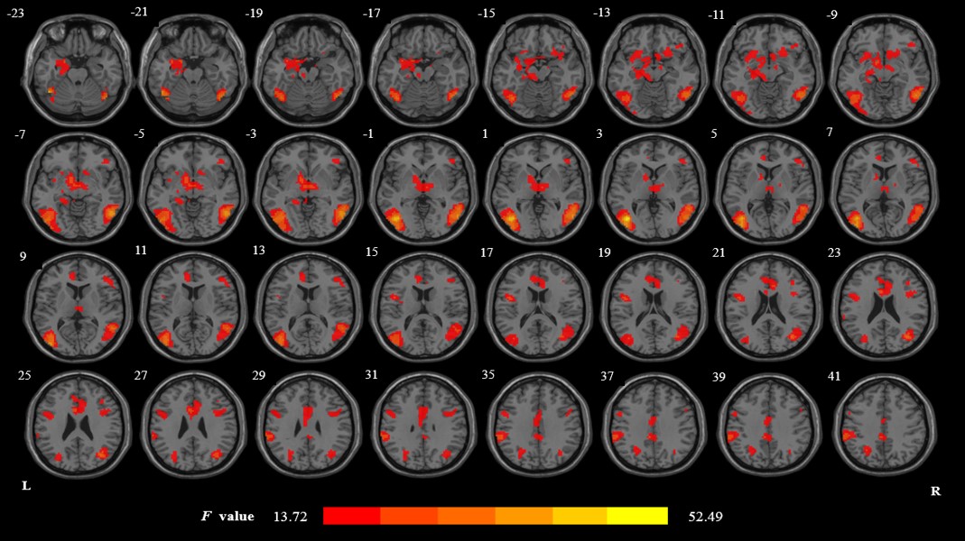
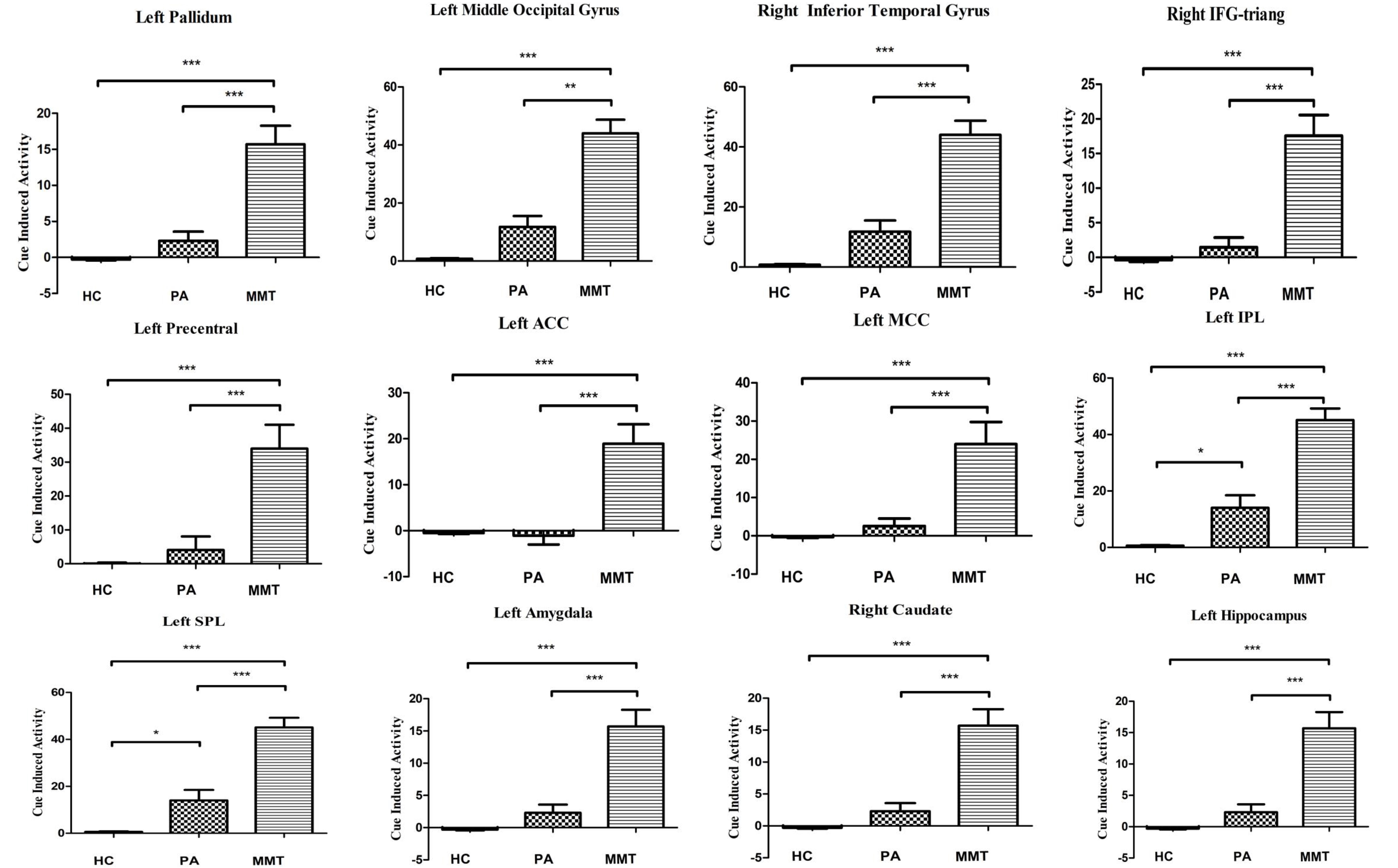
Under heroin-related > neutral cues, MMT, PA and HC group in drug cues have a statistically significant difference in the following brain areas (P<0.05, GRF correction): the limbic area, reward-related brain area, visual spatial attention area, conditioned memory area, executive control area, somatosensory cortex and auditory cortex (Fig.1 and Fig.2).Post-hoc analysis showed that compared with HC and PA groups, MMT group commonly demonstrated significantly higher brain responses during exposure of heroin-related cues in above regions. However, there was no difference between PA and HC group (Fig.3 ). No difference in cue induced craving between MMT and PA groups was found.Discussion and Conclusion
In previous reports, little research was compared one year PA with one year MMT, and little known which therapeutic method is help patients to get good withdrawal effect during this time [3] [4]. Here, we obtained new results about this. We know that the four classical neural circuits associated with addiction include reward circuit, motivation/drive circuit, memory and learning circuit and cognitive control circuit [5]. Building on professor Volkow’s classical circuitry, as the study progressed, it was found that some brain regions such as Auditory cortex regions, visuospatial-attention regions. They have participated in the formation of addiction and enriched the theoretical system of addiction research.Our data showed that, compared with PA group, the MMT group demonstrated higher cue-induced activation in the mesolimbic regions, reward-related brain regions, visual spatial attention regions, executive control regions, conditioned memory regions, somatosensory cortex and auditory cortex regions. The findings suggest that MMT and PA have different effect on patterns of brain response to heroin related cues in heroin-dependent individuals, by contrast, PA is more beneficial for the heroin-dependent patients to lower the salience value of drug related cues, in turn to reduce relapse risks.Acknowledgements
We thank Mr. Xinhai Wu for contributions to the recruitment of heroin-dependent subjects.References
1. Volkow, N. D., & Boyle, M.Neuroscience of Addiction: Relevance to Prevention and Treatment. Am J Psychiatry.2018;175(8):729-740.
2. Xiao, Z., Lee, T., Zhang, J. X. ,et al. Thirsty heroin addicts show different fMRI activations when exposed to water-related and drug-related cues. Drug Alcohol Depend.2006;83(2):157-162.
3. Qiang Li, Yarong Wang, Yi Zhang et al. Craving correlates with mesolimbic responses to heroin-related cues in short-term abstinence from heroin: An event-related fMRI study. Brain Res.2012;21(8):1469:63-72.
4. Xuan Wei,Wei Li, Wei Wang, et al. Assessing drug cue-induced brain response in heroin dependents treated by methadonemaintenance and protracted abstinence measures. Brain Imaging and Behavior 2019,2.doi:10.1007/s11682-019-00051-55.
5. Volkow, N. D., Wang, G. J., Fowler, J. S., et al. Addiction: beyond dopamine reward circuitry. Proceedings of the National Academy of Sciences.2011,108(37):15037–15042.
Figures