3915
Large-scale network abnormality in bipolar disorder: A multimodal meta-analysis of resting-state functional connectivity and VBM1Medical Imaging Center, First Affiliated Hospital of Jinan University, Guangzhou, China, 2Department of Radiology, Six Affiliated Hospital of Sun Yat-sen University, Guangzhou, China, 3Department of Applied Psychology, Guangdong University of Foreign Studies, Guangzhou, China, 4MR Research, GE Healthcare, Beijing, China
Synopsis
The present study is the first meta-analysis to integrate these diverse seed-based whole-brain resting-state functional connectivity (rs-FC) and voxel-based morphometry (VBM) results in bipolar disorder (BD) that exhibits abnormal connectivity within and between brain networks involved in internally oriented attention, processing of emotion or salience, goal-directed regulation of these functions, gating information, and sensorimotor processing. These findings motivate a large-scale neurocognitive model in which network abnormality is tightly linked to deficits in maintaining the integrated self (DN), processing of emotion (AN) or salience (VAN), and goal-directed regulation (FN) in BD.
Introduction
BD is one of the most debilitating mental disorders. The incidence is as high as 4-5%, and about 90% patients will relapse.1 However, due to the clinical characteristics of BD, this high incidence is often not recognized, resulting in inadequate treatment, huge medical costs, and high co-morbidity. In recent years, brain imaging and neuropathological studies have demonstrated structural and functional brain changes in BD. Such changes has been greatly advanced by attempts to define the distinct brain regions, their interregional connectivity, and their specific function and/or physiology which affect the large-scale brain systems.2-4 While, changes in large-scale systems in BD are reflected by intrinsic disorganized network communication, which, in turn, have been demonstrated for several modalities, but a robust conclusion has not yet been obtained.Methods
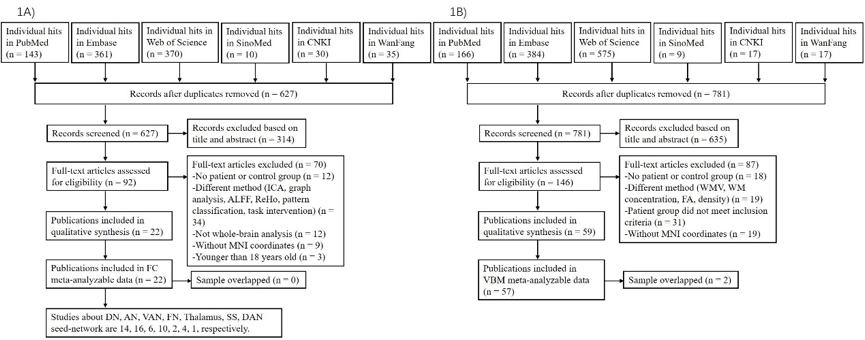
Whole-brain seed-based rs-FC and VBM studies comparing individuals with BD and healthy controls (HCs) (published before May 3, 2019) were retrieved from multiple electronic databases (PubMed, Embase, Web of Science, SinoMed, Chinese National Knowledge Infrastructure (CNKI), and WanFang). Then, coordinates of seed regions of interest (ROI) and between-group effects were extracted and coded. Seed ROIs were categorized into seed networks by their location within an a priori template. Multilevel kernel density analysis (MKDA) was used to identify brain networks in which BD was linked to hyper-connectivity or hypo-connectivity with each a priori network and the overlap between dysconnectivity and GMV changes.Results
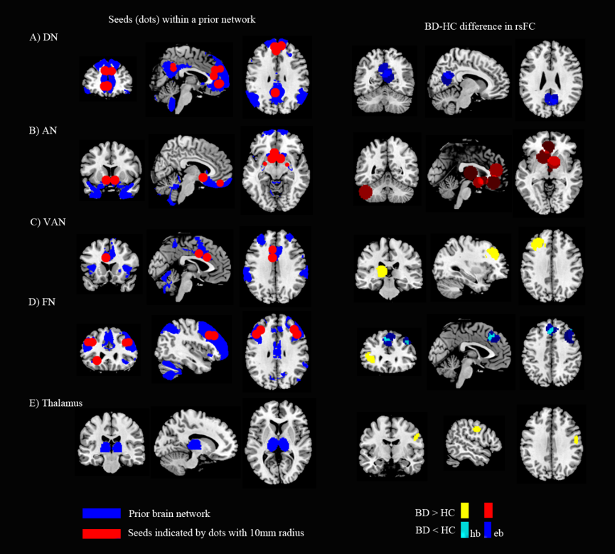
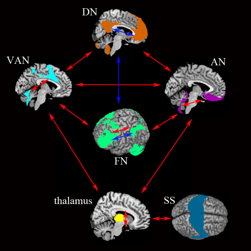
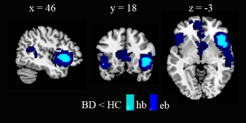
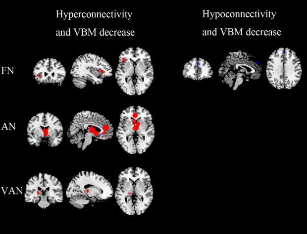
Twenty-two seed-based rs-FC publications (929 individuals with BD and 976 HCs) and 57 VBM publications (2277 individuals with BD and 2590 HCs) were included in the meta-analysis (Fig. 1). Our results showed that BD was characterized by hypo-connectivity within the DN (self-related thought), hyper-connectivity within the AN (emotion processing), VAN (processing of salience), and TN (gating information), and hypo- and hyper-connectivity within the FN (external goal-directed regulation). Additionally, hyper-connectivity between the AN and DN, AN and FN, AN and VAN, AN and TN, FN and VAN, VAN and TN, VAN and DN, and TN and SS (involved in sensory and auditory perception). The only instance of hypo-connectivity between-network in BD was observed between the FN and DN (Fig. 2, Fig.3). Decreased GMV in BD was found for DN, AN, VAN, FN, TN, and SS (Fig. 4). Finally, dysconnectivity and GMV reductions converged in the insula, ACC, mPFC, caudate, thalamus, and superior frontal gyrus (SFG) (Fig. 5).Discussion and Conclusion
To the best of our knowledge, this is the first meta-analysis to comprehensively illustrate disconnection and structure perturbations in large-scale brain networks in individuals with BD. More specifically, reduced connectivity within the DN, FN, increased connectivity within the AN, VAN, FN, and TN, , and imbalanced connectivity among the networks involved in internal thought (DN), processing of emotion (AN), salience processing (VAN), goal-direction regulation (FN), gating information (TN), and sensorimotor processing (SS) may underlie BD biases toward salient information because of the difficulty in differentiating self-produced and external world stimuli and deficits in emotional perception and regulation. Multimodal conjunction analysis found that dysconnectivity in BD converged with reduced GMV in insula, ACC, mPFC, SFG, caudate and thalamus, meaning that dysconnectivity was substantial in these regions. These findings motivate a large-scale neurocognitive model in which network abnormality is tightly linked to deficits in maintaining the integrated self (DN), processing of emotion (AN) or salience (VAN), and goal-directed regulation (FN) in BD.Acknowledgements
No acknowledgement found.References
1. Hirschfeld RM, Vornik LA. Bipolar disorder--costs and comorbidity. AM J MANAG CARE. 2005;11(3 Suppl):S85-90.
2. Brady RJ, Masters GA, Mathew IT, et al. State dependent cortico-amygdala circuit dysfunction in bipolar disorder. J Affect Disord. 2016; 201:79-87.
3. Sani G, Chiapponi C, Piras F, et al. Gray and white matter trajectories in patients with bipolar disorder. BIPOLAR DISORD. 2016;18(1):52-62.
4. Altamura AC, Maggioni E, Dhanoa T, et al. The impact of psychosis on brain anatomy in bipolar disorder: A structural MRI study. J Affect Disord. 2018; 233:100-9.
Figures