3902
APT combined DKI for differential diagnosing endometrial carcinoma from benign endometrial lesions1First affiliated hospital of dalian medical university, Dalian, China, 2Philips Healthcare, Beijing, China
Synopsis
Amide proton transfer (APT) combined diffusion kurtosis imaging (DKI) imaging technology have been preliminarily applied in the diagnosis of cervical diseases. However, there is no study on the differentiation of endometrial carcinoma and endometrial benign lesions with APT combined DKI. In this examine, we used APT combined DKI to identify endometrial carcinoma and endometrial benign lesions.
INTRODUCTION
Endometrial carcinoma is a malignant tumor of epithelium. Benign endometrial lesions mainly include endometrial polyps and endometrial hyperplasia, it is sometimes difficult to distinguish from endometrial carcinoma on the conventional sequences of MRI due to similar signals of the two diseases on conventional sequences of MRI. APT imaging technology can provide the information of high resolution free protein and amino compound proton of peptide in vivo to reflect the distribution of protein in the tumor and aid in the differential diagnosis and treatment[1], providing more detailed information for the diagnosis. Previous studies have shown that APT technology can be used to study variation of amide proton transfer signal intensity of the menstrual cycle in the normal uterus[2], and it has also been preliminarily applied to identify cervical squamous carcinomaand evaluate its differentiation grade[3].DKI adapts a kurtosis based model to depict the non-Gaussian diffusion process, which could be caused by the presence of different barriers in cellular complex structures (e.g. cell membranes and organelle compartments). DKI have been used widely, such as assessing the response to treatment in hypervascular hepatocellular carcinoma[4], assessing diagnostic accuracy with DKI in patients with breast lesions[5]. In this study, we investigated the value of APT combined DKI in the differential diagnosis ofendometrial carcinoma and endometrial benign lesions.METHODS
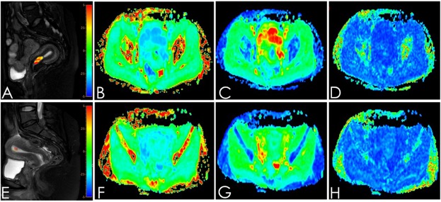
The data of 7 patients with endometrial carcinoma and 6 patients with benignendometrial lesions (5 cases of endometrial polyps and 1 case of endometrial hyperplasia) confirmed by operation and pathology were retrospectively analyzed. All patients received 3.0T MRI examination before surgery (including the APT-weighted free-breathing 3D turbo-spin-echo sequence in the Axial orientation, where the TR/TE = 6500/8 ms, FOV = 130 mm3, voxel size = 2.0×2.0 mm3, Slice Thk=7.0, Scan Duration=5min59s; DKI: b =0, 1000 and 2000 s/mm2, in 15 directions). APT values and and DKI parameters(MD, FA) of both groups were measured by two observersusing double-blind methods. ROIs (diameter≥1.0cm) were placed on the three adjacent layers of the largest cross section of the lesions (avoid heterogeneous areas).Average value for these parametersof the three layerswas calculated and recorded. Intra-group correlation coefficient (ICC) was used to test the consistency of the two observers' measurements.The difference in these parameters between the two groups was analyzed by independent-samples T test, and the diagnostic efficacy was evaluated by ROC analysis.The ROC curves of all the parameters were drew and analyzed. Use the Logistics regression parameters of diagnostic efficacy for the these parameter association after combining the parameters step by step.RESULTS
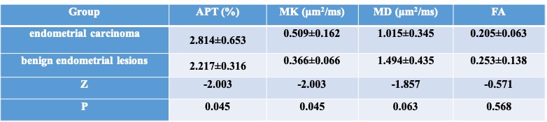
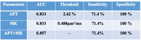
The ICC values of the APT and DKI parameters were all greater than 0.75 in the three groups, exhibiting an amenable consistency. The APT value and MK ofendometrial carcinomawere significantly higher (p < 0.05) than benign endometrial lesions, while the MD and FA values of endometrial carcinoma were lower than benign endometrial lesions(p >0.05) (Table 1). The area under the ROC curve of APT value was 0.833, When APT value ≥ 2.62 , the sensitivity and specificity were 71.4% and 100%. The area under the ROC curve of MK value was 0.833, When MK value ≥ 0.488 , the sensitivity and specificity were 71.4% and 100%.When ATP combined MK, the area under the ROC curve was 0.857, the sensitivity and specificity were 71.4% and 100%.(Table 2)DISCUSSION AND CONCLUSIONS
APT imaging combined with DKI has a high value in the differential diagnosis of endometrial carcinoma and 6 patients with benignendometrial lesions. Among the single parameter, the diagnostic efficacy of APT value and MK value were high (AUC, sensitivity and specificity of APT value were: 0.833, 71.4% and 100%; AUC, sensitivity and specificity of MK value were: 0.833, 71.4% and 100%). When APT value was combined with MK value, the diagnostic efficacy was significantly improved (AUC: 0.857; sensitivity: 92.3%; specificity: 100%). Carcinoma consists of proliferative cells which produce a large amount of free protein, leading to increased APT weighting, or it may be the tumor angiogenesis that resulted in higher APT value[6]. APT value can effectively distinguish endometrial carcinoma from benign endometrial lesions. MK value is the most representative parameter of DKI, it was the average value of multiple b values and gradient directions in the same direction. Its size depends on the complexity of the organizational structure. The more complex the structure is, the more significant the deviation of the diffusion motion of water molecules from the normal distribution is, and the larger the MK value is. Previous literature indicated that DKI has been used for assessment of uterine tumors[7]So, the combination of APT imaging and DKI had a better diagnostic performance.Acknowledgements
No acknowledgementReferences
[1] He YL, Li Y, Lin CY, et al. Three-dimensional turbo-spin-echo amide proton transfer-weighted MRI for cervical cancer: a preliminary study. J Magn Reson Imaging 2019. DOI: 10.1002/jmri.26710
[2] Zhang S, Sun H, Li B, et al. Variation of amide proton transfer signal intensity and apparent diffusion coefficient values among phases of the menstrual cycle in the normal uterus: A preliminary study. Magn Reson Imaging 2019. 63:21-28.
[3] Meng N, Wang J, Sun J, et al. Using amide proton transfer to identify cervical squamous carcinoma/adenocarcinoma and evaluate its differentiation grade. Magn Reson Imaging 2019. 61:9-15.
[4] Suo S, Cao M, Zhu W, et al. Stroke assessment with intravoxel incoherent motion diffusion-weighted MRI. Nmr in Biomedicine, 2016, 29(3):320-328.
[5] Du J, Li K, Zhang W, et al. Intravoxel Incoherent Motion MR Imaging: Comparison of Diffusion and Perfusion Characteristics for Differential Diagnosis of Soft Tissue Tumors. Medicine, 2015, 94(25).
[6] Li B,SunH, ZhangS, et al. The utility of APT and IVIM in the diagnosis and differentiation of squamous cell carcinoma of the cervix: A pilot study. MagnReson Imaging 2019. 63:105-113.
[7] Dia Aliou Amadou, Hori Masatoshi, Onishi Hiromitsu, Application of non-Gaussian water diffusional kurtosis imaging in the assessment of uterine tumors: A preliminary study. Plos One, 12(11):e0188434.
Figures