3897
Use of functional correlation tensors for correlating white matter fMRI and brain structure1Engineering Science, Simon Fraser University, Burnaby, BC, Canada, 2SFU ImageTech Lab, Health Science and Innovation, Surrey Memorial Hospital, Fraser Health, Surrey, BC, Canada, 3Cummings School of Medicine, University of Calgary, Calgary, AB, Canada, 4Biomedical Physiology and Kinesiology, Simon Fraser University, Burnaby, BC, Canada, 5Faculty of Applied Science, Simon Fraser University, Burnaby, BC, Canada, 6Djavad Mowafaghian Centre for Brain Health, University of British Columbia, Vancouver, BC, Canada
Synopsis
White matter functional activity is a neglected area of research and key component for understanding the brain’s ability to adapt and learn. Participants completed a fine motor task during functional scans. DTI images were also collected for structural comparison. Functional correlation tensors were computed to examine local functional signal synchronicity. Strong agreement was found between the functional anisotropy maps and the structural anisotropy maps. Functional correlation tensors substantiate white matter functional response and identify a novel link between structure and function.
Introduction
There is a rapidly growing body of research investigating white matter (WM) functional activity1. However, the development of a robust fMRI method for analyzing WM integrity has been neglected. An emerging technique, called functional correlation tensors (FCT), is being developed to investigate functional tractography of the brain. The pattern of hemodynamic fluctuations in each voxel are correlated with its adjacent neighbours to assess synchrony. This creates a matrix of 26 inter-voxel correlations, which can be used to create a “local spatio-temporal correlation tensor”2. The linear nature of WM tracts have been shown to exhibit an associated anisotropy in the correlation of hemodynamic fluctuations2,3. This opens a novel avenue for investigating functional WM connections. Compared to diffusion tensor imaging (DTI) that has been widely used to understand structural WM integrity, FCT can further detect WM connections of the brain at work2. Combining these MRI techniques has the potential to reveal nuanced information on the relationship between brain structure and function, providing critical information to sensitively identify a functional brain change. Here, we used fMRI data from participants trained on a motor learning task to investigate functional WM condition and motor –learning induced alteration using FCT. We also linked the FCT results to that of structural WM connections using DTI.Methods
A motor learning task was used to investigate brain function4,5. Twelve healthy, right-handed participants trained at home daily over a two-week period with a baseline, mid-point, and end-point scan. The participants guided an MRI compatible cursor through a visual trail as quickly and accurately as possible. The task was completed with both the dominant and the non-dominant hand during training and scanning. Data were acquired using a 3T Phillips Ingenia MRI. Functional MRI images were acquired using an FFE single-shot GRE-EPI sequence. The acquisition parameters were as follows: TR = 2000 ms, TE = 30 ms, and flip angle = 90°. fMRI data were preprocessed using FSL; scans were brain extracted, motion and slice time corrected, high pass filtered at 100s, and registered to the MNI152 template. Spatial smoothing was not applied. The preprocessed fMRI data and a brain mask were used to compute FCTs. A more detailed explanation of how FCTs are computed can be found in Ding et al. 20132 and Zhou et al. 20183. DTI data were acquired using a single shot EPI sequence with 32 directions and b0 of b800. DTI data was eddy current corrected then preprocessed and registered to standard space using FSL’s tract-based spatial statistics tool. The resulting FCT fractional anisotropy (FA) maps were analyzed, which, like DTI FA, is a measure of the local anisotropy of a voxel. For FCT this represents the anisotropy of local BOLD signal synchronicity. Tissue masks were computed to analyze WM and gray matter (GM) FCT FA individually.Result
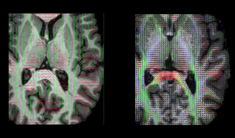
The averaged FCT FA, registered to a standard template and computed across all participants showed clear patterns, as seen in Fig 1 (left panel). Similarly, patterns were observed in the registered and average DTI FA for all participants (right panel). The correlation coefficient between DTI FA and FCT FA was R = 0.68 (p < 0.00001), indicating a strongly significant correlation between the functional and structural modalities. As expected, WM FCT FA was significantly greater than GM FCT FA (p < 0.00001) across all participants. The principle directions of local correlation for FCT and DTI are represented in Fig 2, which showed similar adherence to WM tracts.Discussion
In this study we applied FCT, a mathematical analysis to conduct a deeper investigation into whole brain functional connections. FCT revealed that GM exhibited higher isotropy, resulting in spherical tensors, whereas WM tensors are more anisotropic and smaller. This is confirmed in the existing literature on FCT2,3,6. In Fig. 1 the average fMRI FCT exhibits similar FA patterns and similar motor-learning associated changes as DTI; greater FCT FA values correspond with the WM tracts in the brain. Fig 2 shows that not only is the FCT FA in WM tracts higher, but the principle direction of FCT correlation complements the direction of brain architecture as well. This agreement between DTI and FCT demonstrates a compelling correlation between structure and function. These results substantiate earlier reports of WM BOLD signal1,7,8. The results also suggest that while GM generates a much stronger BOLD signal, local correlations are a key metric for detecting and mapping WM function. Previous work has shown that the hemodynamic response in WM can be highly variable and significantly different than GM7,8. By investigating WM BOLD signal without a hemodynamic response function, the sensitivity to WM functional response may be increased.Conclusion
FCTs have potential to significantly improve monitoring sensitivity to WM fMRI response and may improve our ability to study brain functional connections. A better understanding of functional organization of the brain may help create a key framework for integrating structural and functional information.Acknowledgements
This study received support from the Natural Sciences and Engineering Research Council Discovery Grant #206875 and from the Surrey Hospital & Outpatient Center Foundation under Grant FH2017-277 001.References
[1] J. R. Gawryluk, E. L. Mazerolle, and R. C. N. D’Arcy, “Does functional MRI detect activation in white matter? A review of emerging evidence, issues, and future directions,” Front. Neurosci., vol. 8, no. 8 JUL, pp. 1–12, 2014.
[2] Z. Ding, A. T. Newton, R. Xu, A. W. Anderson, V. L. Morgan, and J. C. Gore, “Spatio-temporal correlation tensors reveal functional structure in human brain,” PLoS One, vol. 8, no. 12, 2013.
[3] Y. Zhou et al., “Functional MRI registration with tissue-specific patch-based functional correlation tensors,” Hum. Brain Mapp., vol. 39, no. 6, pp. 2303–2316, 2018.
[4] M. V. Sale, L. B. Reid, L. Cocchi, A. M. Pagnozzi, S. E. Rose, and J. B. Mattingley, “Brain changes following four weeks of unimanual motor training: Evidence from behavior, neural stimulation, cortical thickness, and functional MRI,” Hum. Brain Mapp., vol. 38, no. 9, pp. 4773–4787, 2017.
[5] L. B. Reid, M. V. Sale, R. Cunnington, J. B. Mattingley, and S. E. Rose, “Brain changes following four weeks of unimanual motor training: Evidence from fMRI-guided diffusion MRI tractography,” Hum. Brain Mapp., vol. 38, no. 9, pp. 4302–4312, 2017.
[6] Z. Ding et al., “Visualizing functional pathways in the human brain using correlation tensors and magnetic resonance imaging,” Magn. Reson. Imaging, vol. 34, no. 1, pp. 8–17, 2016.
[7] M. Li, A. T. Newton, A. W. Anderson, Z. Ding, and J. C. Gore, “Characterization of the hemodynamic response function in white matter tracts for event-related fMRI,” Nat. Commun., vol. 10, no. 1, p. 1140, Dec. 2019.
[8] M. J.
Courtemanche, C. Sparrey, X. Song, A. MacKay, and R. C. N. D’Arcy, “Detecting
white matter activity using conventional 3 Tesla fMRI: An evaluation of
standard field strength and hemodynamic response function,” Neuroimage,
pp. 145–150, 2018.
Figures