3888
How far PFT reconstruction technique can improve spatial resolution in radial fMRI acquisition: An experimental study1School of Cognitive Science, Institute for Research in Fundamental Sciences (IPM), Tehran, Iran (Islamic Republic of), 2Biomedical Engineering, Amirkabir University of Technology, Tehran, Iran (Islamic Republic of)
Synopsis
Radial acquisition along with Polar Fourier Transform (PFT) reconstruction allows to retrospectively choose the image pixel-size. We experimentally investigated how this selected pixel-size was related to spatial resolution in fMRI studies. The functional contrast to noise ratio (CNR) was considered as a measure to assess whether the improvement in apparent spatial resolution is real or just resulted from interpolation. In an fMRI study on 9 subjects, SSFP raw data was reconstructed by PFT technique with four pixel areas. Results showed that the CNR improvement stopped at the boundaries of reduced-FOV, where the spacing in azimuthal and radial directions are equal.
Purpose
Balanced SSFP with radial acquisition and Polar Fourier Transform (PFT) reconstruction has been suggested for fMRI applications to investigate neural activity with higher spatial specificity and resolution.1, 2 Since the PFT reconstruction is performed in the polar coordinates, the pixel size in the azimuthal direction has a range, as it is proportional to the distance from the center.3 After conversion to the Cartesian coordinates, the pixel size should be selected from that range. In this study, we experimentally investigated how (and where) this pixel size represents the actual spatial resolution in the polar reconstruction technique. One measure that distinguishes between the real improvement of spatial resolution and interpolation is the functional contrast as it drops with degradation of resolution through partial volume effect.4 Hence, we used the functional contrast to noise ratio (CNR) as a validation measure to assess the improvement in spatial resolution of the data, reconstructed by PFT with different pixel sizes.Method
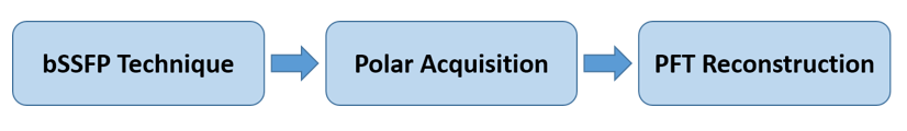
Here, we performed SSFP-fMRI experiments on healthy subjects and reconstructed each raw data with four selected pixel sizes. The CNR was then calculated for equal active areas of each subject to assess the improvement in spatial resolution. Data acquisition: fMRI studies with visual stimulus task were performed on 9 healthy subjects using bSSFP protocol with following sequence parameters: Acquisition trajectory = radial, TR/TE = 6.12/3.06 ms, squared FOV = 224 mm, number of spoke = 112, flip angle = 30°, slice thickness = 3 mm, volume TR = 4s (for 72 measurements). The data were acquired from 4 coronal slice to cover the occipital lobe, using a 3T MRI Siemens scanner. The task included 6 rest-act blocks (24s-24s) which at the act-state, a rotating checkerboard was presented to the subject. Data reconstruction: PFT reconstructed the bSSFP data in polar coordinates.3 The nearest neighbor interpolation was then applied to convert images to Cartesian grid for visualization. In this step, four pixel sizes including 2×2, 1.4×1.4, 1×1 and 0.7×0.7 mm2 were chosen for Cartesian images. The pipeline to reconstruct high-spatial bSSFP technique is presented in Fig. 1. Analysis pipeline: All the preprocessing and analysis steps were performed using FSL (FMRIB, Oxford Univ.). Motion correction, temporal filtering (cut-off frequency= 120s) and spatial smoothing (FWHM=2×pixel size) were applied on all cases as a preprocessing step. For statistical analysis, a general linear model (GLM) was used as implemented in FSL Feat. For generating activation maps, cluster-based thresholding with z-score=3 & p=0.05 was applied. To calculate the CNR in each active voxel, the absolute difference value between act and rest states of time series for that voxel was divided by its standard deviation in rest condition.Results
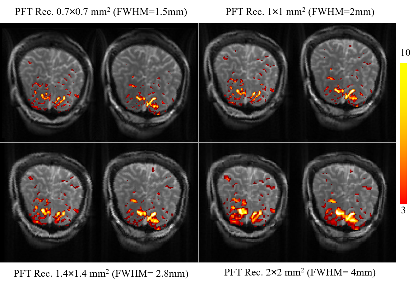
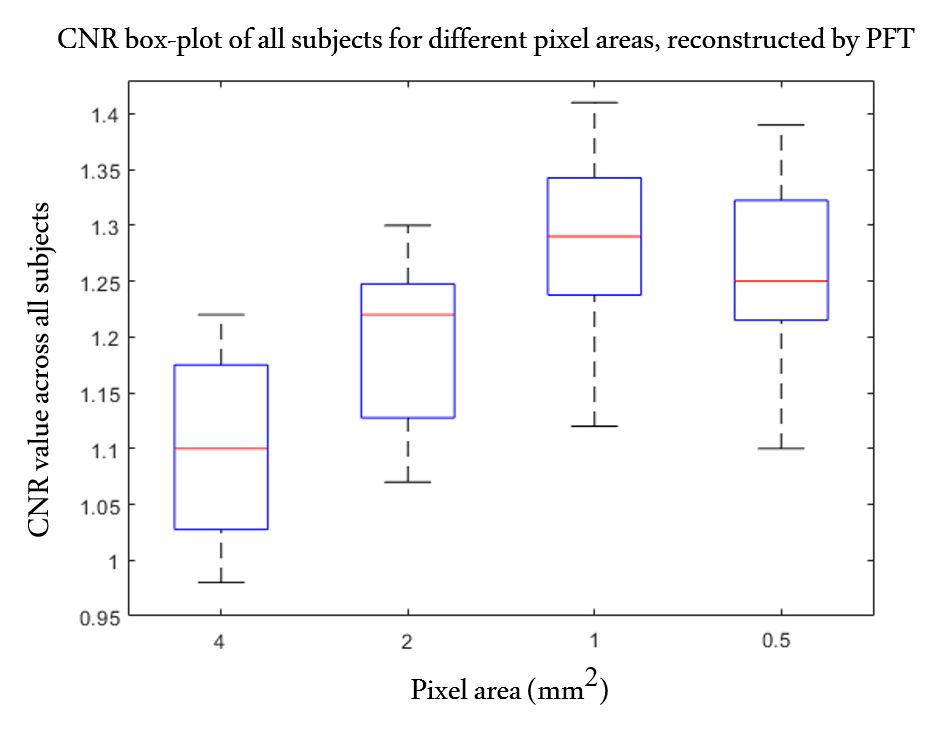
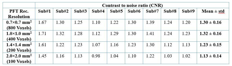
Figure 2 shows the activation maps of PFT reconstructions in four pixel sizes for a representative subject (coronal view). As can be seen in this figure, activities are getting more localized in gray-matter areas as pixel size decreases from 2 to 1 mm. However, almost no significant changes is visually found, comparing activity patterns of two smaller pixel sizes of 1 mm and 0.7 mm. For all subjects, CNR values were calculated for a fixed-area which has the highest activity. In Table 1, the CNR values for all 9 subjects along with their mean and standard deviation for each resolution were presented. According to this table and Fig. 3, mean of CNR across subjects were increased from 2 to 1mm pixel size, but remained almost constant from 1 to 0.7 mm.Discussion and conclusion
Here, we utilized PFT technique to reconstruct data with different pixel sizes. Images were processed and their functional maps were evaluated qualitatively by inspecting how well activity patterns fit in gray-matter. Results were further quantitatively assessed by measuring CNR values for equal active areas. The CNR was increased as the voxel area decreases from 4 to 2 and similarly from 2 to 1 mm2, a result that would not be expected in a simple interpolation (Table 1 & Fig. 3). Such an improvement in CNR, therefore, should be attributed mostly to the decrease of partial volume effect as observed in high resolution studies.4 Nevertheless, the CNR improvement stopped at 1 mm, which is the resolution in readout (radial) direction. This can be explained by the concept of reduced FOV (rFOV) whose boundary is determined by equal spacing in azimuthal and radial directions.5 In this study the number of spokes were chosen in a way that the rFOV encompasses the brain portion of the image. In conclusion, we experimentally investigated the limit for improving the resolution by using PFT reconstruction for radially acquired fMRI data at 3T.Acknowledgements
No acknowledgement found.References
1. Shafiei, B. & Moghaddam, A. N. (2018). Radial acquisition and PFT reconstruction allow for retrospective selection of spatial resolution in fMRI studies. International Society for Magnetic Resonance in Medicine (ISMRM 2018), France, Paris.
2. Malekian, V., Shalikar, Sh., Rastegar, F. & Moghaddam, A. N. (2019). SSFP-fMRI: Radial reading helps to Improve Temporal SNR. International Society for Magnetic Resonance in Medicine (ISMRM 2019), Canada, Montreal.
3. Golshani, S. & Moghaddam, A. N. (2017). Efficient radial tagging CMR exam: A coherent k‐space reading and image reconstruction approach. Magnetic resonance in medicine, 77(4), 1459-1472.
4. Molloy, E.K., Meyerand, M.E. & Birn, R.M. (2014). The influence of spatial resolution and smoothing on the detectability of resting-state and task fMRI. Neuroimage, 86, pp.221-230.
5. Scheffler, K. & Hennig, J. (1998). Reduced circular field‐of‐view imaging. Magnetic resonance in medicine. 40(3), 474-80.
Figures