3884
Evaluation of multiband EPI for T2*-weighted and diffusion-weighted imaging1Brain Imaging Center (BIC), Goethe University Frankfurt, Frankfurt am Main, Germany, 2Max Planck Institute for Empirical Aesthetics, Frankfurt am Main, Germany
Synopsis
The combination of multiband EPI with in-plane parallel imaging yields a total acceleration factor AF=MB*R, MB and R representing the slice and in-plane acceleration factors, respectively. The data quality of T2*-weighted and diffusion-weighted imaging was compared for different AF, using different combinations of MB and R, and for different head coils, providing advice on the optimum experimental set-up. Results suggest that severe artifacts may occur for large MB and R, a low number of slices (SL) and head coils with a reduced number of elements (NE). Suggestions comprise SL≥48, NE≥32, MB≤8 and R≤4.
Introduction
T2*-weighted and diffusion-weighted imaging (DWI) require high spatial resolution at short scan times. The combination of multiband echo-planar imaging (MB-EPI), which excites several slice groups simultaneously [1], with in-plane parallel imaging yields accelerations by the factors MB and R, respectively, without compromising spatial resolution. This study assesses the data quality of T2*-weighted imaging and DWI for different total acceleration factors AF=MB*R, using different combinations of MB and R, and for different head coils, providing advice on the optimum experimental set-up.Methods
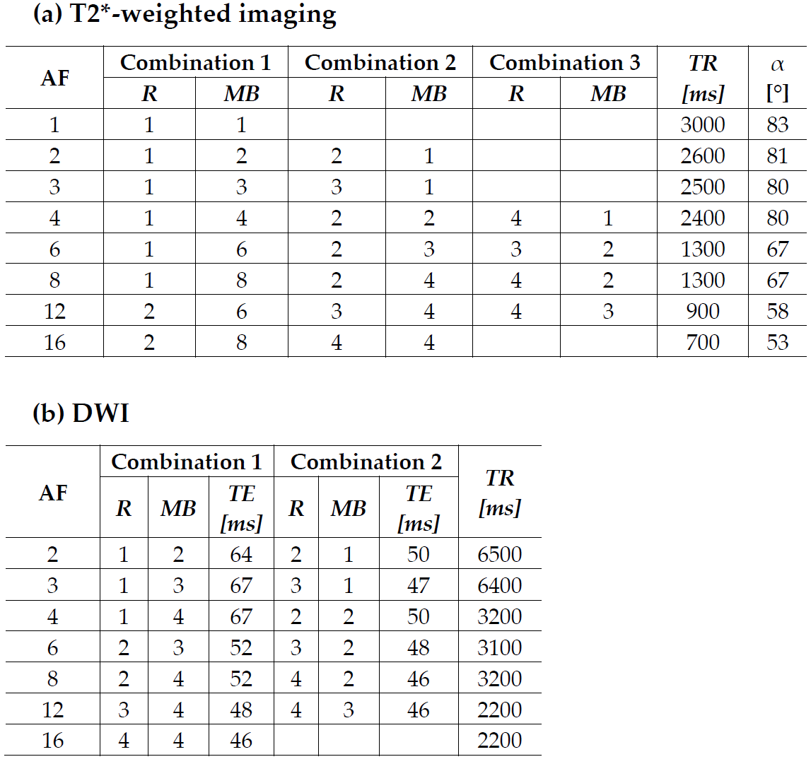
MRI experiments were performed on a 3T whole-body scanner. For T2*-weighted imaging, a body TX coil and three different phased-array RX head coils (20, 32 and 64 channels) were used. All protocols employed the MB-EPI sequence and were first tested in vitro on an agarose gel phantom. In vivo scans were performed on five healthy volunteers who gave written informed consent before participation.T2*-weighted imaging: In-plane resolution: 3mm (FoV=192×192 mm2, matrix-size=64×64), slice-thickness=2mm, interslice gap=1mm, maximum of 48 axial slices, TE=30 ms, readout bandwidth BW=2604Hz/pixel, echo-spacing=490-550µs (depending on MB and R). The different combinations of MB and R, the TR, and the flip-angle α (adjusted to the Ernst angle [2], assuming T1=1.4s) are listed for each AF in table1a.
DWI: Isotropic resolution: 2mm (FoV=224×224mm2, matrix-size=112×112, slice-thickness=2mm, no interslice gap), 72 interleaved axial slices, BW=1940Hz/pixel, echo-spacing=600µs, partial-Fourier=6/8, 30 different directions of diffusion-weighted gradients (DWG), b-value=1000s/mm2. The different combinations of MB and R, TE and TR are listed for each AF in table1b. For distortion correction, two sets of 5 reference images without diffusion weighting were acquired with positive and negative phase-encoding gradients. Fractional anisotropy (FA) maps and principal eigenvectors were calculated according to the literature [3]. For all protocols and head coils, the central slice was located at the magnet's isocenter.
Results
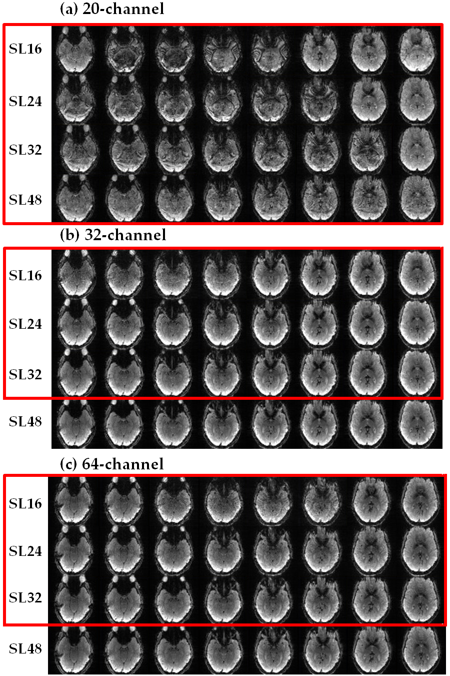
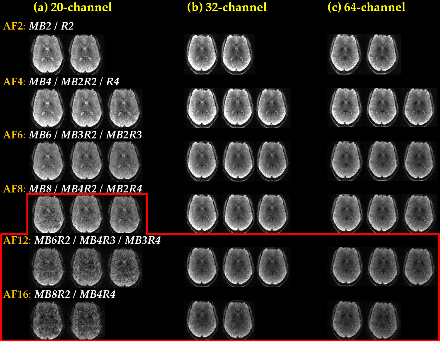
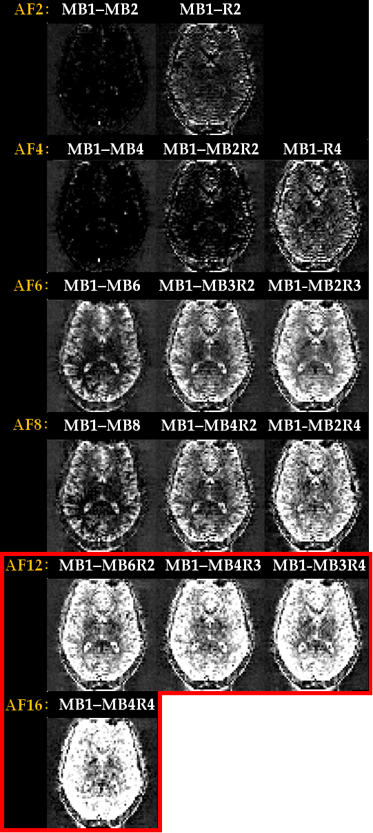
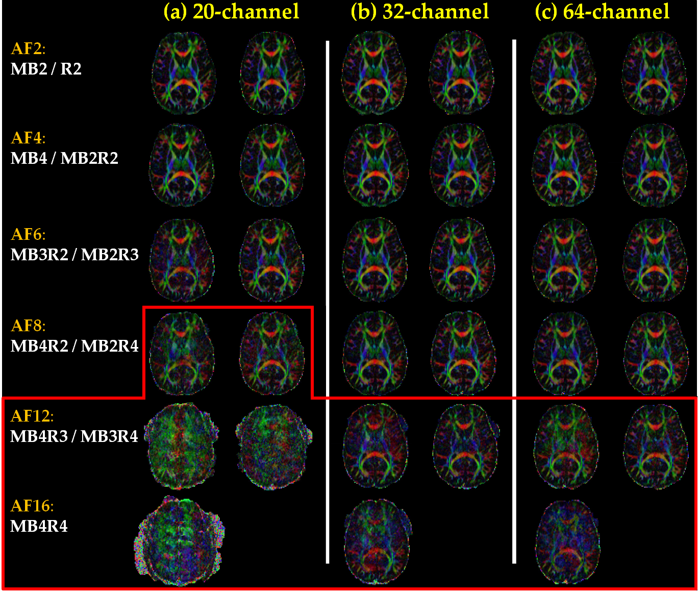
Figure 1 compares T2*-weighted data acquired with MB=8 and R=1 for different head coils (a, b, c) and different numbers of slices (SL, rows), showing for each case the central 8 slices (columns). Artifacts due to signal leakage between simultaneously excited slices are reduced by increasing SL and/or the number of coil elements (see data inside red frames). Best results are achieved with SL=48, in particular for 32 and 64 coil channels (b and c, bottom rows). Figure 2 compares T2*-weighted data (central slice only) acquired with different AF (rows) for different head coils (a, b, c) and different combinations of MB and R for each AF at constant SL=48. Artifacts and signal losses occur for large AF (data inside red frame). Best results are achieved for AF≤6 (20-channel) and AF≤8 (32- and 64-channel). Figure 3 shows for T2*-weighted data (central slice only) differences between a non-accelerated reference (MB=1, R=1) and data acquired with different AF, showing in each case different combinations of MB and R. All data were acquired with the 64-channel coil and SL=48. Signal differences are lowest for AF≤4 and moderate for 6≤AF≤8 with R=1. Severe discrepancies arise for AF≥12 (red frame). Figure 4 shows FA maps (central slice only), the color code denoting the direction of the diffusion tensor's principal eigenvector, for different head coils (a, b, c), different AF (rows) and two different combinations of MB and R for each AF (columns). Severe artifacts (red frame) arise for AF≥8 (20-channel coil) and AF≥12 (32- and 64-channel coils).Discussion/Conclusion
In this study, T2*-weighted and diffusion-weighted images (3 mm and 2 mm spatial resolution, respectively), acquired with different acceleration factors MB and R, were compared. Data show that severe artifacts may occur for large MB, a low number of slices and head coils with a reduced number of elements. The results suggest that great care should be taken when choosing optimum MB and R values for a specific protocol. Suggestions comprise a large number of slices (SL≥48), a large number of coil array channels (≥32), moderate MB (≤8) and low R (≤4).Acknowledgements
No acknowledgement found.References
[1] Feinberg DA, Setsompop K (2013). Ultra-fast MRI of the human brain with simultaneous multi-slice imaging. Journal of magnetic resonance, 229, 90-100.
[2] Ernst RR, Bodenhausen G, Wokaun A (1987). Principles of nuclear magnetic resonance in one and two dimensions. Oxford: Clarendon Press; pp. 124–125.
[3] Shrestha M, Hok P, Nöth U, Lienerth B, Deichmann R (2018). Optimization of diffusion-weighted single-refocused spin-echo EPI by reducing eddy-current artifacts and shortening the echo time. Magnetic Resonance Materials in Physics, Biology and Medicine, 31(5), 585-597.
Figures