3880
Artefact reduction in simultaneous EEG-fMRI: improving current practice.1Faculty of Medicine, Dentistry and Health Sciences, University of Melbourne, Melbourne, Australia, 2Florey Institute of Neuroscience and Mental Health, Melbourne, Australia
Synopsis
EEG recorded during fMRI is subject to artefact many times greater than neuronal events of interest, therefore, artefact removal methods are crucial for accurate EEG-fMRI studies. This work systematically reviews all novel artefact reduction methods (1998-2018), as well as the use of artefact reduction methods (2016-2018). Results show that whilst there are many published artefact reduction methods, contemporary studies overwhelmingly use only a few established methods. It is recommended that: 1. Artefact reduction techniques are adequately reported, 2. Novel software is robust to help adoption by others, and 3. Commercial EEG-fMRI vendors consider including additional hardware for recording artefact.
Background
Simultaneous EEG-fMRI is a multimodality imaging method useful for understanding brain dynamics. Its use is growing in neuroscience research - from fewer than 20 papers published per year in the early 2000s to around 80 papers per year more recently [1]. This growth is largely due to the wide range of research questions that can be investigated using simultaneous EEG-fMRI: from better understanding of epilepsy & other brain disorders, to investigating normal behaviour such as decision making or sleep onset.Scalp electroencephalography (EEG) is a widely available non-invasive technique that can detect brain activity with up to a millisecond precision, however its spatial resolution is poor and it can be less sensitive for brain structures located far from the scalp. Simultaneous acquisition of fMRI with scalp EEG provides better spatial localisation of brain activity than could be achieved with scalp EEG alone. For example, in epilepsy, simultaneous EEG-fMRI is typically used as follows: timing of epileptiform events of interest are first determined from the EEG, and these are then used in an event-related general linear model to generate fMRI maps of statistically significant activation associated with EEG events.
The biggest challenge for simultaneous EEG-fMRI is that EEG recorded during fMRI is subject to artefact many times greater than neuronal events of interest [2]. Artefacts seen on EEG include: interference from the magnetic gradient switching that occurs during fMRI acquisition (gradient artefact), artefacts related to the scanner environment such as lighting or ventilation noise (environmental artefact), and artefact from the subject themselves - due to pulse (ballistocardiogram artefact) or motion (motion artefact) during the fMRI scan. Removal of artefact from the EEG is crucial for accurate and reproducible EEG-fMRI studies.
Rationale and Purpose
Despite the well-known impacts of artefact on EEG-fMRI, and over 20 years of research to develop and improve methods for artefact reduction, we are concerned that the best methods do not seem to be widely adopted. To confirm this, in the present study we have reviewed methods for artefact reduction in EEG-fMRI and provide recommendations for best practice; and we have systematically reviewed recent literature that has used EEG-fMRI, collating data on the current practice in artefact reduction.Methods
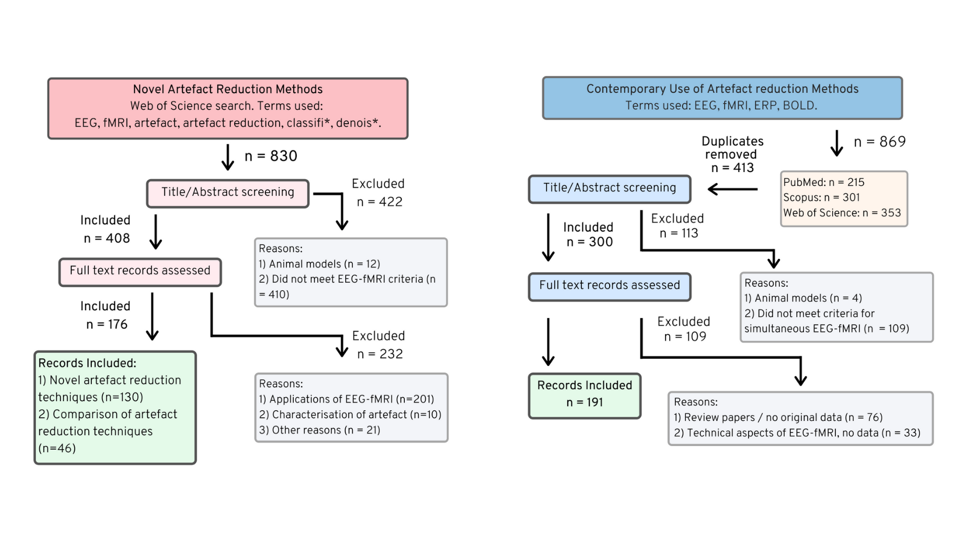
This work presents two systematic reviews (Figure 2):- Novel artefact reduction methods for simultaneous EEG-fMRI (1998-2018), and
- Prevalence of artefact reduction methods in contemporary EEG-fMRI studies (2016-2018).
Results
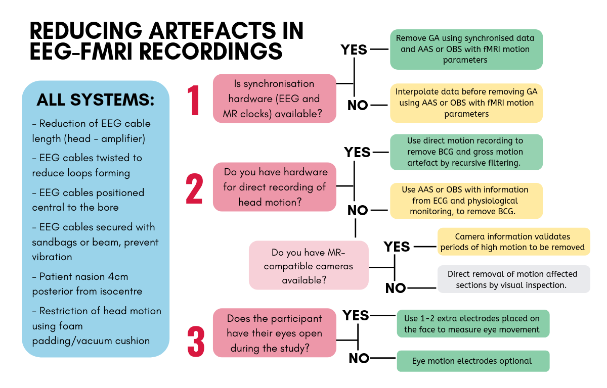
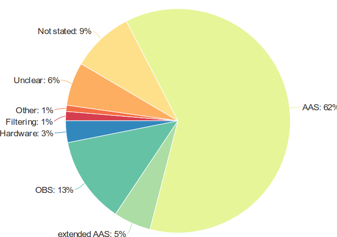
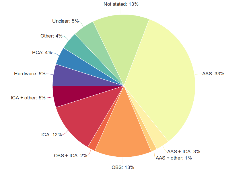
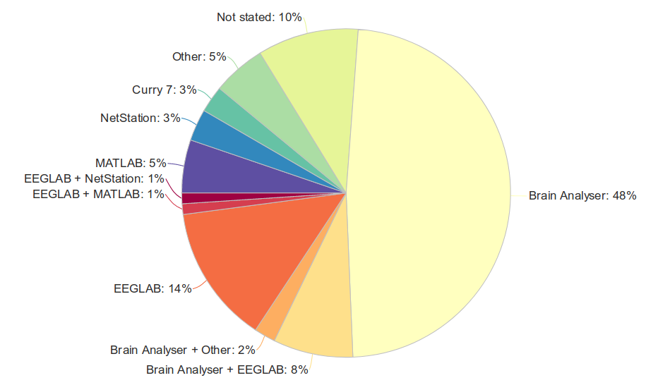
The search results for novel artefact reduction methods showed that whilst there are many published artefact reduction methods (n = 130), there are far fewer papers which independently compare their use on a single dataset (n = 46) (Figure 2). Based on these independent comparisons of data, recommendations for the current best practice in EEG-fMRI artefact reduction were devised (Figure 1). These include minimisation of artefact by optimising the setup of the EEG equipment and subject in the MR room, and use of additional hardware for clock synchronisation, and directly recording artefact on EEG during fMRI. These methods are generally superior to data-driven artefact estimation and filtering methods.The review of contemporary studies showed an overwhelming preference towards just a few longstanding artefact reduction methods, namely Average Artefact Subtraction [4, 2], Optimal Basis Sets [5], or, for ballistocardiogram artefact, Independent Components Analysis [6] (Figures 3 & 4). The commonly used methods are based on literature published over thirteen years ago, with newer methods rarely gaining use outside the group that developed them. Moreover, despite studies showing the benefits of additional hardware to record artefact [7, 8, 9] , uptake of these systems in contemporary studies is very limited (Figures 3 & 4). Many studies used a commercial toolbox for removing artefact from EEG recorded during fMRI (Figure 5), and therefore the options available in each of the toolboxes would play a large part in the artefact reduction method chosen for the study. Alarmingly, almost 15% of contemporary EEG-fMRI studies fail to adequately describe the artefact reduction methods used, raising questions about reproducibility of these studies (Figures 3 & 4).
Conclusions
Whilst EEG-fMRI is a useful technique for understanding brain dynamics, its more widespread adoption would be enhanced by the availability of turn-key implementations of best-practice artefact reduction techniques.We recommended that:
- Users of EEG-fMRI adequately report artefact reduction techniques used,
- Developers of novel artefact reduction techniques ensure their methods are easy to adopt, and
- Commercial EEG-fMRI vendors consider including additional hardware for recording artefact with all systems.
Acknowledgements
MB is supported by the Research Training Program from the Commonwealth Government of Australia and the Fay Marles Scholarship from the University of Melbourne. DFA is supported by fellowship funding from the Australian National Imaging Facility. The Florey acknowledges support from the Victorian Government Operational Infrastructure Support Grant.References
[1] S. Manganas and N. Bourbakis, “A Comparative Survey on Simultaneous EEG-fMRI Methodologies,” in 2017 IEEE 17th International Conference on Bioinformatics and Bioengineering, pp. 1–8, New York: IEEE, 2017.
[2] P. J. Allen, O. Josephs, and R. Turner, “A method for removing imaging artifact from continuous EEG recorded during functional MRI,” NeuroImage, vol. 12, pp. 230–239, Aug. 2000.
[3] R. Abreu, A. Leal, and P. Figueiredo, “EEG-Informed fMRI: A Review of Data Analysis Methods,” Frontiers in Human Neuroscience, vol. 12, 2018.
[4] P. J. Allen, G. Polizzi, K. Krakow, D. R. Fish, and L. Lemieux, “Identification of EEG events in the MR scanner: the problem of pulse artifact and a method for its subtraction,” NeuroImage, vol. 8, pp. 229–239, Oct. 1998.
[5] R. K. Niazy, C. F. Beckmann, G. D. Iannetti, J. M. Brady, and S. M. Smith, “Removal of FMRI environment artifacts from EEG data using optimal basis sets,” NeuroImage, vol. 28, pp. 720–737, Nov. 2005.
[6] G. Srivastava, S. Crottaz-Herbette, K. M. Lau, G. H. Glover, and V. Menon, “ICA-based procedures for removing ballistocardiogram artifacts from EEG data acquired in the MRI scanner,” Neuroimage, vol. 24, pp. 50–60, Jan. 2005.
[7] R. A. J. Masterton, D. F. Abbott, S. W. Fleming, and G. D. Jackson, “Measurement and reduction of motion and ballistocardiogram artefacts from simultaneous EEG and fMRI recordings,” NeuroImage, vol. 37, pp. 202–211, Aug. 2007.
[8] J. N. van der Meer, A. Pampel, E. J. W. Van Someren, J. R. Ramautar, Y. D. van der Werf, G. Gomez-Herrero, J. Lepsien, L. Hellrung, H. Hinrichs, H. E. M ̈oller, and M. Walter, “Carbon-wire loop based artifact correction outperforms post-processing EEG/fMRI corrections—A validation of a real-time simultaneous EEG/fMRI correction method,” NeuroImage, vol. 125, pp. 880–894, Jan. 2016.
[9] K. Hermans, J. C. d. Munck, R. Verdaasdonk, P. Boon, G.
Krausz, R. Prueckl, and P. Ossenblok, “Effectiveness of Reference Signal-Based
Methods for Removal of EEG Artifacts Due to Subtle Movements During fMRI
Scanning,” IEEE Transactions on Biomedical Engineering, vol. 63, pp.
2638–2646, Dec. 2016.
Figures