3879
Simultaneous multi-slice spiral-out acquisitions for high resolution perfusion fMRI at 7T1Maastricht Brain Imaging Centre (MBIC), Faculty of Psychology and Neuroscience, Maastricht University, Maastricht, Netherlands, 2A. A. Martinos Center for Biomedical Imaging, Massachusetts General Hospital, Charlestown, MA, United States, 3Skope MRT, Zürich, Switzerland
Synopsis
At ultra-high fields, high-resolution and large coverage functional perfusion mapping with Arterial Spin Labeling (ASL) becomes challenging due to shorter T2* requiring shorter read-out times and B0 inhomogeneity causing geometric distortions. We address these challenges with a single-shot, highly undersampled, short-TE spiral-out trajectory, and simultaneous multi-slice sampling with incoherent CAIPIRINHA for increased slice coverage. Spiral ASL acquisitions at short TE allows functional perfusion mapping with higher SNR compared to conventional EPI acquisitions, showing potential for high-resolution perfusion fMRI applications at ultra-high field.
INTRODUCTION
Perfusion fMRI, acquired with Arterial Spin Labeling (ASL) technique, has been proposed as an attractive alternative to BOLD due do its higher spatial specificity1. However, perfusion fMRI suffers from much lower contrast-to-noise ratio (CNR) than BOLD. This might be mitigated by the use of ultra-high field (UHF) imaging, which poses the following challenges that must be overcome: (i) the short T2* causes fast signal decay limiting the available readout window and induces BOLD signal contamination; and (ii) the higher B0 field inhomogeneity at UHF causes strong geometric distortions. To address the first, we propose spiral acquisitions with highly undersampled short-TE spiral-out trajectory, and simultaneous multi-slice (SMS) sampling with incoherent CAIPIRINHA for increased slice coverage. The effect of field inhomogeneity is addressed by incorporating separately acquired off-resonance maps into the image reconstruction. Similarly, the resulting gradient wave-form is obtained by magnetic-field monitoring and considered in the reconstruction.METHODS
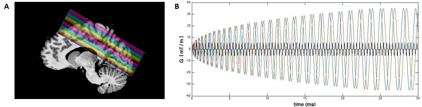
A high-resolution simultaneous multi-slice (SMS) spiral ASL sequence was created in-house based on an SMS-EPI implementation with FAIR QUIPSS II labeling module using a tr-FOCI inversion pulse2. Data were acquired on a Siemens Magnetom 7T scanner (Siemens Healthineers) equipped with a 32-channel head coil (Nova Medical). The single-shot spiral read-out parameters were as follows: FoV=200x200mm2, 30 slices with 1.25x1.25x1.2mm3 voxels, 100% slice gap, SMS-factor 2, TE=2ms, TI1/TI2/TR= 700/1800/2400 ms, flip angle=65o, variable density spiral trajectory with in-plane undersampling factor of 2.2 as described in3, readout duration 30ms, dwell time 2.5ns, gradient amplitude 29 mT/m, slew rate 132 T/m/s. Receive coil sensitivity and B0 off-resonance maps were obtained for image reconstruction using a multi-echo GRE sequence. The k-space trajectory with the corresponding slice angulation was measured on phantom using a Skope 16-channel field-camera. The trajectory and imaging data were merged before reconstruction with the i-Skope image production software (Skope MRT, Zurich, Switzerland)4. For comparison, conventional ASL with SMS EPI were acquired with matched parameters (29.5ms EPI readout, TE=16ms, 6/8 partial Fourier, GRAPPA=3, echo spacing=0.74ms). With each sequence, two 9-minute functional runs were acquired with a block-design combined flickering checkerboard and finger-tapping task (12/15 TR activation/rest). Data of the two runs were motion corrected using FSL and averaged before the statistical analysis was carried out in FSL-FEAT (without spatial smoothing). Two regions-of-interest (ROIs) covering motor and visual cortices were defined based on z-scores thresholded at 1.5. Time courses were extracted from these ROIs and averaged across all trials.RESULTS & DISCUSSION
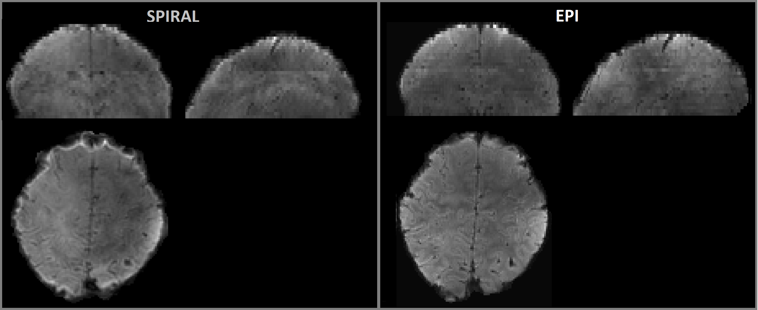
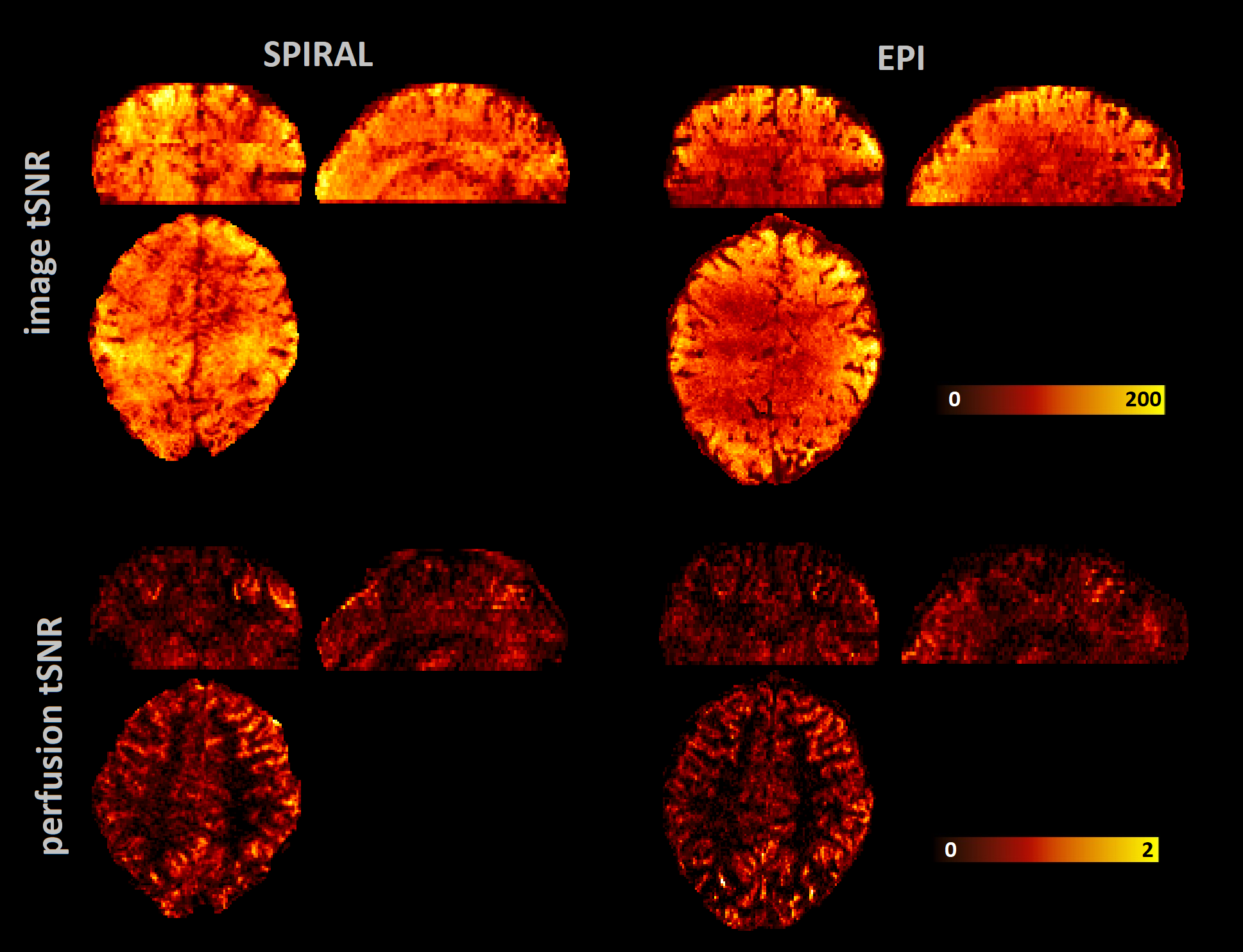
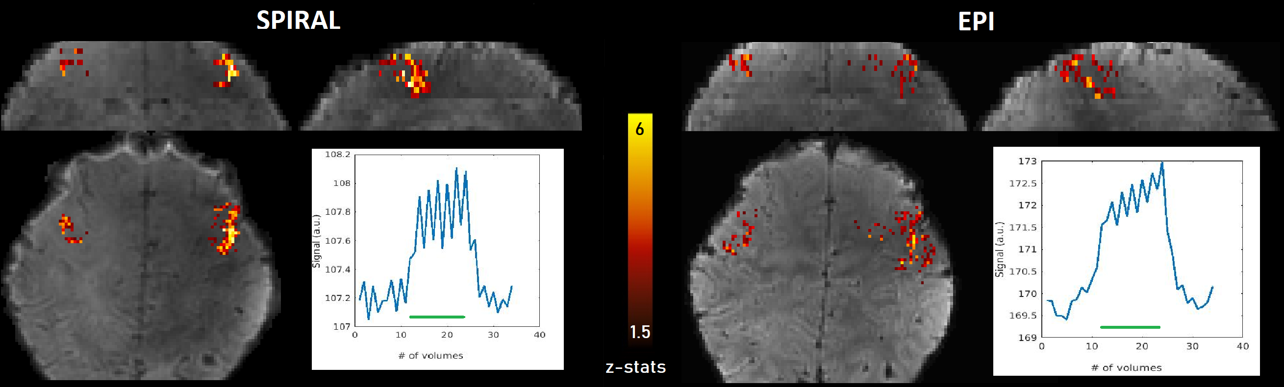
Figure 2 shows reconstructed raw spiral images with the expected proton density contrast due to the very short echo time. The short echo time translated into higher image and perfusion tSNR than observed in the EPI data (figure 3). Spiral acquisitions also resulted in higher z-scores in the motor cortex than the EPI data, for the same region (figure 4). Spiral time courses from the activated voxels show that spiral data were much less affected by BOLD contamination. Although the EPI configuration was optimized for short-TE ASL2, at this resolution requirement the functional response exhibits very strong T2* effects that dominate the perfusion changes. The spiral-based images, however, are dominated by the perfusion changes, allowing for robust detection of functional perfusion modulation even in this relatively short experimental duration. The proposed sequence shows potential for perfusion-based laminar fMRI, albeit limited to the regions such as motor cortex where the cortex is thicker.Acknowledgements
Acknowledgements Funding support: LH NWO VENI 016.198.032, BAP, DK VIDI 016.178.052, BAP and SK R01 MH111444/MH/NIMHReferences
1- Silva AC, Lee SP, Iadecola C, Kim SG. Early temporal characteristics of cerebral blood flow and deoxyhemoglobin changes during somatosensory stimulation. J Cereb Blood Flow Metab. 2000;20:201–206.
2- Ivanov D, Poser BA, Huber L, Pfeuffer J, Uludağ K.Optimization of simultaneous multislice EPI for concurrent functional perfusion and BOLD signal measurements at 7T. Magn Reson Med. 2017;78(1):121-129.
3- D.H. Kim, E. Adalsteinsson, and D. M. Spielman, “Simple analytic variable density spiral design,” Magn Reson Med. 2003;50(1):214–219.
4- Wilm BJ, Barmet C, Pavan M, Pruessmann KP. Higher order reconstruction for MRI in the presence of spatiotemporal field perturbations. Magn Reson Med. 2011 Jun;65(6):1690-701.
Figures