3876
Functional line-scanning in humans with ultra-high spatiotemporal resolution: reconstruction and BOLD sensitivity assessment
Luisa Raimondo1, Tomas Knapen1,2, ĺcaro A.F. de Oliveira1, Xin Yu3,4, Serge O. Dumoulin1,5, Wietske van der Zwaag1, and Jeroen C.W Siero1,6
1Spinoza Centre for Neuroimaging, Amsterdam, Netherlands, 2VU University, Amsterdam, Netherlands, 3Max Planck Institute for Biological Cybernetics, Tuebingen, Germany, 4Athinoula A. Martinos Center for Biomedical Imaging, Massachusetts General Hospital and Harvard Medical School, Charlestown, SC, United States, 5Experimental and Applied Psychology, VU University, Amsterdam, Netherlands, 6Radiology, University Medical Centre Utrecht, Utrecht, Netherlands
1Spinoza Centre for Neuroimaging, Amsterdam, Netherlands, 2VU University, Amsterdam, Netherlands, 3Max Planck Institute for Biological Cybernetics, Tuebingen, Germany, 4Athinoula A. Martinos Center for Biomedical Imaging, Massachusetts General Hospital and Harvard Medical School, Charlestown, SC, United States, 5Experimental and Applied Psychology, VU University, Amsterdam, Netherlands, 6Radiology, University Medical Centre Utrecht, Utrecht, Netherlands
Synopsis
We present initial results of line-scanning fMRI in humans. The potential of this technique lies in the combination of both high spatial and temporal resolution while sacrificing spatial coverage outside the region of interest. We reached a 250 μm resolution along the line direction with a temporal resolution of 200 ms. Coil sensitivity profiles and the average tSNR per channel were used to optimize the line reconstructions. We obtained similar BOLD sensitivity compared to standard 2D GE-EPI BOLD and high spatial specificity for a visual task. Hence, we demonstrate the feasibility of ultra-high spatiotemporal resolution in humans using line-scanning.
Introduction
Neurons with similar properties cluster together into sub-millimeter columnar and laminar structures, moreover neural activity occurs at millisecond resolution. Advances in fMRI approaches increase either spatial or temporal resolution but never both. Instead, line-scanning fMRI in rodents1 can achieve very high resolution across cortical depth (50 μm) and time (50 ms), by sacrificing volume coverage and resolution along the cortical surface. This high spatiotemporal resolution can also allow us to isolate microvessel responses and to characterize the distribution of blood flow and laminar fMRI profiles across cortical depth. Here, we present the first human line-scanning implementation and results. First, we will focus on the evaluation of the quality of the saturation pulses, followed by the description of optimal coil combination for the reconstruction and finally we will demonstrate the sensitivity of line-scanning fMRI in a comparison with standard 2D GE-EPI BOLD fMRI using a visual task.Methods
We scanned five healthy volunteers at 7T MRI (Philips) with a 32 channel receive head coil (Nova Medical). Line-scanning data acquisition used a modified 2D gradient-echo sequence: line resolution=250μm, TR/TE=200/13-22ms, 520 timepoints, flip angle=16°, array size=720, line thickness=2.5mm, in-plane line width=4mm, fat suppression using SPIR. Two saturation pulses (5 ms pulse duration) suppressed the signal outside the line of interest. We computed the outer volume suppression quality as the ratio signal along the line and outside the line. The phase-encoding in the direction perpendicular to the line was turned off2. The line was positioned along the right-left axis, crossing the visual cortex (Figure 1). We acquired functional data using a block design in 6 runs. Visual stimuli were 20 Hz flickering checkerboard, presented for 10 s on/off. Reconstruction was performed offline (MatLab, Gyrotools). We combined multi-channel coil data in four different ways: 1) sum-of-squares (SoS), 2) tSNR-weighted SoS, 3) coil sensitivity weighted SoS (csm), 4) tSNR and coil sensitivity weighted SoS (tSNR+csm). Resulting tSNR was used to select the best coil combination. Functional data were analyzed using a GLM approach and t-statistic values were computed to detect active voxels. We also compared line-scanning data to a standard 2D GE-EPI BOLD acquisition with: 1x1 mm2 in-plane spatial resolution, TR/TE=200/22 ms, 600 dynamics, flip angle=30°, FOV=176x176 mm2, SENSE factor 3, partial Fourier=0.8. Here, we averaged the 2D BOLD timeseries data in the region of interest coinciding with the line before computing t-statistics, resulting in an activation profile along the line. Following manual coregistration and averaging of line-scanning data every 4 voxels (in order to match the different spatial resolutions of the two acquisitions), we also calculated the correlation between the t-statistic values of line-scanning and 2D GE-EPI BOLD.Results
The achieved undesired signal suppression outside the relevant cortical area was (97±0.4)% (Figure 1).Figure 2 shows the tSNR for the four different coil combinations. The weighted combination of both tSNR and csm outperformed the other variants in terms of final tSNR. This coil combination was selected for subsequent line-scanning reconstructions. Line-scanning data averaged over 6 runs is shown in Figure 3a, for a representative subject. The average line signal intensity profile through the occipital lobe is shown in Figure 3b.
Figure 4a shows the t-statistic values overlaid on the anatomical scan. Note the good spatial correspondence between the positive BOLD t-statistic values and the grey matter ribbon, indicated with white arrows. Figure 4b shows the time course of an example active voxel along with the GLM.
Finally, in Figure 5 the comparison between the line-scanning (left column) and standard 2D GE-EPI BOLD (right column) shows a high similarity in BOLD sensitivity expressed by the t-statistic line profile. Good correlation (R=0.75±0.17) was found between the t-statistic values for the two acquisitions.
Discussion & Conclusion
We report first line-scanning fMRI results in humans with very high spatiotemporal resolution and show similar BOLD sensitivity to standard 2D GE-EPI BOLD. Adequate outer volume suppression can be achieved with saturation pulses. A coil combination including coil sensitivity maps and tSNR/channel for the reconstruction furnishes a promising temporal stability, since the resulting tSNR values are comparable to sub-milllimeter 3D imaging and sufficient for BOLD signal detection3, relatively to voxel size. Note that for the current line-scanning data processing no temporal filtering was applied. Future experiments will examine physiological noise contributions and its removal. Moreover, further development will investigate improvements in signal suppression outside the line of interest (2D spatial excitation, spin-echo beam excitation using orthogonal 90-180° pulses) and increased SNR using surface coil arrays4.Overall, the line-scanning fMRI technique seems very promising due to its potential in detecting evoked BOLD responses with sub-millimeter and sub-second resolution. Potential applications for line-scanning are microvessel function measurements in clinical research on cerebrovascular diseases but also fMRI at the mesoscopic scale such as cortical lamina5.
Acknowledgements
This study was supported by the Royal Netherlands Academy of Arts and Sciences Research Fund 2018 (KNAW BDO/3489) and the Visiting Professors Programme 2017 (KNAW WF/RB/3781) granted to the Spinoza Centre for Neuroimaging.References
- Yu X et al. Deciphering laminar-specific neural inputs with line-scanning fMRI. Nature Methods. 2013; 10.1038/nmeth.2730.
- Siero J.C.W et al. BRAIN 2019, #10538.
- Van der Zwaag W et al. Temporal SNR Characteristics in Segmented 3D-EPI at 7T. MRM. 2012; 10.1002/mrm.23007.
- Peridou N et al. Pushing the limits of high-resolution functional MRI using a simple high-density multi-element coil design. NMR in Biomedicine. 2012; 10.1002/nbm.2820.
- Petridou N
and Siero J.C.W. Laminar fMRI: What can time domain tell us? NeuroImage.
2019; 10.1016/j.neuroimage.2017.07.040.
Figures
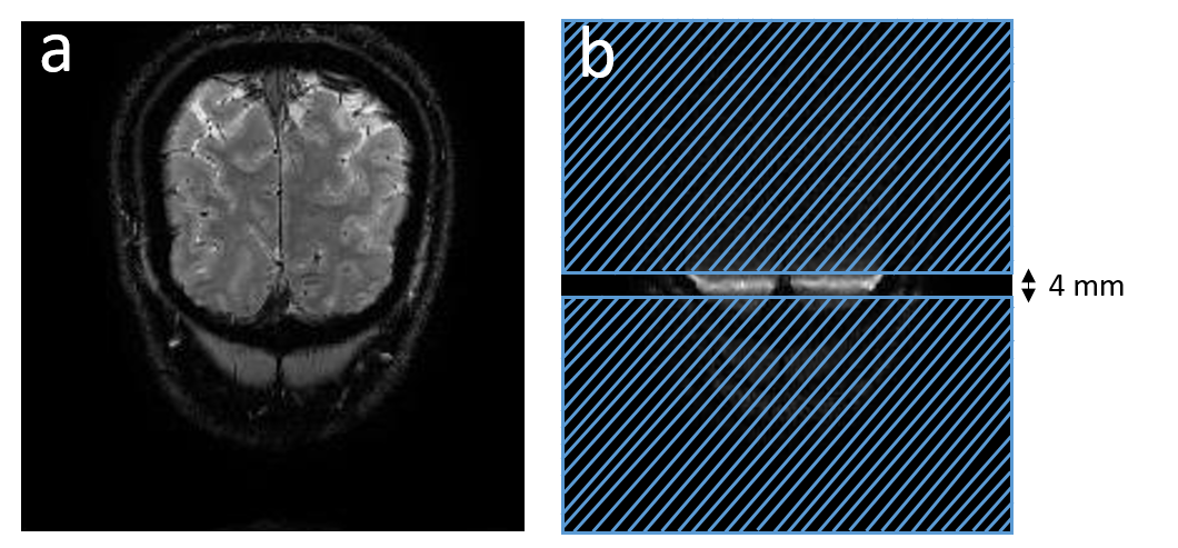
Figure1: (a) Acquired
slice and (b) outer volume suppression: placement of saturation slabs to
suppress unwanted signal outside the line of interest, depicted by the gap
(4mm) between the saturation slabs, in right/left direction across visual
cortex. 97% suppression outside the region of interest was achieved.
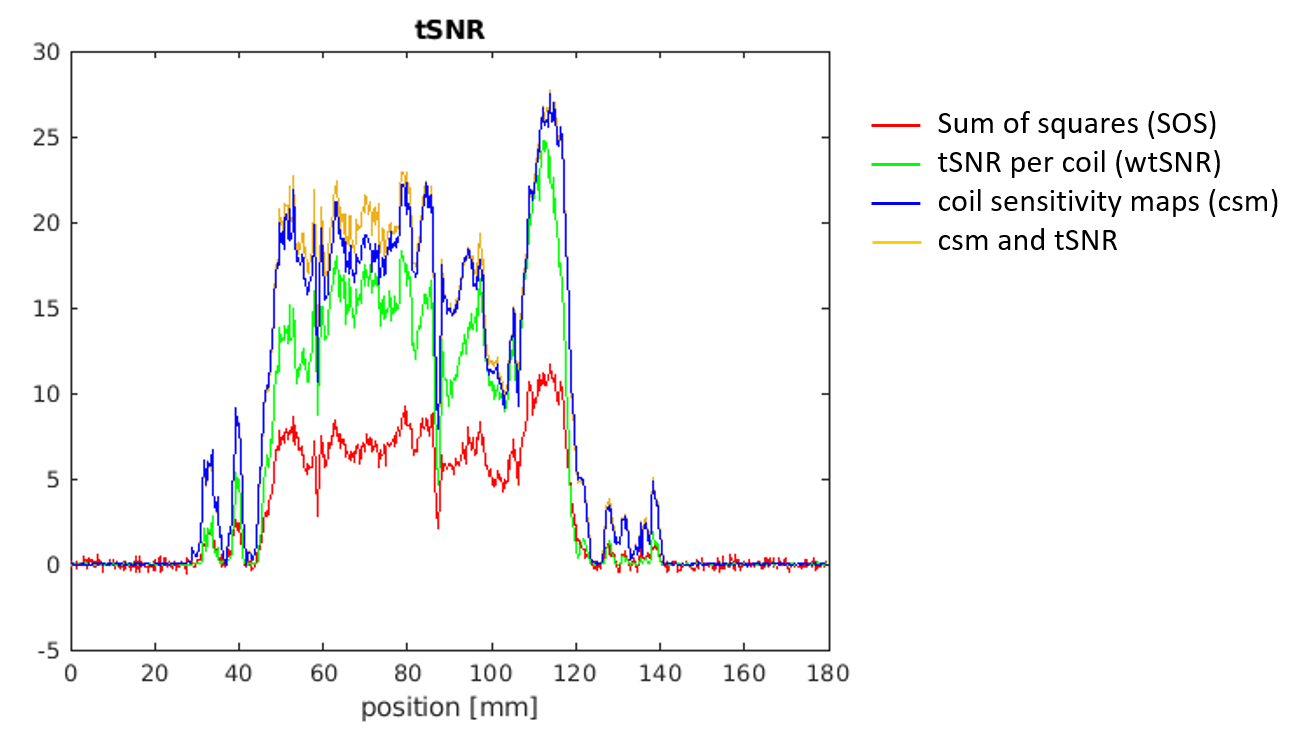
Figure2: tSNR for different coils
combination: sum of squares (red curve, SOS) and weighted combinations using
tSNR per single channel (green curve, tSNR per coil), synthetic coil
sensitivity maps (evaluated from data acquired with the phase-encoding
direction on and saturation pulses (csm)) and merged combination of the
previous two methods (csm and tSNR).
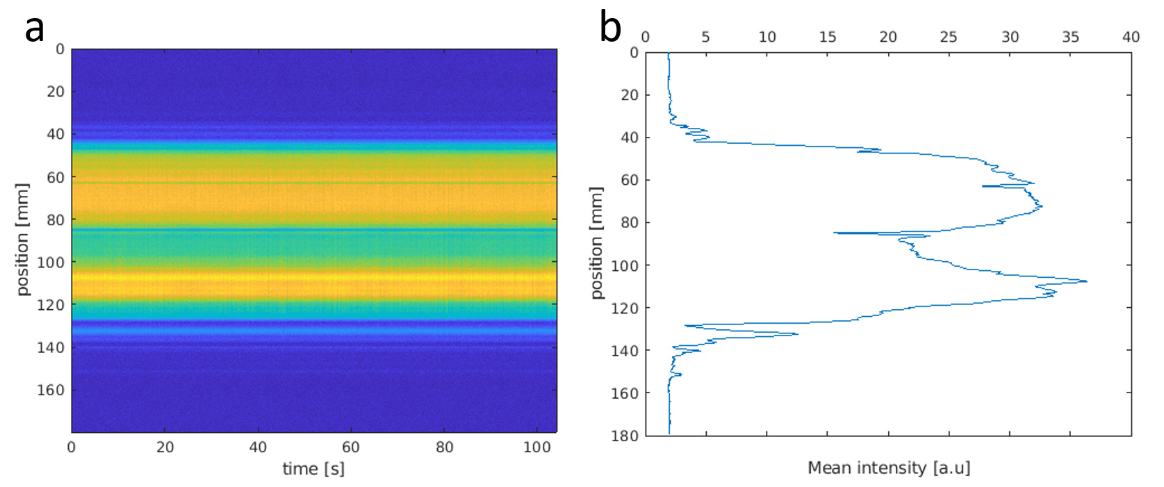
Figure3: (a) line-scanning data, average
over 6 runs and (b) mean intensity signal over the line.
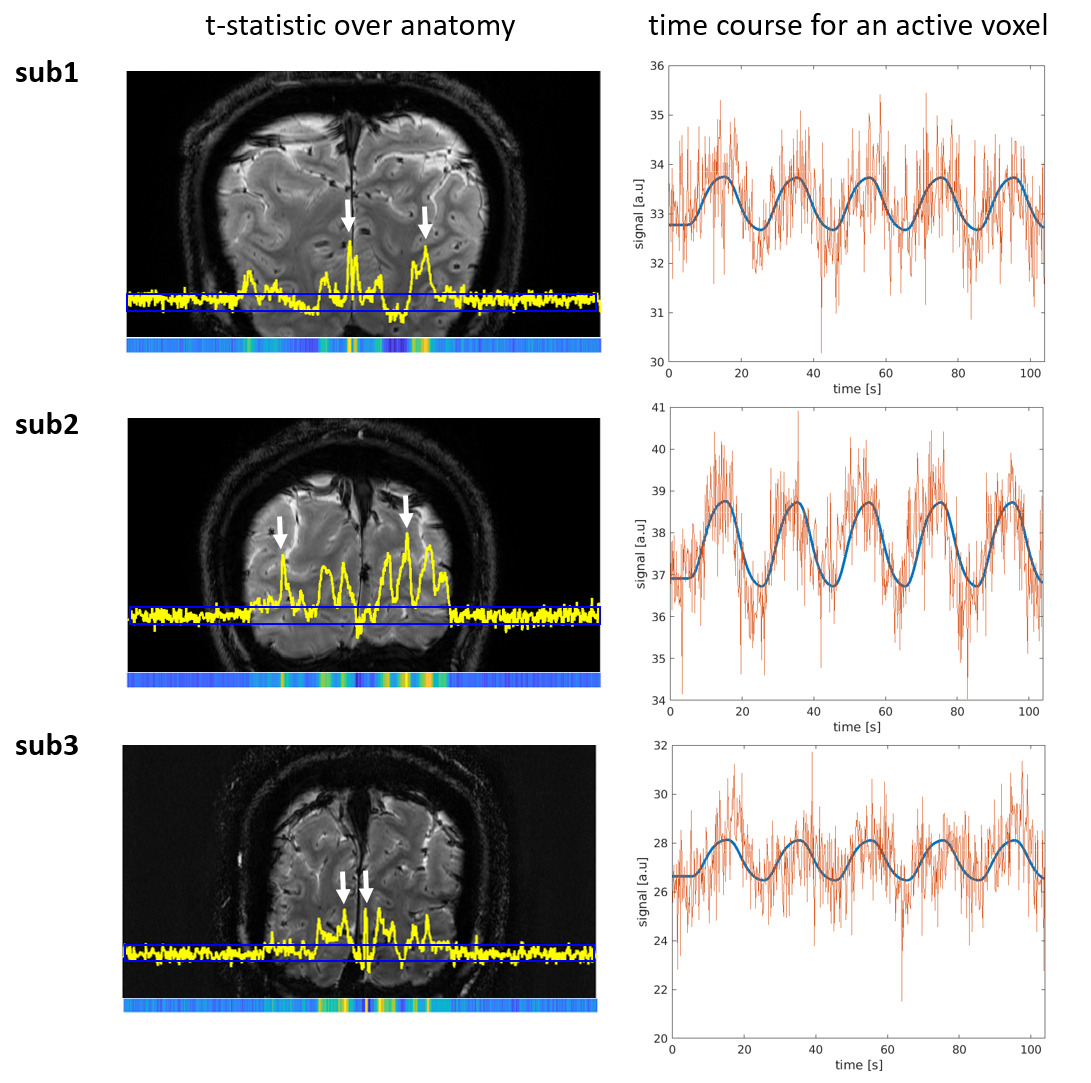
Figure4: (Left column) t-value (plot in
yellow and colormap on the bottom) superimposed on the anatomical scan for the
acquired slice. The position of the line is indicated with a blue box. White
arrows underline voxels with highest activity. (Right column) time course for
an active voxel with the raw time-series in orange and the predicted responses
in blue.
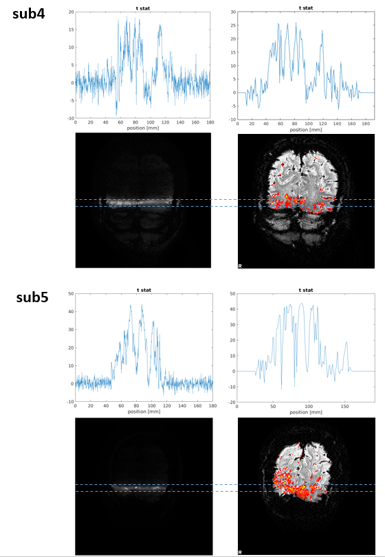
Figure5: (Left column) t statistic for
line-scanning acquisition and (Right column) t statistic for 2D GE-EPI BOLD for
the mean signal over the same line of interest.