3874
Sensitivity of Dynamic ASL and Resting-state BOLD in Patients with Bipolar Disorder1Computer Science, State University of New York at Binghamton, Binghamton, NY, United States, 2Psychiatry, Beth Israel Deaconess Medical Center & Harvard Medical School, Boston, ME, United States, 3Psychiatry, University of Texas Southwestern Medical Center, Dallas, TX, United States, 4Psychology and Neuroscience, University of Georgia, Athens, GA, United States, 5Psychiatry, Yale University, New Haven, CT, United States, 6Radiology, Beth Israel Deaconess Medical Center & Harvard Medical School, Boston, ME, United States
Synopsis
The effect sizes of bipolar disorder (BD) on low frequency fluctuation (LFF) and functional connectivity (FC) using dASL and rsBOLD imaging were evaluated in forty-five subjects (19 BD patients, 26 control). dASL showed significant increase of LFF and FC in BD, while rsBOLD did not show any difference. dASL demonstrated significantly higher effect sizes compared to rsBOLD, which lead to decreases of 39% and 49% in sample size for LFF and FC measures respectively. These findings support that dASL is more sensitive to BD than rsBOLD and therefore may offer advantages in reducing costs for clinical trials of BD therapies.
Introduction
Resting-state blood-oxygen-level dependent (rsBOLD) imaging has been predominantly used to study the low frequency fluctuation (LFF) [1] and functional connectivity (FC) [2] of brain and found to provide valuable information in neurological and psychiatric studies [3]. Dynamic arterial spin labeling (dASL) has recently demonstrated its capability in detecting the abnormal LFF and FC in bipolar disorder (BD) [4]. It is worth asking whether dASL can offer comparable sensitivity of LFF and FC in BD compared to rsBOLD. Due to heavy suppression of background tissues, the dASL signals were shown minimally contaminated with physiological noise, such as cardiac pulsations and respiratory motions [5]. We hypothesized that dASL offers high sensitivity in LFF and FC in BD. Here, we directly compared the rsBOLD and dASL imaging for their effect sizes of BD on the LFF and FC measures.Methods
Forty-five subjects (19 BD patients, 26 controls) from the Boston site of the multi-site Psychosis and Affective Research Domains and Intermediate Phenotypes (PARDIP) study underwent resting-state dASL images acquired with 3D stack of spirals RARE sequence [6] and BOLD images acquired with 2D gradient-echo echo planar imaging (EPI) on a General Electric (GE) 3 Tesla scanner using an 8-channel head coil receive array. Pseudo-continuous arterial spin labeling (PCASL) was used for ASL labeling with 2s labeling duration, 1.8s post-labeling delay, and less than 0.3% of background signals [7]. Twenty-seven 3D ASL whole-brain volumes and a reference volume were collected in 9 minutes. Two hundred and forty rsBOLD image volumes were collected in 8 minutes. BOLD volumes were slice-timing corrected, regressed out white matter signal, CSF signal, rigid-body motion and linear trend corrected, and then filtered with band-pass filter [0.01, 0.08] Hz. ASL volumes were not performed with any of the preprocessing steps. Both BOLD and ASL volumes were registered to standard space using T1-weighted MPRAGE images as intermediate images. LFF maps were calculated for rsBOLD and dASL. FC maps were calculated for rsBOLD and dASL with an anterior cingulate cortex (ACC) seed. For rsBOLD and dASL separately, LFF maps and FC maps were compared between BDs and controls using two-sampled t tests on a voxel-by-voxel basis using SPM8. A voxel-level p-value threshold of 0.001 was used. The clusters with cluster-level p value of 0.05 were reported significant. Because of no significant difference shown in rsBOLD with the voxel-level p value of 0.001, we increased the voxel-level p value to 0.01, in steps of 0.005 increase, until we found a significant cluster. The effect size of each method was evaluated using the regional average over the significant clusters from the respective method. The effect size of BD was defined as the group mean difference between BDs and controls, divided by the pooled standard deviation. To understand how reliable the effect sizes using dASL and rsBOLD are, one thousand random permutations were performed on the LFF and FC maps by keeping the same number of BDs and controls. The effect size was evaluated over the significant clusters (defined for each method separately).Results & Discussion
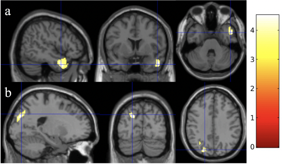
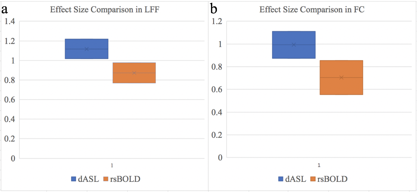
With the voxel-level p value of 0.001, dASL showed an increased LFF in BD (Fig. 1a, cluster-level p < 0.001, cluster size = 352) and an increased FC in BD (Fig. 1b, cluster-level p = 0.005, cluster size = 235), while rsBOLD did not show a significant change of either LFF or FCC in BD. rsBOLD showed an increased LFF in BD (Fig. 2a, cluster-level p = 0.005, cluster size = 599) with voxel-level p value of 0.01 and an increased FC (Fig. 2b, cluster-level p =0.047, cluster size = 1547) with the voxel-level p value of 0.035. dASL and rsBOLD detected different BD-affected regions when a significance threshold is reduced for rdBOLD. However, a large BSNIP study applied rsBOLD on 180 BD subjects and detected abnormal LFF in the similar temporal pole region (as shown in Fig. 1a detected using dASL), indicating largely reduced sensitivity in rsBOLD relative to dASL on our sample size. The effect sizes of BD were 1.1178 0.0993 using dASL vs. 0.8722 ± 0.1047 using rsBOLD in LFF (Fig. 3a), and 0.9934 0.1187 using dASL vs. 0.7053 0.1498 using rsBOLD in FC (Fig. 3b). For 90% of power and type I error rate of 5%, the increases in ASL effect sizes lead to decrease of 39% and 49% in sample size for LFF and FC measures respectively. With random permutations, the effect sizes of BD were significantly higher (p < 0.0001) using dASL (1.1112 ± 0.2673) than using rsBOLD (0.9216 ± 0.1577) in LFF measure, and significantly (p < 0.0001) higher using dASL (1.0050 ± 0.2623) than using rsBOLD (0.7400 ± 0.1208) in FC measure.Conclusion
The study demonstrates dASL is more sensitive to bipolar disorder in LFF and FC measures compared to rsBOLD. dASL may offer great advantages in reducing the sample size required for future clinical trials of BD therapies.Acknowledgements
No acknowledgement found.References
1. Zang, Y.F., et al., Altered baseline brain activity in children with ADHD revealed by resting-state functional MRI. Brain Dev, 2007. 29(2): p. 83-91.
2. Biswal, B., et al., Functional connectivity in the motor cortex of resting human brain using echo-planar MRI. Magn Reson Med, 1995. 34(4): p. 537-41.
3. Lv, H., et al., Resting-State Functional MRI: Everything That Nonexperts Have Always Wanted to Know. AJNR Am J Neuroradiol, 2018. 39(8): p. 1390-1399.
4. Dai, W., et al., Abnormal perfusion fluctuation and perfusion connectivity in bipolar disorder measured by dynamic arterial spin labeling. Bipolar Disord, 2019.
5. Zhao, L., et al., Global Fluctuations of Cerebral Blood Flow Indicate a Global Brain Network Independent of Systemic Factors. J Cereb Blood Flow Metab (In Press), 2017.
6. Dai, W., et al., Quantifying fluctuations of resting-state networks using arterial spin labeling perfusion MRI. J Cereb Blood Flow Metab, 2016. 36(3): p. 463-73.
7. Maleki, N., W. Dai, and D.C. Alsop, Optimization of background suppression for arterial spin labeling perfusion imaging. Magma, 2012. 25(2): p. 127-33.
Figures