3873
Distortion- and Resolution-Matched T1w-Like Anatomy for Investigating Depth-Dependent Activity in Submillimeter-Resolution fMRI at 7T1Brain Function Analysis and Imaging Lab, CiNet, NICT, Osaka, Japan, 2Graduate School of Frontier Biosciences, Osaka University, Osaka, Japan
Synopsis
We demonstrate the reconstruction of isotropic submillimeter-resolution distortion- and resolution-matched (DRM) T1w-Like anatomy from the image inversion of T1-maps obtained as five slice-shifted anatomical volumes covering the whole brain. The proposed anatomy avoids the distortion-mismatch between function and anatomy resulting in a high alignment with the functional data, necessary for activity localization in submillimeter-resolution fMRI. We use the same pulse sequence for acquiring both functional and DRM anatomical images with aligned slice acquisitions except differing in parameters unrelated to distortions. Moreover, the proposed anatomy allows the generation of cortical surface for investigating depth-dependent activity in isotropic submillimeter-resolution fMRI analysis.
Introduction:
Distinguishing cortical layers and investigating depth-dependent BOLD [1] responses in isotropic submillimeter-resolution fMRI [2] require the use of a high-contrast distortion- and resolution-matched (DRM) anatomy. Based on our earlier work of DRM anatomical MR image acquisitions for T1w-Like image reconstruction [3,4], we present here the results of isotropic submillimeter-resolution fMRI on a T1w-Like whole-brain anatomical image reconstructed from the image inversion of DRM T1-maps. These T1-maps are computed from slice varying multi-shot inversion recovery-prepared echo-planar imaging (msIR-EPI) volumes [5, 6]. The same pulse sequence was used for acquiring both functional and DRM anatomical images with aligned slice acquisitions except differing in parameters unrelated to distortions. The reconstructed T1w-Like anatomy in the native EPI space offers high alignment necessary for activity localization providing benefits for studying voxel-wise dynamics in isotropic submillimeter-resolution fMRI. Moreover, the proposed T1w-Like anatomy allows the generation of cortical surface for investigating depth-dependent responses [7].Materials & Methods:
An adult human brain was scanned using the BISEPI sequence [2] on a Siemens MAGNETOM 7T scanner with a 32-channel phased-array head coil to obtain 144 fMRI volumes at a spatial resolution of voxel-wise 0.7 mm isotropic. The functional scan (#Segments = 3, GRAPPA = 2, #Shots in block = 24, TR/TE/FA = 1000 ms /22.2 ms / 50°, #Slices = 15, PF = 6/8) covering the region of interest in primary motor cortex was acquired in a bilateral finger tapping block-design task 12s (ON) / 12s (OFF). Functional scan was followed by five slice-shifted anatomical volumes at a spatial resolution of voxel-wise 0.7 mm isotropic using 2D msIR-EPI (#Segments = 6, TR/TE/FA = 3000 ms /22.2 ms/ 90°, Slices/Averages = 35x5/2, Slice thickness = 0.7 mm, Spacing = 3.5 mm, PF = 6/8) with variable inversion recovery for each slice generated by an IR-sweep with varying inversion time (TI) across slices (TI=100-2400ms) [6]. The obtained anatomical volumes were used to generate T1-maps after T1-fitting and combined and processed in Matlab to generate whole-brain T1w-Like DRM anatomy based on image inversion [4]. The obtained native EPI space T1w-Like anatomy was post-processed in BrainVoyager for generating cortical surface after AC-PC transformation. Mid-GM surface was generated and the functional data at four distinct depths moving from GM-WM interface to GM-pial boundary were sampled and overlaid on this mid-GM surface. fMRI pre-statistics processing includes mean intensity correction, motion correction, and high-pass filtering with GLM-Fourier 2 sines/cosines and Gaussian FWHM of 2 data points, with no spatial smoothing. The generalized linear model analysis was applied with stimulation ON/OFF as a binary regression variable in BrainVoyager 21.2.Results and Discussion:
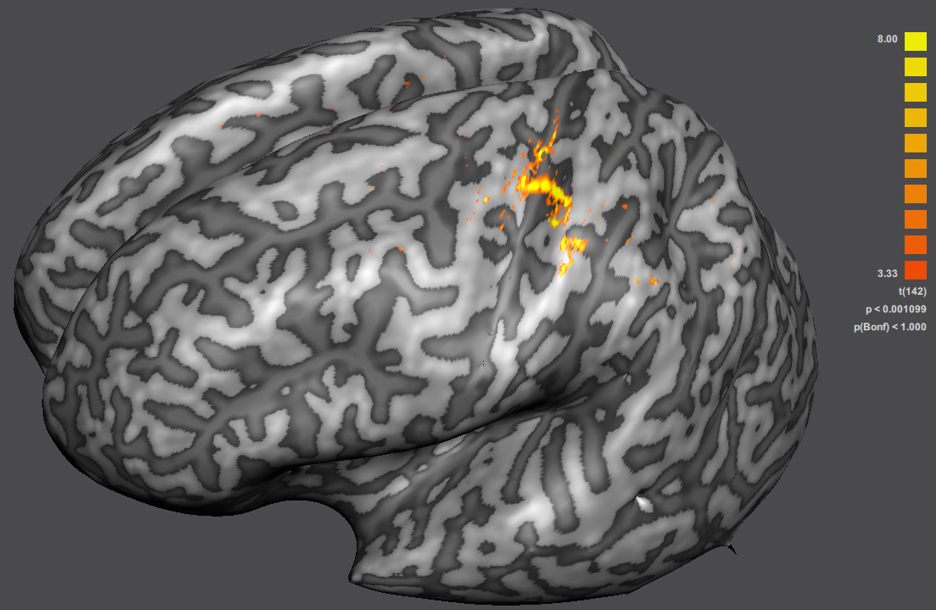
Figure 1 shows fMRI BOLD activity overlaid on the obtained T1w-Like DRM anatomy based on image inversion [4]. As shown here, the fMRI BOLD activity at 0.7 mm isotropic resolution follows the ribbon of the grey matter structures on the reconstructed T1w-Like DRM whole-brain anatomy. Figure 2 shows the mid-GM cortical surface generated from this anatomy where fMRI BOLD activity is sampled at distinct cortical depths moving from the GM-WM interface (left-most) to the GM-pial boundary (right-most). Figure 3 shows overlaid fMRI BOLD activity as the multiple-selection of all depth maps on the mid-GM cortical surface.Conclusion:
DRM T1w-Like anatomy in the native EPI space [3, 4] alleviates the problem of distortion mismatch that exists between function and T1w anatomy. Moreover, it improves the accuracy of activity localization and interpretation of the results in submillimeter-resolution fMRI studies. In this study, we reconstructed T1w-Like whole-brain DRM anatomical slices from the image inversion of T1-maps computed from slice varying 2D msIR-EPI [5, 6] acquired at 0.7 mm isotropic resolution. The reconstructed T1w-Like DRM anatomy demonstrates a strong alignment with the acquired functional data, necessary for studying voxel-wise dynamics in submillimeter-resolution fMRI. The proposed T1w-Like whole-brain anatomy allows the generation of a cortical surface, useful for investigating layer-specific functional responses in a region of interest.Acknowledgements
This study was supported in part by Japan Society for the Promotion of Science (JSPS) Grants-in-Aid for Scientific Research “KAKENHI” (Grant Numbers JP26282223 and JP26350471 and JP19K08244).References
[1] Ogawa S, Lee TM, Kay AR, Tank DW. Brain magnetic resonance imaging with contrast dependent on blood oxygenation. Proc Natl Acad Sci USA. 1990;87(24):9868-9872. [2] Liu G, Shah A, Ueguchi T. Block-Interleaved segmented EPI for voxel-wise high-resolution fMRI studies at 7T. Proceedings of the International Society for Magnetic Resonance in Medicine Joint Annual Meeting ISMRM-ESMRMB. 2018;5450. [3] Shah A, Ueguchi T, Liu G. Distortion-Matched Anatomical Imaging using Inversion Recovery-Prepared EPI for high-resolution fMRI. 24th Annual Meeting of the Organization for Human Brain Mapping. 2018;1727. [4] Shah A, Liu G, Ueguchi T. EPI based distortion- and resolution-matched T1-Like anatomy for submillimeter-resolution fMRI at 7T. 25th Annual Meeting of the Organization for Human Brain Mapping. 2019;2591. [5] Turner R, Panchuelo RS, Mougin O, Francis S. Multi-shot inversion recovery EPI with SMS excitation for high spatial resolution T1-mapping. 25th Annual Meeting of the Organization for Human Brain Mapping. 2019;2627. [6] Panchuelo RS, Turner R, Mougin O, Francis S. A 2D multi-shot inversion recovery EPI (MS-IR-EPI) sequence for high spatial resolution T1-mapping at 7T. Proceedings of the International Society for Magnetic Resonance in Medicine. 2018;60. [7] Shah A, Liu G, Ueguchi T. Investigating Depth-Dependent Activity in Submillimeter-Resolution fMRI at 7T with Native- Space Transformed T1w MPRAGE Anatomy. ICMRI. 2019;SS03-08.Figures