3861
The difference of left and right side after chronic pontine infarction based on resting-state fMRI1GE Healthcare, MR Research China, Beijing, Zheng Zhou, China
Synopsis
The resting state functional images of 53 patients with chronic pontine infarction and 35 healthy controls were collected, and A and B were used to evaluate the difference and sensitivity of functional information integration between single brain regions and hemispheres.It was associated with clinically relevant cognitive function scores.Finally, the contribution of the left and right sides to the difference was evaluated.The results showed that the difference and contribution of the right infarction group were significantly higher than that of the left infarction group.In the future, functional integration between the two hemispheres will be further studied.
Introduction
In recent years, various studies devoted to studying the early recovery and evaluation of the disease after infarction have developed rapidly in functional and structural imaging techniques, and further details are being explored, such as the difference between left and right infarcts and the overall sample.Whether there are sensitivity differences between monomeric elements or regional brain regions and functional integration or information communication between cerebral hemispheres in chronic phase. Toward this end,the purpose of this study is to 1) sensitivity of VMHC parameters to brain functional imaging studies;2) left and right side differences after pontine infarction;3) the correlation between different brain regions and clinical motor cognition scores.Materials and Methods
53 patients who were first-onset stroke patients showed deficits in the assessment of clinical motor cognition scores (29 on the left and 24 on the right) and 35 age-matched healthy controls were recruited.All MRI examinations were performed at a 3.0 Tesla GE Discovery 750 with an 8-channel phased array head coil. The resting-state fMRI (TR/TE= 2000/30, FA = 90°, FOV = 240 mm × 240 mm, reconstruction matrix = 220mm × 220mm, slice thickness = 4mm, gapof slice=0.5mm, number of slices, =32,Time point=180)with 3DT1 image were acquired.The scanning time was 6 mins 20s.All standardized individual values of regional Homogeneity (Reho) and the voxel mirrored homotopic connectivity (VMHC) were obtained after preprocessing .The difference were compared respectively between groups (G1=normal G2=pon stroke in the left G3=pon stroke in the right) by one-way analysis of variance (ANOVA). using the GRF method with a corrected threshold of p < 0.05.And the correlation between the different brain regions and statistically significant clinical motor cognition scores was then analyzed.
Results
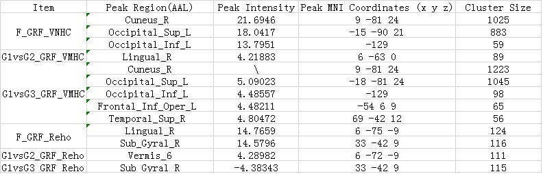
There were two cluster show increased value between the normals and the all strokes (F=G2&G3) in Reho,the Vermis_6 (AAL) was significantly higher in the left side group (G1vsG2) and Sub_Gyral_R (AAL) was lower in the right side group (G1vsG3) . While,There cluster (Cuneus_R(AAL)、Occipital_Sup_L (AAL)、Occipital_Inf_L (AAL) exhibited enhanced by comparing normal controls with stroke patients in VMHC. Lingual_R (AAL) was higher in the left side group (G1vsG2) and Cuneus_R (AAL)、Occipital_Sup_L (AAL)、Occipital_Inf_L (AAL)、Frontal_Inf_Oper_L (AAL)、Temporal_Sup_R (AAL) were higher in the right side group (G1vsG3). Notely, the right side group manifest much more higher alter and much more cluster changed which were less small. As shown in the figure1 and figure2 .There are also lessons from correlation analysis : ADL score was only negatively correlated with Sub_Gyral_R ; NIHSS score was negatively correlated with Occipital_Sup_L (AAL)、Occipital_Inf_L (AAL)、 lingual_R (AAL),、Vermis_6 (AAL); Short-term memory (score of RAVLT_short) was positively correlated with both values of ReHo and VMHC, while long-term memory (score of RAVLT_long) was only positively correlated with Occipital_Sup_L (AAL) and Frontal_Inf_Oper_L (AAL) in VMHC.
Conclusion
The right infarct group appeared to be more similar or significantly different from the overall sample than the left group, which may be associated with the dominant hemisphere.This may provide reference for clinical symptom progression and prognosis. Whether the left occipital lobe's role in interhemispheric information integration and the mental activity of the temporal lobe are really related to limb functional activity needs to be verified by larger samples and related structural studies.The long-term memory association results seem to explain that synaptic building of information storage mechanisms that require bilateral hemispheric coordination has little to do with single voxel or brain region.Acknowledgements
No acknowledgement found.References
No reference found.Figures
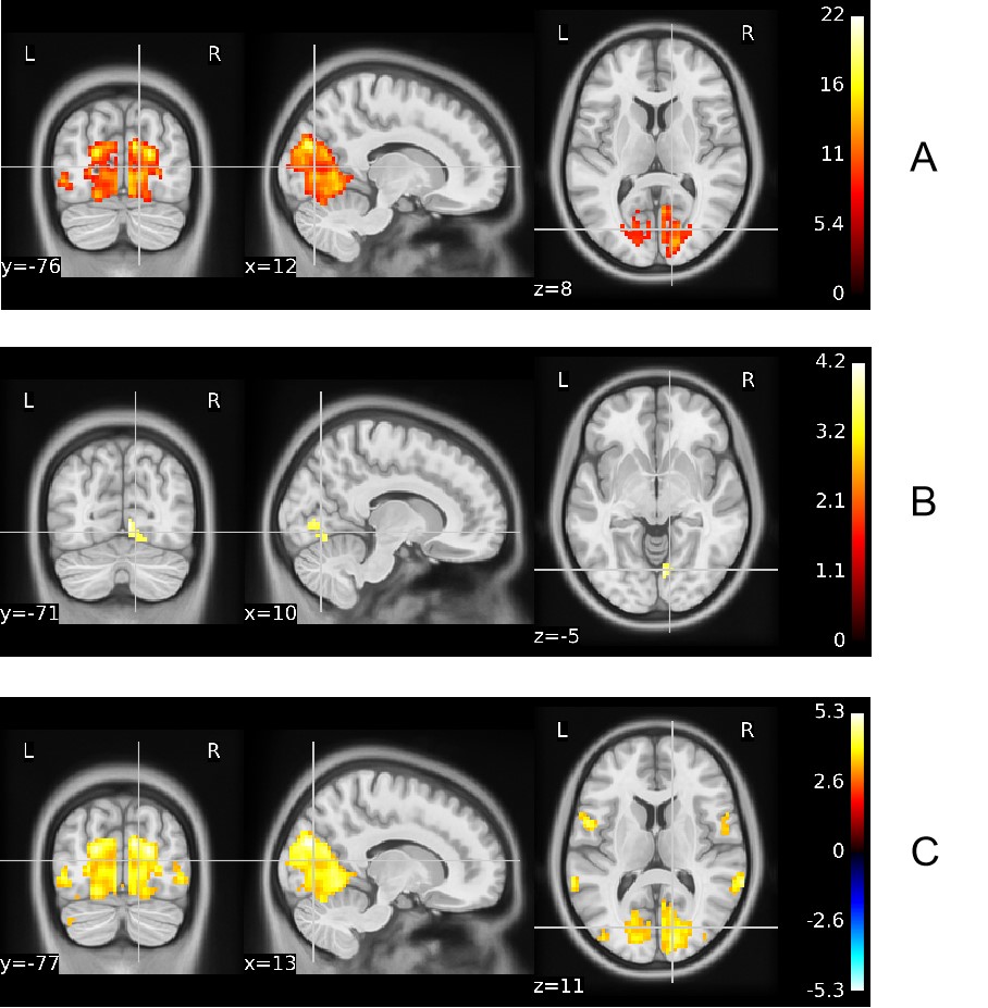
A:F_GRF_VMHC B:G1vsG2_GRF_VMHC C:G1vsG3_GRF_VMHC
Warm and cold color bars indicate an increase in warm colors/decrease in cool colors
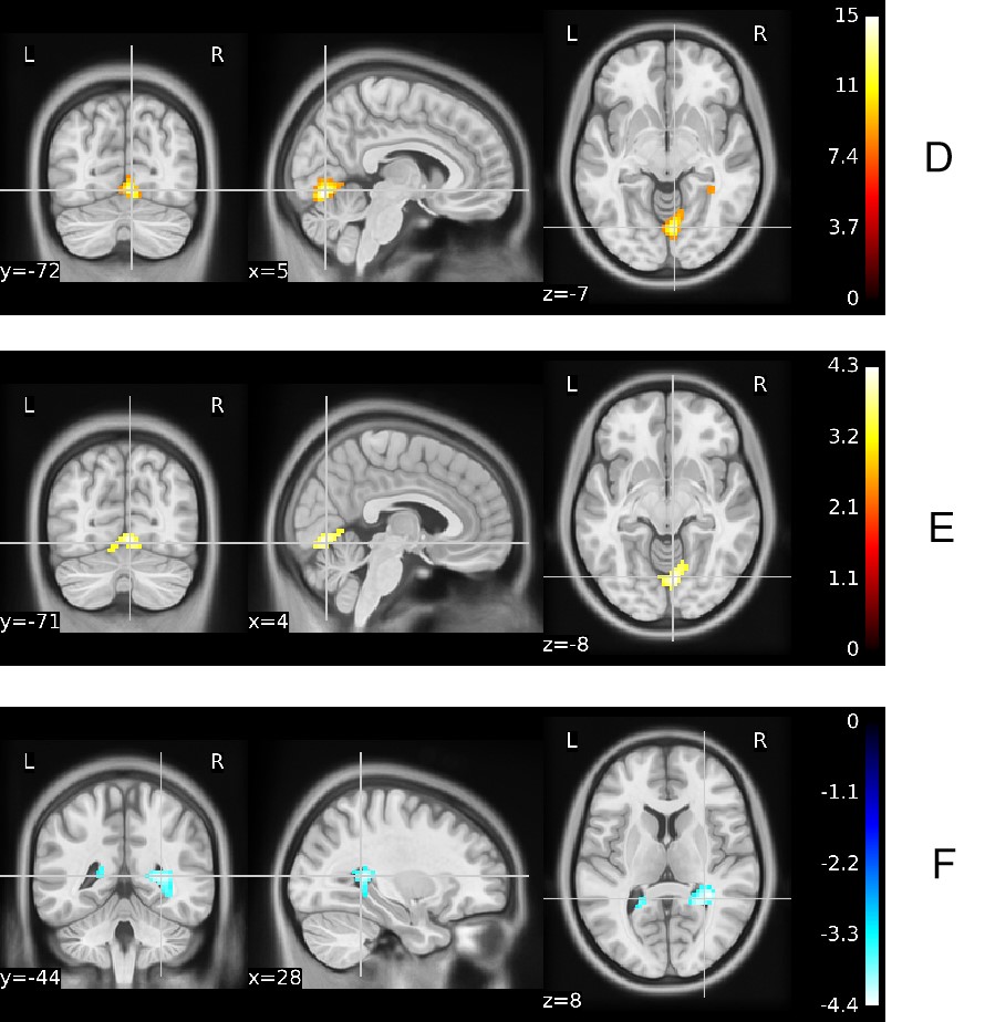
D:F_GRF_ReHo E:G1vsG2_GRF_ReHo F:G1vsG3_GRF_ReHo
Warm and cold color bars indicate an increase in warm colors/decrease in cool colors