3859
Cerebral hemodynamic responses of subjective cognitive decline evoked by loaded N-back tasks1Center for MRI Research, Peking University, Beijing, China, 2Department of Neurology, XuanWu Hospital of Capital Medical University, Beijing, China, 3Center of Alzheimer’s Disease, Beijing Institute for Brain Disorders, Beijing, China, 4National Clinical Research Center for Geriatric Disorders, Beijing, China
Synopsis
Subjective cognitive decline (SCD) is a high risk for Alzheimer’s disease. Most of the existing studies made their observations during the resting state of the brain. This study compared the responses of SCD and normal controls (NC) evoked by loaded N-back tasks. A load-dependent effect is clearly seen in both groups. CBF, as compared to other parameters, shows higher sensitivity in detecting the difference between SCD and NC subjects. Our results may help understand the physiological mechanisms and develop early diagnostic strategies of SCD.
Introduction
Subjective cognitive decline (SCD) is a high risk for Alzheimer’s disease. The physiological mechanisms of SCD is largely unknown. Most of the existing studies made their observations during the resting state of the brain. How the SCD group performs differently from the normal controls (NC) during functional tasks is less studied. This study compared the responses of SCD and NC evoked by loaded N-back tasks. Relevant physiological parameters, including cerebral blood volume (CBV), cerebral blood flow (CBF), blood oxygen-level dependent (BOLD) and cerebral metabolic rate of oxygen (CMRO2) were obtained. The results of this study help clarify the underlying hemodynamic mechanisms of SCD.Methods
15 NC (8 females) and 23 SCD subjects (14 females) participated in the study. All subjects complete the standardneurological cognitive tests and do not have psychiatric or neurological diseases which may cause memory impairmentand no abuses of alcohols and drugs. The SCD subjects were included based on the SCD diagnostic framework suggested by the Subjective Cognitive Decline Initiative[1]. All subjects completed the required cognitive tests. Table 1 shows the biographical and behavioral data of the two groups.All subjects participated block-designed 0-back, 1-back, 2-back and 3-back working memory tasks. The experimental designs were adopted from a Zou et al.[2]. English letters were replaced by Arabic numbers for the consideration of elderly Chinese participants. Followed by the N-back tasks, each subject underwent a hypercapnic challenge during which the subject breathed a gas mixture (5% CO2, 20% O2, 75%N2) through a non-rebreather mask.
A pulse sequence which simultaneously acquires CBV, CBF and BOLD-weighted signals[3] was implemented on a 3.0 T GE MR750 system. A single oblique slice passing through the frontal and parietal cortices was imaged. The exact location of the slice was determined via a training task in the beginning of the scan. Acquisition parameters, procedures for preprocessing and data analysis were adopted from Lin et al.[4]
At the individual level, the δCBV, δCBF, δBOLD and δCMRO2 evoked by 1-, 2- and 3-back with respect to 0-back in the frontal cortex of each subject was obtained. At the group level, holding sex, age and education as covariates, a general linear model was applied to compare the above parameters between the two groups, and a partial correlation test was performed to relate the imaging results with the behavior data. P < 0.05 was considered statistically significant.
Results
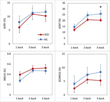
The group averages of δCBV, δCBF, δBOLD and δCMRO2 evoked by 1-, 2- and 3-back, with respect to 0-back, in the frontal cortex were displayed in Figure 1. In general, all parameters show a load-dependent effect, where 1-back caused minimum signal changes. Among all parameters, δCBF evoked by 3-back, with respect to 0-back, was significantly lower (p = 0.04) in the SCD group compared to the NC group. None of the other parameters showed statistical differences between the two groups. In addition, δCBF at 3-back was found to be significantly correlated with the MoCA (p = 0.04) and AFT (p = 0.03) scores of the subjects, although neither MoCA nor AFT shows significant differences between the two groups.Discussion and Conclusion
This study explored the differences in the hemodynamic responses in the frontal cortex evoked by loaded N-back tasks between SCD and NC subjects. A load-dependent effect is clearly seen in both groups. CBF, as compared to other parameters, shows higher sensitivity in detecting the difference between SCD and NC subjects. Our results may help understand the physiological mechanisms and develop early diagnostic strategies of SCD.Acknowledgements
No acknowledgement found.References
[1] JESSEN F, AMARIGLIO R E, VAN BOXTEL M, et al. A conceptual framework for research on subjective cognitive decline in preclinical Alzheimer's disease [J]. Alzheimers Dement, 2014, 10(6): 844-52.
[2] ZOU Q, GU H, WANG D J J, et al. Quantification of Load Dependent Brain Activity in Parametric NBackWorking Memory Tasks using Pseudo-continuous ArterialSpin Labeling (pCASL) Perfusion Imaging [J]. J Cogn Sci, 2011, 12(2): 127-210.
[3] YANG Y, GU H, STEIN E A. Simultaneous MRI acquisition of blood volume, blood flow, and blood oxygenation information during brain activation [J]. Magn Reson Med, 2004, 52(6): 1407-17.
[4] LIN A L, FOX P T, HARDIES J, et al. Nonlinear coupling between cerebral blood flow, oxygen consumption, and ATP production in human visual cortex [J]. Proc Natl Acad Sci U S A, 2010, 107(18): 8446-51.

