3833
30-day Reliability Assessments between Cortisol, Vigilance and Brain Activity1Taipei Medical University, Taipei, Taiwan, 2National Taiwan University, Taipei, Taiwan, 3Quanta Computer Inc., Taoyuan, Taiwan, 4Chang Gung University, Taoyuan, Taiwan, 5Graduate Institute of Mind, Brain and Consciousness, Taipei Medical University, Taipei, Taiwan
Synopsis
To estimate the reliability of task-fMRI, we conducted a repeated measure fMRI study of psychomotor vigilance task once per day and 30 days in total. Salivary cortisol and sleep duration were taken into consideration based on previous study. However, we found dramatic within-subject variability in the brain activity of dACC and primary motor cortex. The cortisol showed insignificant association with brain activity, but sleep duration affected the motor and temporal cortices. Such finding suggests that the PVT-based fMRI showed large variability across different days, which might be associated with the deviations of sleep duration.
Introduction
The within-subject reliability is the foundation of functional MRI studies for group analysis. However, recently the circadian rhythm has been reported from behavior patterns, hormone levels, cognitive functions and even the neuroimaging.1,2 Similarly, the circalunar rhythm (i.e., the cycle of a month) was reported using electroencephalography 3, challenging the presumption of fMRI reliability across time. Chen et al reported recently that attention and somatomotor networks showed high intra-individual variability of resting-state fMRI within one month 3, but the repeated-measure reliability assessment was not applied to task performances. Therefore, we hypothesized that the fMRI variability in attention task can be contributed from the cortisol level, as assessed in circadian rhythm.2 Therefore, we conducted a repeated-measure fMRI scans of psychomotor vigilance task (PVT) and collected saliva cortisol/sleep duration to assess their reliability and correspondence across 30 days.Methods
We recruited 8 young adults for participating the fMRI experiment for consecutive 30 days. Each day we recorded the daytime activity and sleep regularity of the participants using wrist actigraphy. The participant came to the MRI center at 5-8 pm on each day and they were asked to collect saliva sample for cortisol assessment and to perform PVT in the scanner. During the PVT task, the participant was asked to press button as soon as possible when they saw a red square appearing at the center of the screen (random inter-trial interval between 1 to 7 sec, 72 trials in total). The fMRI data were acquired at a 3T Prisma scanner at National Taiwan University using EPI sequence: TR/TE/FA=2000 ms/30 ms/90°, 33 slices with thickness of 3.4 mm, matrix: 64×64, and FOV=240×240 mm2. After standard preprocessing steps, the brain activation map was calculated using the general linear model for each day. The salivary cortisol concentration was measured with a commercial immunoassay kit after the recommended procedure. The statistical analysis included one-sample t-test and Pearson’s correlation analysis.Results
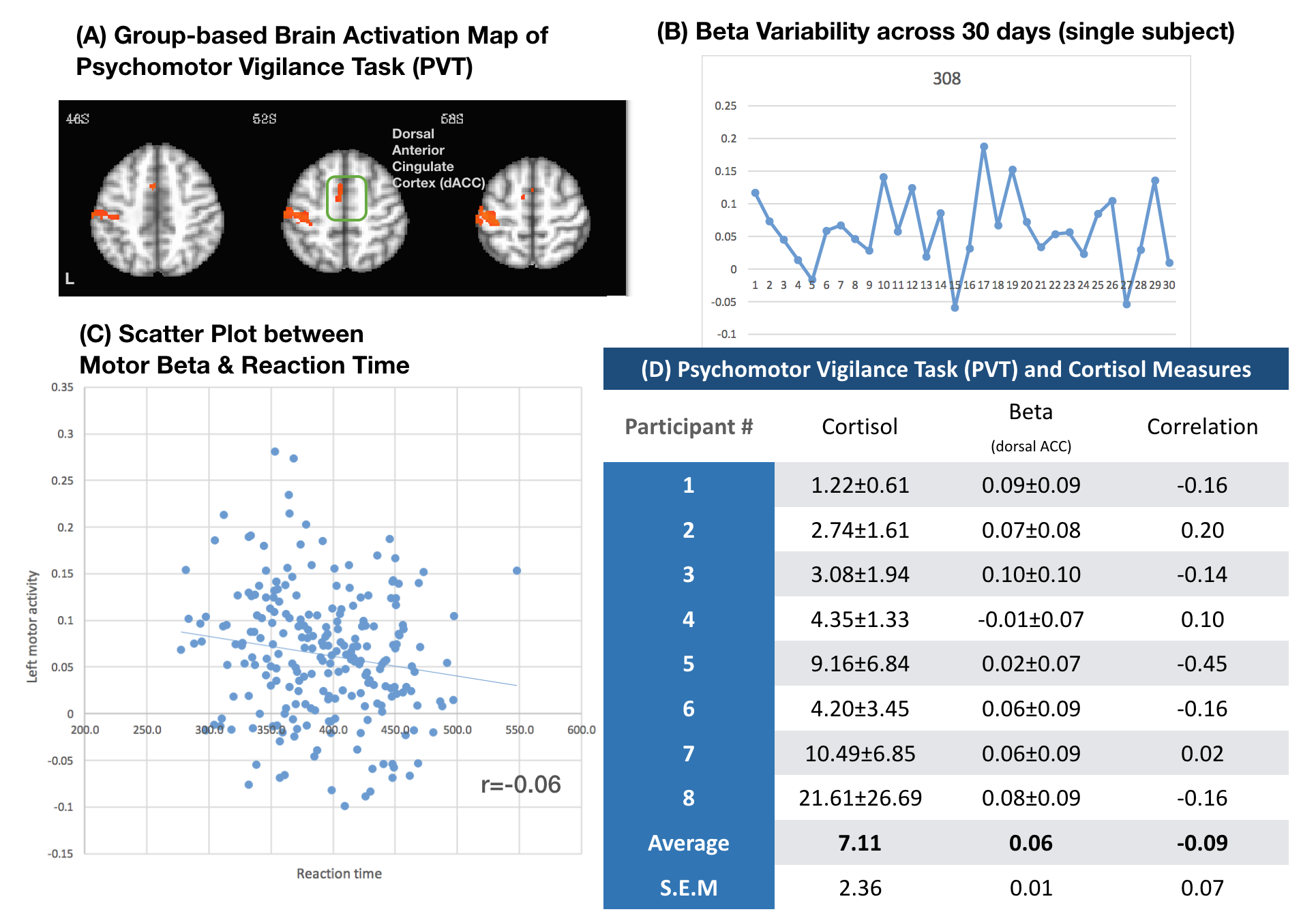
Figure 1A shows the brain activation of dorsal anterior cingulate cortex (dACC) in PVT, and the dACC activity showed weak negative correlation with the PVT reaction time (Fig.1B, r = -0.06). However, the dACC activity seemed unstable across 30 days (Fig. 1C represents a data from single participant), which could be further revealed by the large cross-day standard deviation in the Fig.1D. In biological level, the correlation between dACC and salivary cortisol showed between-individual differences and resulted in a negative correlation (r = -0.09) (Fig.1D). At last, we found that the sleep duration had significant association with the middle temporal gyrus (r = 0.16, p = .013) and the primary motor cortex (r = 0.13, p = .04) in the vigilance task.Discussion and Conclusion
The repeated-measure results indicated the dramatic intra-individual variability of the PVT-based brain activity within 30 days. Previously, Hodkinson et al. presented a significant negative correlation between the salivary cortisol and the ACC perfusion2, but the cortisol-brain association was not observed in the brain activity of PVT (including dACC and the associated brain areas in the dorsal attention network). The finding implied that the baseline perfusion might be modulated by cortisol level, but the brain activity did not. Instead, some brain regions might be affected by the sleep duration of previous day. Other biological factors may be involved to mediate the brain activity or underlying functional connectivity, which awaits further investigations to disclose the reliability across different days.Acknowledgements
This research was supported by Taiwan Ministry of Science and Technology (MOST 108-2321-B-038-005-MY2 and MOST 105-2628-B-038-013-MY3).References
1. Cheeseman JF, Fewster RM, Walker MM. Circadian and circatidal clocks control the mechanism of semilunar foraging behaviour. Sci Rep. 2017;7(1):3780.
2. Hodkinson DJ, O’Daly O, Zunszain PA, et al. Circadian and homeostatic modulation of functional connectivity and regional cerebral blood flow in humans under normal entrained conditions. JCBFM. 2014; 34, 1493-1499.
3. Chen B, Xu T, Zhou C, et al. Individual Variability and Test-Retest Reliability Revealed by Ten Repeated Resting-State Brain Scans over One Month. PLoS One. 2015;10(12):e0144963.
Figures