3825
Multislab multiband 3D EPI for simultaneous high spatial and temporal resolution at 7T1CMRR, University of Minnesota, Minneapolis, MN, United States
Synopsis
Ultra-high field scanners allow recording BOLD images with unprecedented spatio-temporal resolution. These highly precise measurements permit studying the human brain at the mesoscale level, investigating the roles of some of the most fundamental units of neural computations, namely, layers and columns. These submillimeter recordings are however SNR-starved and the ideal choice of sequence remains to be determined. Here, we show that by using a multislab multiband 3D EPI approach we can achieve simultaneously high spatial and temporal resolution for sub-millimeter fMRI applications at 7T. We compare this approach to the more common strategies of 3D EPI and 2D MB EPI.
Introduction
With the growing availability of ultra-high field scanners (UHF - i.e. >= 7 Tesla) and the development of accelerated and highly efficient sequences, such as multiband accelerated 2D (Moeller 2010) or highly accelerated 3D approaches 1-3, researchers are trading the gains in SNR accompanying UHF fMRI to record BOLD images with unparalleled spatial (e.g. 0.8 mm isotropic 4,5) and temporal (e.g. <1000 ms TR 6,7) resolutions. These highly precise measurements in space and time can, at least in principle, aid in bridging the gap between invasive animal electrophysiology and non-invasive human recordings. Scientists can therefore tackle research questions, such as the functional organization of human cortical layers and columns, which had previously been elusive. The debate over the optimal protocol to study the functional profile of cortical layers and columns in humans remains wide open. 2D gradient echo (GE) protocols are still extremely popular because of their ease of use and the familiarity of investigators with the approach. Even at high magnetic fields, sub-millimeter fMRI studies are SNR-starved and, by consequence, temporal SNR (tSNR) starved. It has been reported that 3D approaches can offer significant tSNR advantages, provided that physiological noise is not dominating 1,8. On the other hand, because the required resolution, number of slices, and subsequent TR and flip angles can vary, and consequently the dominant noise regime (i.e. thermal vs. physiological), the optimal sequence choice remains to be determined. As such, we explore an alternative hybrid (2D/3D) EPI pulse sequence strategy, which allows more intrinsic flexibility in the protocol choices. That is, we employ a multi-slab 3D-EPI approach with simultaneous multi-slab capabilities for a high spatial-temporal resolution fMRI application at 7T. This strategy has been proposed in a single shot EVI context to achieve high temporal resolution 9,10, whereas here we use a fully segmented 3D EPI readout per simultaneous slabs (achieving both high spatial and temporal). With this approach we are able to use identical 2D multiband acquisition and reconstruction strategies that are routinely implemented for whole brain lower field, lower resolution fMRI and resting state studies. More importantly, unlike in a pure 2D or 3D EPI approach, a different parameter space is available - number of slices per slab, slab thickness, corresponding TR and flip angle and slab MB factor. These can be adjusted and optimized for a specific spatial-temporal resolution and/or volume coverage. Here, we evaluate the feasibility of such a multi-3D slab multi-band approach for sub-millimeter fMRI applications at 7T and compare it with the more conventional approaches of 2D MB and 3D-EPI.Methods
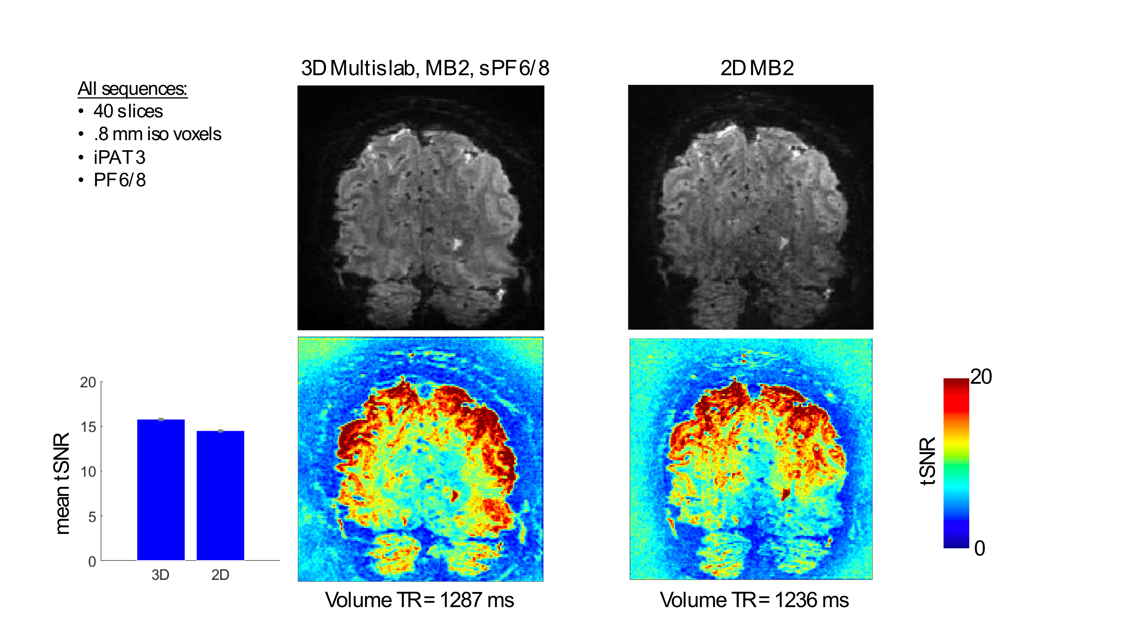
Functional images were acquired using a 3D (single or multi-slab) and a 2D (MB/no MB) GRE EPI sequence on a 7 Tesla scanner with a standard Nova coil (32 TRx). For this case example, the starting requirement was to achieve 0.8 mm isotropic resolution over an ~ 3cm z-FOV (or 40 (after any oversampling) - 0.8 mm slices) while also trying to achieve the shortest TR (around 1 sec.). Stimuli and paradigm: while in the scanner, participant viewed flickering (6 Hz) gratings, centrally positioned on a gray background. The stimuli consisted of 2 conditions: a target and a surround (see figure 2). Stimuli were presented using a standard block design. Analysis: we computed tSNR maps and compared these across sequences (figure 1). We further computed classic GLM analysis to derive b weights summarizing the BOLD activation triggered by our visual conditions and assess its quality and extent. First, we aligned all runs within and across sequences. Then, we used the first runs of each sequence to compute the linear contrast btarget > bsurround to identify the retinotopic representation of the target in V1. These two runs, used to localize our region of interest, were excluded from subsequent analyses, which were confined within the retinotopic representation of the target within right V1. After manually segmenting the cortex, we parcellated the cortical ribbon into 6 equivolume depths, ranging from 10% to 90% distance from the Pial surface.Results
Our results demonstrate that the multiband multi-slab EPI sequence yielded tSNR that was comparable or better to the 2D MB counterpart, depending on location in the brain. While tSNR was somewhat comparable along the cortex, the major difference was observed in the middle of the brain, with the 3D multiband multislab outperforming the 2D. Laminar BOLD profiles were comparable across sequences (figure 2).Conclusion
Here, we show that by using a multislab multiband 3D EPI approach we can achieve a comparable regime (in terms of VAT and coverage) relative to that of 2D GRE EPI for this high-resolution application. While tSNR is extremely relevant for fMRI sensitivity, further data analyses are required to determine whether this translates into improved detection power for a given fMRI paradigm. The approach presented here could have significant implications for laminar fMRI pulse sequence strategies, especially those aiming to achieve simultaneously high spatial and temporal resolutions and those where the contrast to noise is limiting. Further optimizations, (i.e. different number of slabs, slices/slab, slab MB factor, TR, etc.) have the promise of improving high resolution fMRI studies, facilitating a better understanding of the function of cortical layers and columns in humans.Acknowledgements
P41 EB027061
P30 NS076408
References
1. Huber, L. et al. Ultra-high resolution blood volume fMRI and BOLD fMRI in humans at 9.4T: Capabilities and challenges. Neuroimage 178, 769-779, doi:10.1016/j.neuroimage.2018.06.025 (2018).
2. Poser, B. A. & Setsompop, K. Pulse sequences and parallel imaging for high spatiotemporal resolution MRI at ultra-high field. Neuroimage 168, 101-118, doi:10.1016/j.neuroimage.2017.04.006 (2018).
3. Stirnberg, R. et al. Rapid whole-brain resting-state fMRI at 3 T: Efficiency-optimized three-dimensional EPI versus repetition time-matched simultaneous-multi-slice EPI. Neuroimage 163, 81-92, doi:10.1016/j.neuroimage.2017.08.031 (2017).
4. Huber, L. et al. High-Resolution CBV-fMRI Allows Mapping of Laminar Activity and Connectivity of Cortical Input and Output in Human M1. Neuron 96, 1253-1263 e1257, doi:10.1016/j.neuron.2017.11.005 (2017).
5. Muckli, L. et al. Contextual Feedback to Superficial Layers of V1. Curr Biol 25, 2690-2695, doi:10.1016/j.cub.2015.08.057 (2015).
6. Vizioli, L., Yacoub, E. Probing temporal information in fast-TR fMRI data during attention modulations. ISMRM (2018).
7. Lewis, L. D., Setsompop, K., Rosen, B. R. & Polimeni, J. R. Fast fMRI can detect oscillatory neural activity in humans. Proc Natl Acad Sci U S A 113, E6679-E6685, doi:10.1073/pnas.1608117113 (2016).
8. Lutti, A., Thomas, D. L., Hutton, C. & Weiskopf, N. High-resolution functional MRI at 3 T: 3D/2D echo-planar imaging with optimized physiological noise correction. Magn Reson Med 69, 1657-1664, doi:10.1002/mrm.24398 (2013).
9. Chen, L., Feinberg, D. Simultaneous Multi-Slab Echo Volume Imaging: comparison in sub-second fMRI. ISMRM (2013).
10. Kishore
Vakamudi, K., Moeller, S., Ramanna, S., Yoshimoto, A., Yacoub, E., Otazo, R.,
Syed,A.A., and Posse, S. Enhancing Spatial-Temporal Resolution in Simultaneous
Multi-Slab Echo Volumar Imaging. ISMRM (2018).
Figures