3823
Study of the Spinal Cord BOLD functional response1CNR-Nanotec, Roma, Italy, 2fondazione Santa Lucia, Roma, Italy, 3centro fermi, Roma, Italy, 4CNR-Nanotec, piazzale Aldo Moro 5, Italy
Synopsis
Spinal Cord Functional Magnetic Resonance Imaging (fMRI) techniques are widely exploited for the study of brain activation. Similar approaches have been repeatedly attempted in the spinal cord, but spinal cord fMRI has not yet affirmed as habitual tool for the assessment of spinal cord function. One of the reasons is that the features of the functional contrast are still partially unknown. In the present work, we study the relationship between the intensity of the motor stimulation (performing a controlled motor task with the dominant hand ) and the amplitude of the functional response in healthy
INTRODUCTION
Among different imaging methods, functional Magnetic Resonance Imaging (fMRI) represents the most promising tool for non-invasive investigation of human spinal cord functions [1-3] and dysfunctions, such as those following traumatic injury as well as neurodegenerative disease [4-5]. Moreover, scfMRI can be potentially applied to clinical environment to increase the power of early diagnosis and help the development and evaluation of new therapies. However, in spite of these potentialities, the utilization of scfMRI in both research and clinical settings [6] is widely under-exploited, due to either challenges in acquiring good quality data, or the lack of availability of dedicated tools of analysis. In addition, the exact features and even the biophysical origins of the functional response are still unclear. In the present work, we performed a controlled motor task (graded isometric force) of the right dominant hand, and we parametrically studied the relationship between stimulation strength and functional response in heathy subject.METHODS
We acquired axial and sagittal functional images at 3T from the spinal cord of forty-six healthy subjects (all right-handed with a mean age of 40 years) while performing a block-design isometric motor task consisting of 5 cycles of 30s/30s rest/task epochs. We implemented and optimized a scfMRI data analysis pipeline built around the Spinal Cord Toolbox (SCT)[7]. The pipeline was applied to functional (gradient echo EPI) images acquired on a Philips Achieva 3T MR scanner using a neurovascular coil array with the following sequence parameters: TE/TR = 25/3000 ms, Flip angle = 80°, FOV = 192x144x104 mm (sagittal) or 140x140x143 mm (axial), acquisition Matrix = 128x128x35 (sagittal) or 96x96x34 (axial), resolution = 3x1.5x2 mm (sagittal) or 1.5x1.5x3 mm (axial). Anatomical reference images were acquired using T1-weighted gradient echo sequence (TE/TR 5.89/9.59 ms, flip angle 9°, FOV = 240x240x192 cm , resolution 0.75x0.75x1.5mm ).RESULTS
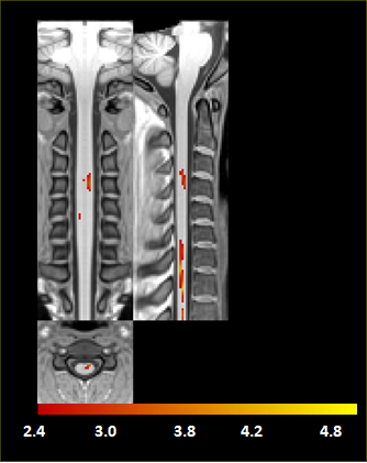
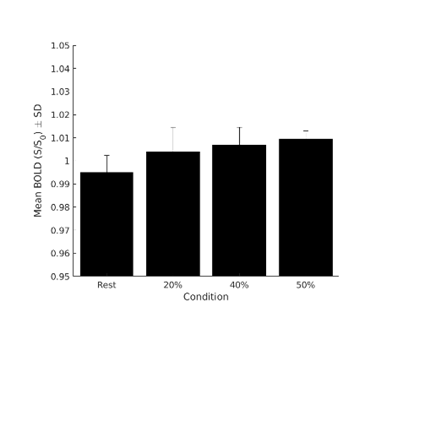
The activation maps (threshold at p<0.001 uncorrected) in the PAM50 tamplate space, using the recorded force data acquired, is reported in figure 1. Positive signal changes were principally detected at C3–C4 vertebral levels and C5-C7, which is consistent with studies employing motor stimuli of the handIn figure 2 we report the mean BOLD change as a function of the grip strength. The results obtained suggest a parametric dependence of functional response in the spinal cord on the stimulation strength in an isometric motor task.
DISCUSSION
A parametric dependence of functional response in the spinal cord on the stimulation strength in an isometric motor task is reported. Such a dependence is important to understand the physiological origin of the response. In addition, this result is of great importance for the SC-fMRI application in neuroradiology, and in particular for the assessment and follow-up of spinal injuries, pain, and neurodegenerative diseases (e.g multiple sclerosis), as well as in the development and evaluation of new therapies.CONCLUSIONS
We implemented and optimized a scfMRI data analysis pipeline built around the Spinal Cord Toolbox (SCT). We acquired axial and sagittal functional images at 3T from the spinal cord of forty-six healthy subjects performing an isometric motor task. Using our SCT-based pipeline, we substantially improved motion correction and image registration. Furthermore, we found task-induced activations with a high level of statistical significance.Acknowledgements
This research was financially supported by The Italian Ministry of Health Young Researcher Grant 2013 (GR-2013-02358177).References
1) Maieron, M., Iannetti, G.D., Bodurka, J., Tracey, I., Bandettini, P.A., and Porro, C.A. (2007). Functional responses in the human spinal cord during willed motor actions: evidence for side- and rate-dependent activity. J Neurosci 27, 4182-4190
2) Stroman, P.W., Coe, B.C., and Munoz, D.P. (2011). Influence of attention focus on neural activity in the human spinal cord during thermal sensory stimulation. Magn Reson Imaging 29, 9-18.
3) Weber, K.A., 2nd, Chen, Y., Wang, X., Kahnt, T., and Parrish, T.B. (2016). Lateralization of cervical spinal cord activity during an isometric upper extremity motor task with functional magnetic resonance imaging. Neuroimage 125, 233-243.
4) Filippi, M., Rovaris, M., and Rocca, M.A. (2004). Imaging primary progressive multiple sclerosis: the contribution of structural, metabolic, and functional MRI techniques. Mult Scler 10 Suppl 1, S36-44; discussion S44-35.
5) Stroman, P.W., Khan, H.S., Bosma, R.L., Cotoi, A.I., Leung, R., Cadotte, D.W., and Fehlings, M.G. (2016). Changes in Pain Processing in the Spinal Cord and Brainstem after Spinal Cord Injury Characterized by Functional Magnetic Resonance Imaging. J Neurotrauma 33, 1450-1460.
6) Martin, A.R., Aleksanderek, I., Cohen-Adad, J., Tarmohamed, Z., Tetreault, L., Smith, N., Cadotte, D.W., Crawley, A., Ginsberg, H., Mikulis, D.J., and Fehlings, M.G. (2016). Translating state-of-the-art spinal cord MRI techniques to clinical use: A systematic review of clinical studies utilizing DTI, MT, MWF, MRS, and fMRI. Neuroimage Clin 10, 192-238.
7) De Leener B, Levy S, Dupont SM, Fonov VS, Stikov N, Louis Collins D et al. SCT: Spinal Cord Toolbox, an open-source software forprocessing spinal cord MRI data. Neuroimage 2017; 145(Pt A): 24-43.
Figures