3775
Synthetic MRI with T2-based Water Suppression without Loss of Tissue SNR1Department of Radiological Science, Shizuoka College of Medicare Science, Hamamatsu-Shi, Japan
Synopsis
We proposed a modified T2-based water suppression synthetic-MRI technique without loss of tissue SNR, which was introduced by subtraction of heavy-T2W image from standard acquired images. Our water suppression was achieved by subtracting only water portions except for tissue portions. We demonstrated both effects that CSF-PVE artifacts were dramatically suppressed and the tissue SNR was kept to before subtraction in our water suppressed quantitative maps; and thus water suppressed synthetic images of FLAIR and SE provided better gray-white matter contrasts than those for subtracting uniformly.
Introduction
A synthetic MRI (Syn.MRI), that generates several kinds of contrast-weighted images by synthesizing quantitative parameter maps of proton density (PD), T1-longitudinal relaxation time (T1), and transverse relaxation time (T2) [1] obtained using acquired fast-spin echo (FSE) data, is beginning to be used clinically because of the advantages of saving total examination time and the freedom to select MRI acquisition parameters of repetition time (TR), inversion time (TI), and echo time (TE). However, a synthetic T2-weighted (T2W) Fluid-attenuation inversion recovery (FLAIR) introduces hyper intense artifacts at the border zone of tissue and ventricle or the surface of the brain, and it is considered that the cause is partial volume effects (PVE) of brain tissue and cerebral spinal fluid (CSF) [2,3]. To solve those artifacts, several methods were proposed [4,5]. However, those methods were not essential or required complicated procedure.In contrast, a synthetic MRI combined with T2-based water suppression technique (Wsup-Syn.MRI) was proposed [6]. However, in exchange for reducing CSF-PVE artifacts, the tissue SNR was reduced by ~ 1/√2 for the standard technique by subtracting long-TE image from standard images; and also it was difficulty in optimal suppression.
The purpose of this study was to propose and assess a modified Wsup-Syn.MRI without loss of tissue SNR.
Methods
TheoryWhen a unit voxel consists of two components of water and tissue, MR signal in the voxel is based on a two-compartment model [6]. Our proposed water suppression was based on the technique of subtracting additionally acquired long TE data of water signal dominant from the shorter TE data [7]. A processing flow for our proposed technique of water-suppressed synthetic MRI using minimum number of acquired images is shown in Fig. 1 for whole, and the detail of “Water suppression for acquired images” is as follows (Fig, 2).
a) TE-dependent Water Scaling: To obtain optimal signal intensity (S.I.) of water considering T2 decay, TE-dependent scaling factor (α(TEn)) was obtained after measuring a pure water S.I., Sw(TEn) in n’th TE, TEn, followed by divided by a long-TE S.I., Sw(TElong) as: α(TEn)=Sw(TEn)/Sw(TElong).
b) Spatially-dependent Water Masking: Since the S.I. of long-TE image is assumed to be proportional to water volume, Vw , the S.I. after normalizing by the pure water S.I. ( e.g. maximum) in long-TE image is regarded to be a Vw map. A spatially-dependent water mask (Mask) is generated from long-TE image by setting a threshold of Vw (ThVw) followed by applying spatial filter to smooth the boundary, where the ThVw must be decided considering the S.I. of water and tissue noise level.
c) Finally, water suppressed acquired images are obtained by subtraction from the standard image with weighting to Sw(TElong) using α(TEn) and Mask.
MRI Experiments
In MR experiments, the first four data in Fig. 1 were acquired for our proposed Wsup-Syn.MRI. A healthy volunteer study was performed on a MRI (‘Galan 3T ZGO’, Canon Medical Systems corp., Otawara, Japan) with a 32-channel head coil after obtaining informed consent. A fast spin echo sequence was used and the acquisition parameters were: parallel imaging (SPEEDER) of speed-up factor 2, acquisition matrix of 256 x 256, display matrix of 512 x 512 after sinc interpolation, FOV=23cm, slice thickness=5mm, the number of slices was selected at maximum, NAQ=1, TR1=10000ms, TE1=20ms, TE2=100ms, TElong=500ms, TI1=1000ms, an adiabatic inversion pulse was used for IR to reduce B1 inhomogeneity. Quantitative maps and synthetic contrast images of SE and FLAIR were compared between masking subtraction and uniform subtraction.
Results
For the effects of our proposed Wsup-images with masking compared with the Wsup-images without masking, CSF signals were comparably suppressed and the tissue SNRs were increased by 50% by selecting optimal ThVw value (Fig. 3 and Table 1). For the Wsup-synthetic T2W images of SE and FLAIR, signal intensities in CSF portions were reduced almost optimally and the tissue SNRs were kept comparably as the standard synthetic images (Fig. 4). Furthermore, for synthetic-images, the gray-white matter contrast ratio for our proposed masking subtraction was ~5% better than those for spatially uniform subtraction and equivalent to those for the standard (non-subtraction) method. (Table 1).Discussion
We proposed a modified water suppressed synthetic MRI (Wsup-Syn.MRI) technique to optimally reduce the CSF-PVE artifacts in conventional synthetic MRI without loss of tissue SNR by applying a spatial mask to separate water and tissue portions when subtraction, while the CSF-PVE artifacts were almost perfectly reduced and the tissue SNR and GM-WM contrast in quantitative maps and synthetic images of SE and FLAIR were kept. Our proposed Wsup-Syn.MRI is very simple and easily combined to the conventional Syn.MRI technique and has a backward compatibility. Our proposed Wsup-Syn.FLAIR will be easily applied to clinical use. Although T2-base Wsup-technique [7] has still not generally applied clinically despite the advantage of tissue SNR. Since our proposed Wsup-Syn.MRI can compare both easily just by calculation, it is a possibility to widely applied in future.Conclusion
Although further optimization of pulse-sequence and processing techniques, and clinical assessments especially for long T2 lesion, our proposed synthetic MRI technique with T2-based water suppression without loss of tissue SNR will solve the problem of CSF-PVE artifacts in current synthetic FLAIR and is expected to become clinically further useful.Acknowledgements
We express our sincere thanks to Yuki Takai and Ryo Shiroishi in Canon Medical Systems corporation for supporting data acquisition and analysis in this study.References
[1] Warntjes JB et al. Rapid magnetic resonance quantification on the brain: optimization for clinical usage. Magn Reson Med. 2008;60:320–329.
[2] Hagiwara A et al. SyMRI of the Brain: Rapid Quantification of Relaxation Rates and Proton Density, With Synthetic MRI, Automatic Brain Segmentation, and Myelin Measurement. Investigative Radiology. 2017; 52:647–657.
[3] Tanenbaum LN et al. Synthetic MRI for Clinical Neuroimaging: Results of the Magnetic Resonance Image Compilation (MAGiC) Prospective, Multicenter, Multireader Trial. AJNR 2017;38:1103-1110.
[4] Hwang KP et al. 3D isotropic multi-parameter mapping and synthetic imaging of the brain with 3D-QALAS: Comparison with 2D MAGIC. ISMRM, 2018, 26th: #5627.
[5] Gong E et al. Improved Synthetic MRI from multi-echo MRI Using Deep Learning. ISMRM, 2018,26th: #2795.
[6] Kimura T et al. Synthetic MRI with water suppression technique to reduce CSF partial-volume dependent artifacts in brain. ISMRM, 27th 2019: #4867.
[7] Essig M et al. Assessment of cerebral gliomas by a new dark fluid sequence, high intensity REduction (HIRE): a preliminary study. JMRI. 2000;11:506-517.
Figures

Fig. 1 Whole process flow for our proposed technique of water-suppressed synthetic MRI (Wsup-Syn.MRI) using minimum number of acquired images.
Actual images or quantitative maps in each stage were shown in the right side
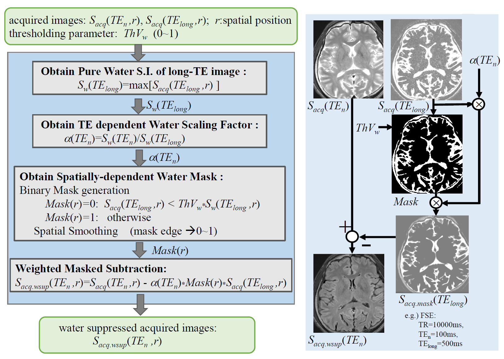
Fig. 2 Detailed process flow for “Water suppression for acquired images” in Fig. 1.
Examples of actual images and quantitative maps in each stage were shown in the right side.

Fig. 3 Images of heavyT2W (TElong=500ms) and T2W (TE2=100ms) as a parameter of masking threshold, ThVw.
Note that the water (CSF) signals were reduced with decreasing ThVw, and ThVw=0.05 is optimal from the views of tissue SNR and water suppression effects.
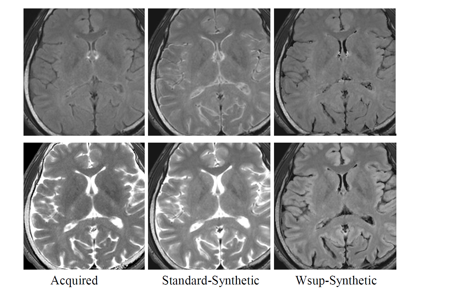
Fig. 4 Comparison of Acquired, Standard-Synthetic, and Wsup-Synthetic images of FLAIR (top) of (TR,TI,TE)[ms]=(10000, 2700,100), and SE (bottom) of (TR,TE)[ms]=(10000,100).
Note that the CSF-PVE artifacts in the Wsup-Synthetic FLAIR image were reduced as in the Acquired FLAIR image while the tissue SNR and gray matter (GM) - white matter (WM) contrast for Wsup-Synthetic FLAIR image were kept as in the Standard-Synthetic FLAIR image.
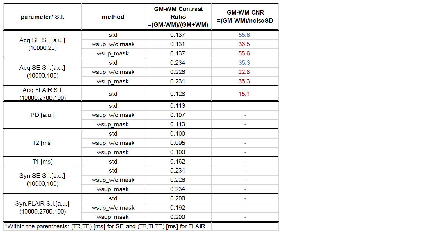
Table.1 Contrast-Ratio (CR) and/or Contrast-Noise-Ratio (CNR) between gray matter(GM) and white matter (WM) in contrast weighted images, quantitative parameters, and synthetic images, each with three methods of standard, without and with T2-based water suppression.
Note that the tissue SNR and gray matter (GM) - white matter (WM) contrast for Wsup-Syn.FLAIR image with masking were kept as in the Standard-Syn.FLAIR image, though those were reduced in the w/o masking. In addition, the GM-WM contrast for Wsup-Syn.SE image was better than for the Wsup-Syn.FLAIR image.