3745
A generalized combined FISP and PSIF MR Fingerprinting with improved T2 quantification1State Key Laboratory of Modern Optical Instrumentation, College of Optical Science and Engineering, Zhejiang University, Hangzhou, China, 2Center for Brain Imaging Science and Technology, Key Laboratory for Biomedical Engineering of Ministry of Education, College of Biomedical Engineering and Instrumental Science, Zhejiang University, Hangzhou, China, 3MR Collaborations, Siemens Healthcare Ltd., Shanghai, China
Synopsis
A generalized combined FISP and PSIF MR Fingerprinting sequence is proposed where either FISP block or PSIF block can be used in each TR. An example sequence pattern shows improved T2 mapping accuracy and its immunity to slice profile imperfection.
Introduction
MR Fingerprinting has been developed to have multiple variants of sequences (bSSFP based, FISP based, SPGR based etc.)[1]-[3] to resolve different problems in simultaneously mapping multiple parameters. Among them, FISP MRF is able to simultaneously obtain T1 and T2 maps without the banding artifact as in bSSFP MRF. While other systematic imperfections like slice profile effects can highly degrade the accuracy and are normally resolved by modeling into dictionary which has been shown for bSSFP MRF and cardiac FISP MRF[4]-[6]. SOIDE MRF has been introduced to improve the T2 accuracy with the introduced PSIF echo[7]. Here a generalized combined FISP and PSIF MRF sequence is proposed and validated with phantom and in vivo exam, and slice profile effect is investigated for both FISP MRF and the proposed MRF.Methods
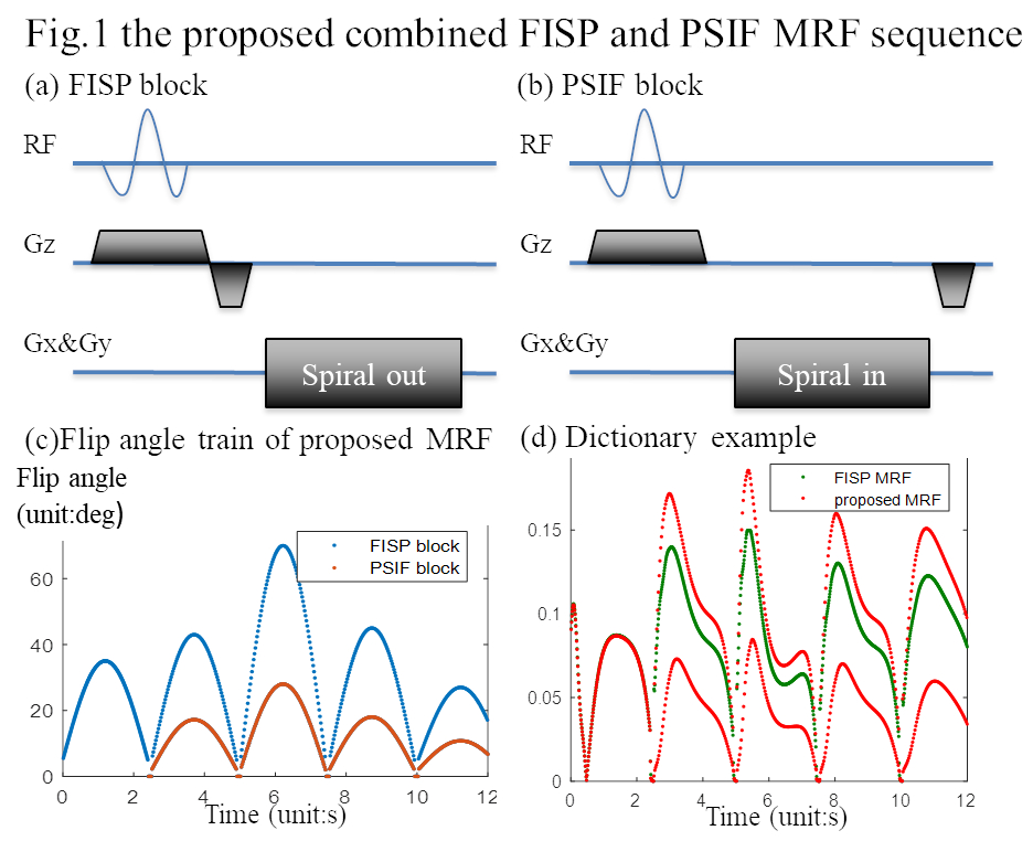
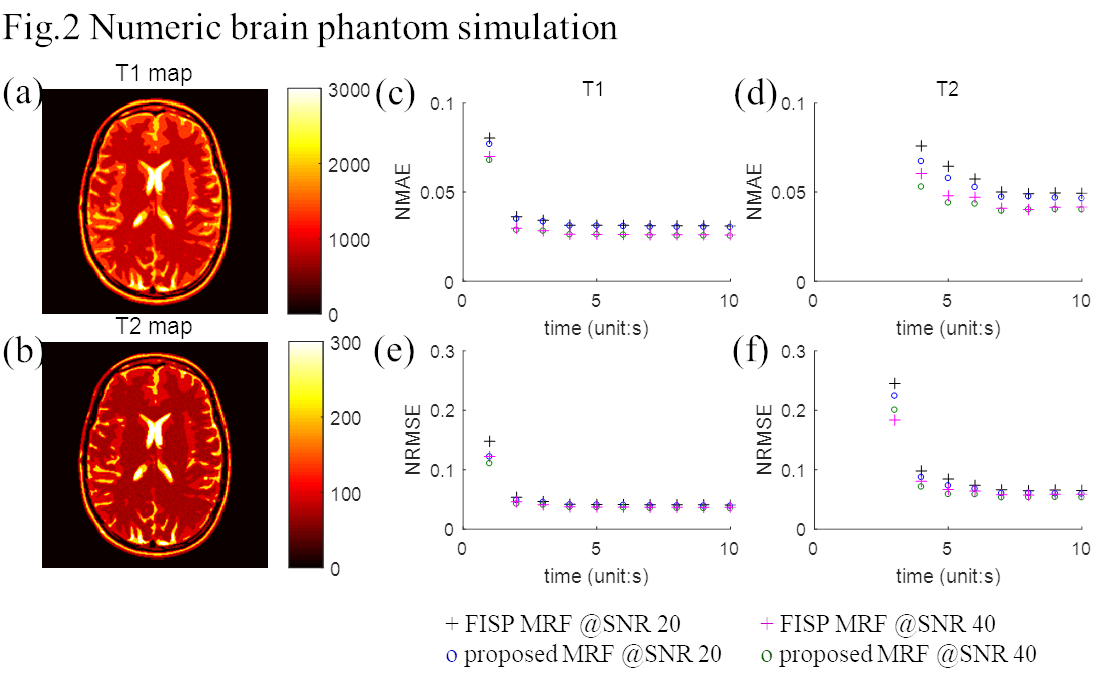
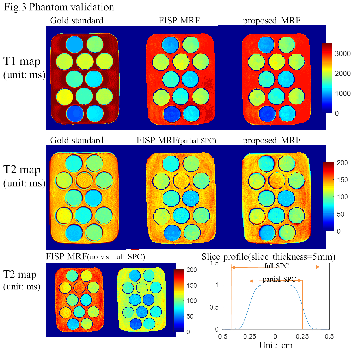
Combined FISP and PSFI MRF sequence consists of conventional FISP blocks and PSIF blocks. FISP block is as in Fig.1(a), and PSIF in Fig.1(b). Either block can be chosen and inserted into any TR which makes it generalized to design. The pattern here is chosen empirically as shown in Fig.1(c) where in the first 2.5s all FISP blocks are used and later interleaved FISP and PSIF blocks are used. The flip angles for PSIF blocks is about 0.4x of the ones for the neighboring FISP blocks. One example dictionary element with WM representative (T1=800ms, T2=70ms) from both FISP MRF and the proposed MRF is shown in Fig.1(d). To validate the performance of the proposed MRF, numeric brain phantom simulation is conducted, which matches the sequence parameter, sampling trajectory and noise distribution as in real exam. Two different level of noise is added where SNR=20 and 40 for mean WM representative signal. After simulation, global statistics are calculated for NMAE(normalized mean absolute error) and NRMSE(normalized root mean square error).Mixed agar and Mncl2 phantom which contains 12 different T1 and T2 value tubes is used for phantom experiment. Three datasets which consist gold standard data, FISP MRF data and the proposed MRF data are acquired. Gold standard T1 acquisition uses the multi TI IR SE sequence and gold standard T2 uses the multi TE SE sequence. The T1 and T2 values are exponential fitted voxel-wise. For FISP MRF and the proposed MRF, tiny golden angle[7] spiral is used in acquisition and gridding reconstruction is for reconstruction. Dictionaries are precalculated with EPG based method[9] and B1+ term is also taken into account. Two additional dictionaries for FISP MRF with full SPC(slice profile correction) and partial SPC which corresponds to the full slice profile area and the given slice thickness area (as shown in Fig.3) are calculated. B1+ maps are additionally measured with the TurboFLASH based method[10].
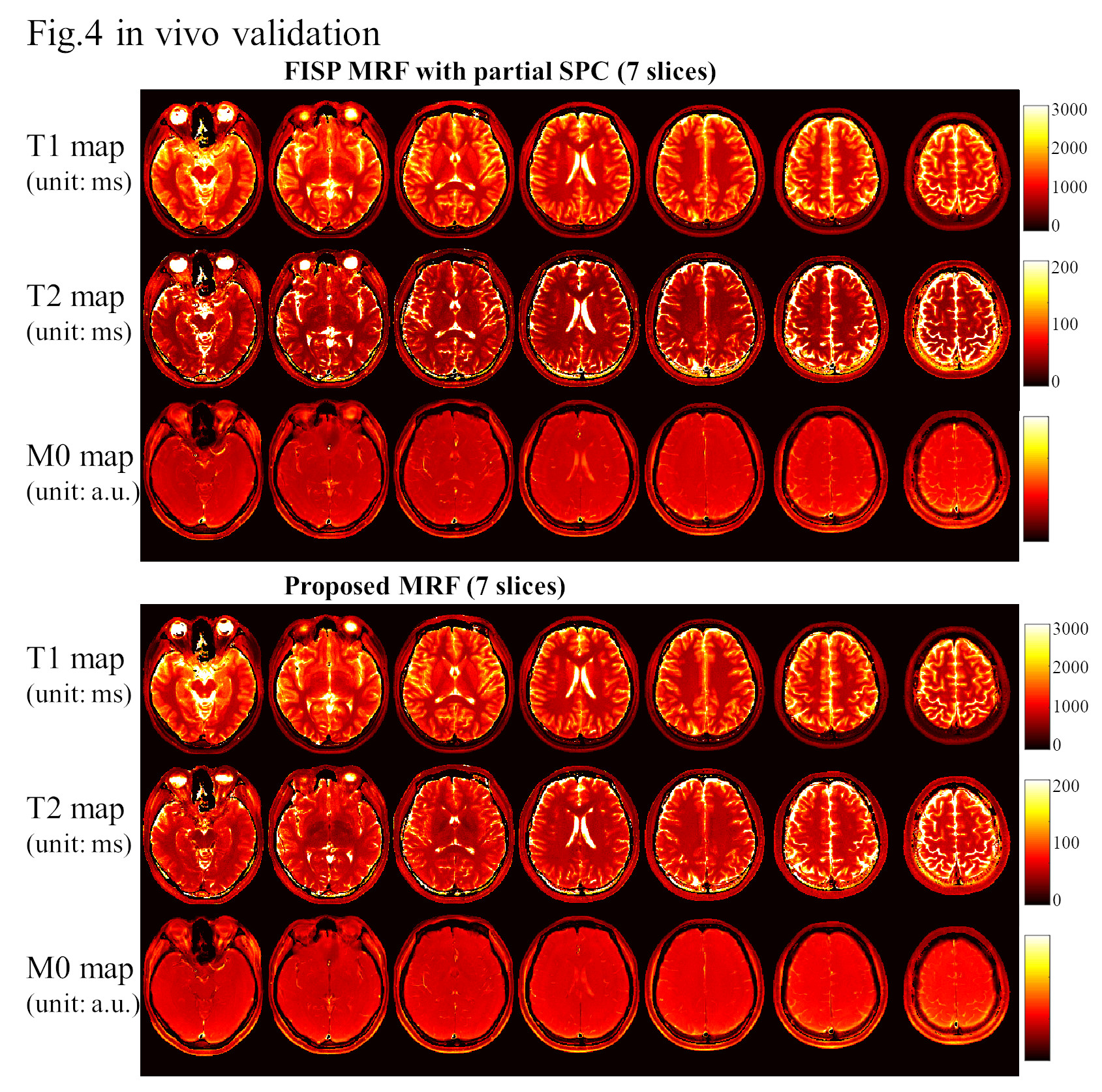
Invivo experiment is also conducted on a healthy volunteer with written consent where 7 slices are acquired for FISP MRF and the proposed MRF. Same acquisition and reconstruction is used in vivo as with the phantom.
All the experiments are conducted on a 3T Siemens Prisma scanner with the 20ch head-neck coil.
Results
Fig.2 shows the simulation result where the NMAE and NMRE of T1 shows not much difference for the two cases, while the NMAE of T2 from the proposed method shows slight improvement with 4-7s/slice acquisition as compared with the FISP MRF.Fig.3 shows the phantom experiment result from 12s/slice acquisition, where the T1 from the 12 tubes has high consistency with the gold standard and the large T1 area shows slightly underestimation. T2 from FISP MRF with partial SPC result show the best consistence with the gold standard result. The T2 from the proposed MRF also show good consistence with the gold standard result, and it is from without SPC.
Fig.4 shows the in vivo results from partial SPC FISP MRF and the proposed MRF at 12s/slice acquisition where 7 slices of T1, T2 and M0 maps are shown.
Conclusions and Discussions
The proposed MRF shows good T1 and T2 quantification accuracy and its immunity to slice profile imperfection. FISP MRF with partial SPC shows best accuracy as compared with without SPC and with full SPC. The proposed sequence pattern is empirically chosen and suboptimal, and further optimization may further improve the mapping accuracy[11].Acknowledgements
This work is supported in part by by Shenzhen Innovation Funding (JCYJ20170816172431715, JCYJ20170818164343304) and National Natural Science Foundation of China (No: 61701436, U1809204, 61525106, 61427807), and by the National Key Technology Research and Development Program of China (No: 2017YFE0104000).References
[1] D. Ma et al., “Magnetic resonance fingerprinting,” Nature, vol. 495, no. 7440, pp. 187–192, 2013.
[2] Y. Jiang, D. Ma, N. Seiberlich, V. Gulani, and M. A. Griswold, “MR fingerprinting using fast imaging with steady state precession (FISP) with spiral readout,” Magn. Reson. Med., vol. 74, no. 6, pp. 1621–1631, 2015.
[3] B. Rieger et al., “Time efficient whole-brain coverage with MR Fingerprinting using slice-interleaved echo-planar-imaging,” Sci. Rep., vol. 8, p. 6667, 2018. [4] T. Hong, D. Han, M. O. Kim, and D. H. Kim, “RF slice profile effects in magnetic resonance fingerprinting,” Magn. Reson. Imaging, vol. 41, pp. 73–79, 2017.
[5] D. Ma et al., “Slice profile and B1corrections in 2D magnetic resonance fingerprinting,” Magn. Reson. Med., vol. 78, no. 5, pp. 1781–1789, 2017.
[6] J. I. Hamilton et al., “Investigating and reducing the effects of confounding factors for robust T 1 and T 2 mapping with cardiac MR fingerprinting,” Magn. Reson. Imaging, vol. 53, pp. 40–51, 2018.
[7] H. Ye et al., “Spiral-out and -in Double Echoes (SOIDE) Magnetic Resonance Fingerprinting with Improved T2 Mapping,” in Proc Int Soc Magn Reson Med, 2017.
[8] K. Haris et al., “Self-gated fetal cardiac MRI with tiny golden angle iGRASP: A feasibility study,” J. Magn. Reson. Imaging, vol. 46, no. 1, pp. 207–217, 2017.
[9] M. Weigel, “Extended phase graphs: Dephasing, RF pulses, and echoes - Pure and simple,” in Journal of Magnetic Resonance Imaging, 2015, vol. 41, no. 2, pp. 266–295.
[10] S. Chung, D. Kim, E. Breton, and L. Axel, “Rapid B1+ mapping using a preconditioning RF pulse with turboFLASH readout,” Magn. Reson. Med., vol. 64, no. 2, pp. 439–446, 2010.
[11] B. Zhao et al., “Optimal experiment design for magnetic resonance fingerprinting: Cramér-rao bound meets spin dynamics,” IEEE Trans. Med. Imaging, vol. 38, pp. 844–861, 2019.
Figures