3726
Dual-Repetition Gradient-Echo Dixon Imaging with High Signal-to-Noise Ratio Efficiency
Holger Eggers1, Christoph Katemann2, and Hendrik Kooijman2
1Philips Research, Hamburg, Germany, 2Philips Healthcare, Hamburg, Germany
1Philips Research, Hamburg, Germany, 2Philips Healthcare, Hamburg, Germany
Synopsis
Dual-repetition gradient-echo sequences are currently not widely used for Dixon imaging, because shorter scan times and better signal-to-noise ratios are usually achieved with their dual-echo counterparts. In this work, the efficiency of dual-repetition gradient-echo sequences is optimized by introducing partial echo sampling to extend the acquisition windows and by applying compressed sensing to reduce scan times. On the example of knee imaging, this is shown to enable high-resolution water-fat imaging with good image quality in reasonable scan times.
Introduction
Dixon imaging with bipolar dual-echo gradient-echo sequences is nowadays routinely included in a broad range of clinical examinations, for example of the abdomen, pelvis, breast, and head and neck.1 The receive bandwidth and echo spacing of these sequences typically increase with spatial resolution, affecting the signal-to-noise ratio (SNR) and water-fat separation. By contrast, dual-repetition gradient-echo sequences permit choosing the receive bandwidth and echo shift independent of spatial resolution, which makes them particularly suited for high-resolution imaging. In the present work, the acquisition duty cycle of these sequences is enhanced by eliminating the dead times commonly introduced to implement the echo shifting. Moreover, scan times acceptable for clinical examinations are demonstrated with these sequences by employing compressed sensing.Methods
A standard three-dimensional (3D) dual-repetition spoiled gradient-echo sequence is illustrated in Fig. 1. Alternately, it samples full echoes at two echo times TE1 and TE2 by shifting the rephasing lobes of the readout gradient and the acquisition windows (AQ). Dead times of ΔTE = TE2 - TE1 are inserted before or after the rephasing lobes to keep the repetition time (TR) constant. Obviously, they decrease the acquisition duty cycle, defined as the ratio of AQ duration and TR. To exploit these dead times, the duration of the rephasing lobes is extended symmetrically by 2 ΔTE and the strength is scaled accordingly. As shown in Figs. 1 and 2, this leads, for the first echo at the shorter TE1, to an overlap with the preceding dephasing lobe with opposite polarity and thus to a reduction in strength or duration of the dephasing lobe. For the second echo at the longer TE2, an overlap with the subsequent spoiling lobe with identical polarity occurs. It requires, in general, a prolongation of the spoiling lobe. The AQ is stretched by ΔTE, stopping later for the first echo and starting earlier for the second echo. At the same time, it is shrunk for the second echo, stopping earlier, to accommodate the longer spoiling lobe. In this way, the proposed sequence samples only partial echoes, with different partial echo factors PE1 and PE2 at TE1 and TE2, respectively. A further optimization, illustrated in Figs. 2 and 3, is obtained by adjusting the duration of the rephasing lobe of the readout gradient and of the AQ for the first echo to the minimum duration of the dephasing lobe of the readout gradient and the minimum duration of the phase encoding and slab selection gradients. Usually, this results in smaller PE1 in the periphery of k-space.Experiments were performed on a 3 T Ingenia scanner (Philips Healthcare, Best, Netherlands) using a knee coil with eight receive channels. Data were acquired on phantoms and in volunteers with the standard and the proposed sequence. Complex single-echo images were individually generated with a compressed sensing reconstruction and were then jointly processed with a two-point Dixon method.2-6 Optionally, a homodyne reconstruction enclosed both.7
Noise measurements were carried out on phantoms with the radiofrequency excitation and any scan acceleration disabled, selecting a resolution of 0.5 x 0.5 x 3.0 mm3 to limit scan times. Relative SNR was predicted by $$rSNR_p=\sqrt{2\left(1-\cos(\theta)\right)/\left(\left(\frac{AQ_F}{AQ_{P_1}}\right)^2+\left(\frac{AQ_F}{AQ_{P_2}}\right)^2\right)},$$ where θ is the dephasing angle of water and fat over ΔTE, and AQF and AQP are the AQ durations in the standard and the proposed sequence, respectively. It was compared to the ratio of standard deviations in a region-of-interest in the complex water image and in the complex single-echo images obtained with the standard sequence.
Results
Predicted and measured relative SNR of six noise measurements are compiled in Tab. 1. They are in excellent agreement and are consistently higher with the proposed sequence, with diminishing gain over the standard sequence towards lower receive bandwidth. Representative examples of knee images are provided in Fig. 4. A field of view of 170 x 170 x 170 mm3 was covered with an acquired resolution of 0.4 x 0.4 x 2.0 mm3 in 3:25 min in this case. Further parameters of the dual-repetition gradient-echo sequence included a flip angle of 12°, a TR/TE1/TE2 of 10.0/4.5/5.5 ms, a minimum PE1/PE2 of 0.87/0.80, and a 4-fold scan acceleration.Discussion
In Dixon imaging, the minimum receive bandwidth is not limited by the fat shift, since the fat shift can be compensated.8 With dual-repetition gradient-echo sequences, it is not restricted by the robustness of the water-fat separation either. High SNR efficiency can thus be achieved by choosing longer AQs and exploiting the resulting longer TRs to increase acquisition duty cycle and steady-state magnetization, respectively. The proposed partial echo sampling further contributes to it. Finally, part of the SNR gain can advantageously be spent on higher scan acceleration to keep scan times reasonable, as previously shown for parallel imaging.9Partial sampling of both echoes allows keeping the fat shift and main field inhomogeneity-induced distortion the same in both repetitions, facilitating an accurate water-fat separation. The SNR gain it provides is limited by the required prolongation of the spoiling gradient towards higher receive bandwidth and by the decreasing ratio of fixed ΔTE to variable AQ durations towards lower receive bandwidth. Further improvements in image quality are expected from applying an integrated reconstruction and water-fat separation.10-11
Acknowledgements
No acknowledgement found.References
1. Eggers H, et al. J Magn Reson Imaging 2014; 40:251-268. 2. Zhao C, et al. Proc ISMRM 2008; 1478. 3. Wu B, et al. Proc ISMRM 2008; 1480. 4. King KF. Proc ISMRM 2008; 1488. 5. Liu B, et al. Proc ISMRM 2008; 3154. 6. Eggers H, et al. Magn Reson Med 2011; 65:96-107. 7. Reeder SB, et al. Magn Reson Med 2005; 54:586-593. 8. Reeder SB, et al. J Magn Reson Imaging 2007; 25:644-652. 9. Weiger M, et al. Magn Reson Med 2005; 53:177-185. 10. Doneva M, et al. Magn Reson Med 2010; 64:1749-1759. 11. Ong F, et al. Magn Reson Med 2018; 80:112-125.Figures

Fig. 1. Sequence diagram of a standard 3D dual-repetition
spoiled gradient-echo sequence. Schematically shown are the readout gradient (GR),
the phase encoding gradient (GP), the slab selection gradient (GS),
the radiofrequency pulses (RF), and the acquisition windows (AQ) for two
consecutive repetition times. The dotted red lines illustrate the suggested extensions
of the rephasing lobes of the readout gradient and of the acquisition windows.

Fig. 2. Sequence diagram of the proposed 3D
dual-repetition spoiled gradient-echo sequence. The suggested extensions entail
a different, partial sampling of the two echoes. The dotted red, blue, and
black lines illustrate a further optimization for the first echo, involving an
adaptation of the readout gradient and of the acquisition
window to the varying phase encoding and slab selection gradients.

Fig. 3. k-space coverage for the first echo (red) and
the second echo (blue) applying the further optimization from Fig. 2.
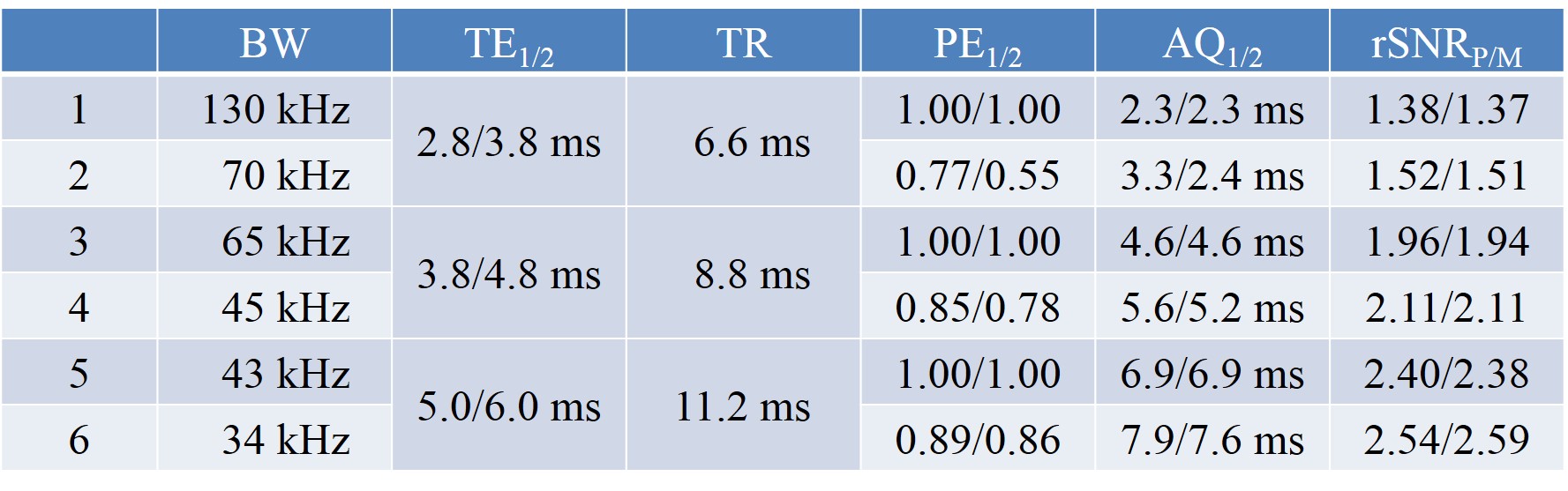
Tab. 1. Selected parameters and results of noise measurements with three corresponding sequences
with full echo (1, 3, 5) and partial echo (2, 4, 6) sampling.
Listed are the receive bandwidth (BW), the echo times (TE), the repetition time
(TR), the partial echo factors (PE), the acquisition window durations
(AQ), and the predicted/measured relative SNR (rSNRP/M).
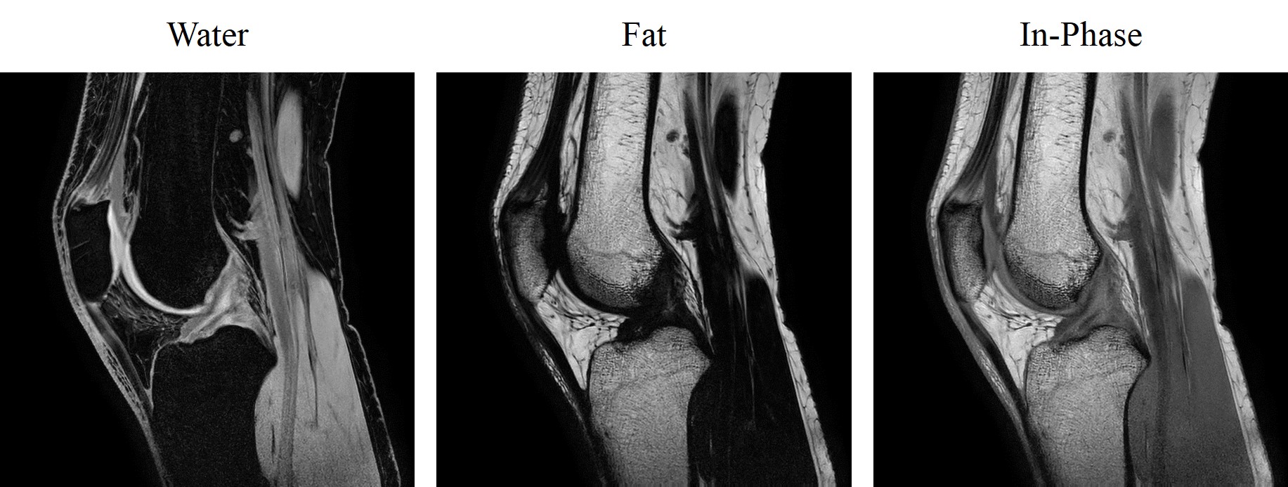
Fig. 4. Sagittal knee images acquired with a spatial
resolution of 0.4 x 0.4 x 2.0 mm3. Shown
are a fat-suppressed or water image (left), a water-suppressed or fat image
(middle), and an in-phase image synthesized with fat shift compensation (right).