3725
Dual Stage Super-Resolution in Quadratic Phase Scrambling Imaging using Iterative Signal Band Expansion and Deep CNN Super-resolution1Utsunomiya University, Utsunomiya, Japan
Synopsis
Spatial resolution of images in phase scrambling Fourier transform imaging can be improved by post-processing signal band extrapolation. However, the improvements is small in the central area of image space. In our research, super-resolution using deep convolutional neural network are applied to temporally resolution improved images through iterative reconstruction. Simulation and experimental results showed that spatial resolution was fairly improved in the central area as well as in the edge of images. Proposed method is applicable to phase varied images by using adequately estimated phase distribution map since signal under-sampling is not utilized in proposed method.
Introduction
The signal obtained in phase scrambling Fourier transform imaging (PSFT) [1] can reconstruct high-resolution images without signal under-sampling which is common in compressed sensing [2,3]. Higher resolution is obtained in the edge of image space, however, resolution improvement is small in the central area. In this work, we consider two stage cascaded resolution improvements; i.e. improvement by iterative processing followed by super-resolution using deep convolutional neural network (CNN).Method
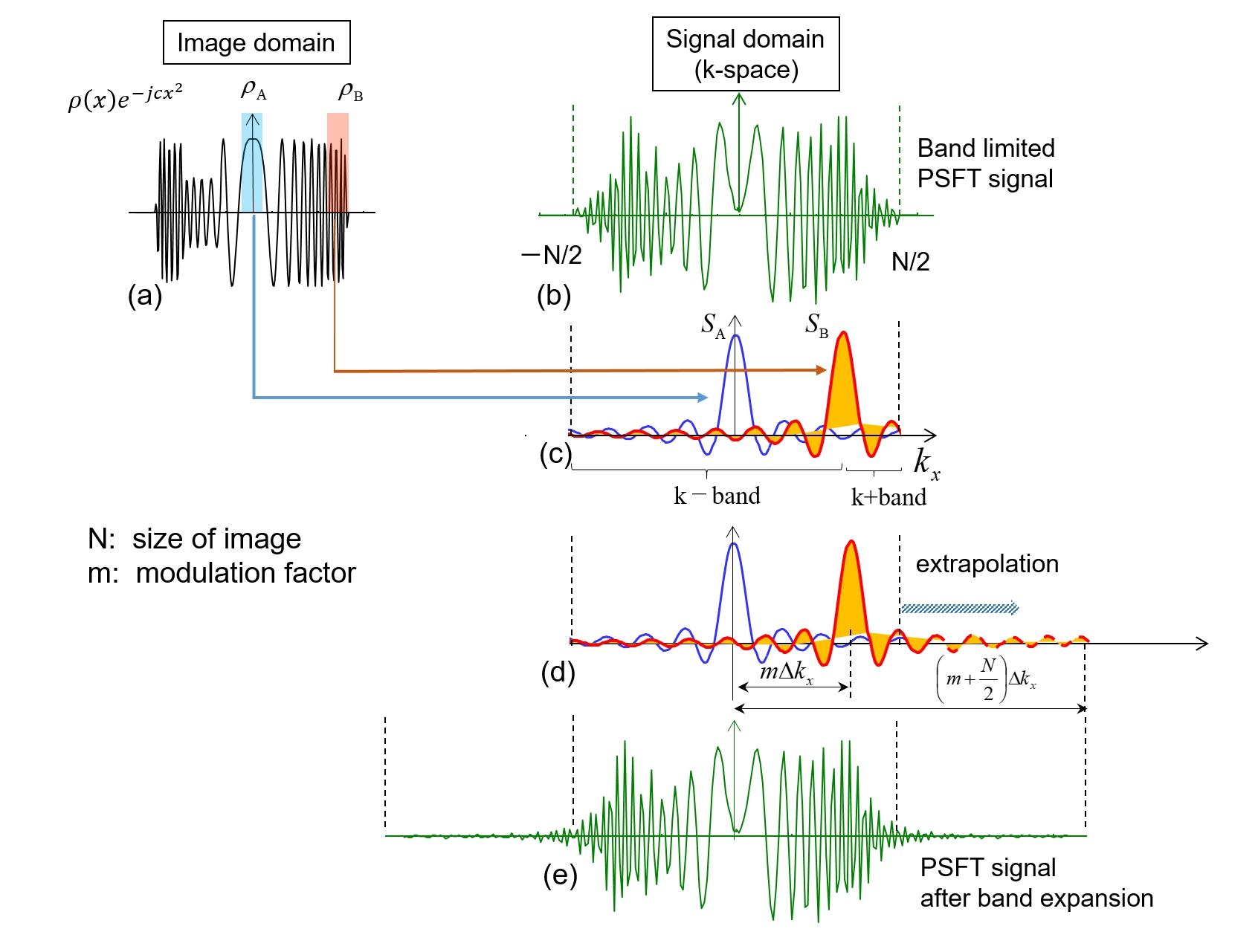
Phase-scrambling Fourier transform imaging (PSFT) is described as Eq.(1),$$v(k_x,k_y)= \int \hspace{-2.0mm} \int^{\infty}_{-\infty} \left\{ \rho(x,y) e^{-j c (x^2+y^2)} \right\} e^{-j(k_x x+k_y y)}dxdy ...(1),$$ $$ = \int \hspace{-2.0mm} \int^{\infty}_{-\infty} \left\{ \rho(x,y) e^{-j \{a_1(x) x+a_2(y) y\} } \right\} e^{-j(k_x x+k_y y)}dxdy ...(2),$$ $$ a_1(x) = c x, a_2(x)=c y ...(3)$$
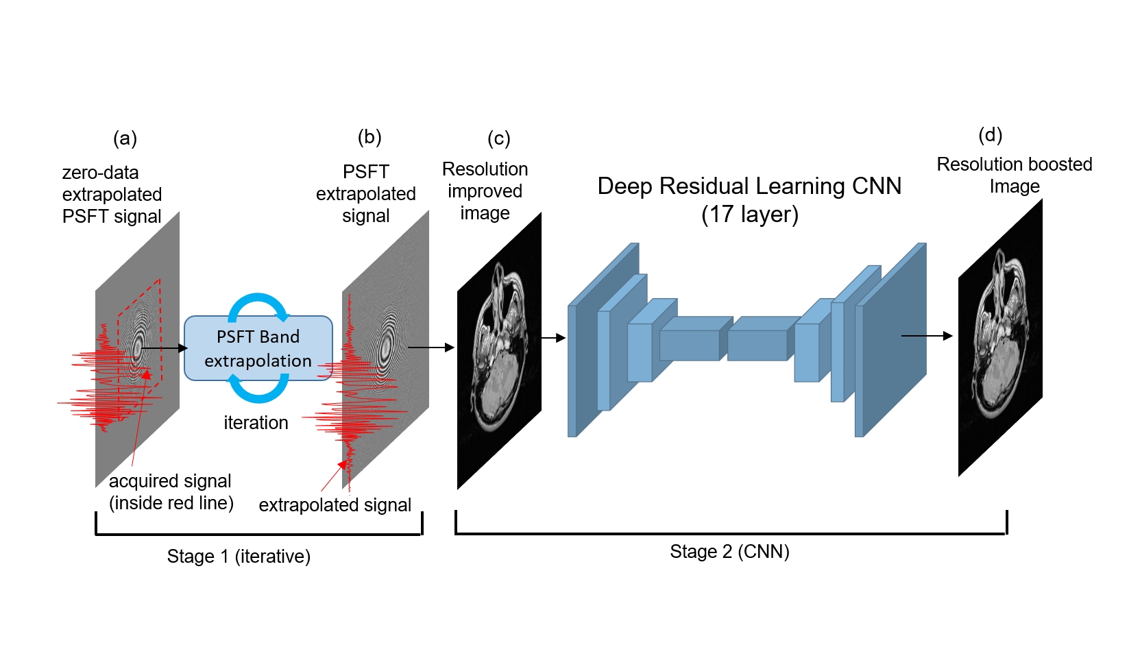
where $$$\rho(x,y)$$$ represents the spin density distribution in the subject, $$$\phi(x,y)$$$ is phase distribution, $$$c$$$ is the coefficient of quadratic phase shifting [1]. Eq.(1) can be rewritten as Eq.(2), where $$$a_1$$$ and $$$a_2$$$ are modulation factor which increase in proportion to $$$x$$$ and $$$y$$$, respectively. Consider small segmented image $$$\rho_B$$$ as shown in Fig.1. Amplitude modulation to $$$\rho_B$$$ makes its Fourier spectrum $$$S_B$$$ shift corresponding to the modulation factor $$$a_2$$$. Spectrum $$$S_B$$$ has an asymmetric frequency band, and the signal can be extrapolated so as to be symmetric with respect to the signal peak using real-value constraint of the image like Half-Fourier imaging. We have proposed a resolution improvement method that does not require estimation of phase distribution by adopting deep CNN, however [4], the resolution improvements is smaller compared to that of iterative reconstruction using properly estimated phase map. In this work, we propose an improved method in which spatial resolution is boosted in two stages. Figure 2 show the schematic of our work. Spatial resolution is improved by iterative reconstruction in the first stage. Iterative reconstruction can extend the PSFT signal as shown in Fig.1, however the resolution improvement is proportional to the distance from the center and therefore resolution improvements is small in the central region. The spatial resolution is boosted by following deep CNN super-resolution stage. Iterative reconstruction require precise phase distribution to correct the phase shift on the image. We estimate phase distribution using acquired k-space signal. Since subsampling is not executed and signal standard symmetrical sampling with respect to the origin of k-space is performed unlike Half Fourier imaging, phase map can be estimated with high accuracy using the acquired signal. We adopted deep residual learning for CNN training [5] which is known as high excellent denoising performances. The depth 17 of CNN was set 17 and corresponding receptive field size was 35x35. Three types of layers were used, (1) Conv+ReLU: for the first layer, 64 filters of size 3 x 3, 2) Conv+BN+ReLU: for layers 2 ~ 16, 64 filters of size 3 x 3 x 64, 3) Conv: for the last layer, 3x3x64 filter were used to reconstruct the output.
Results & Discussions
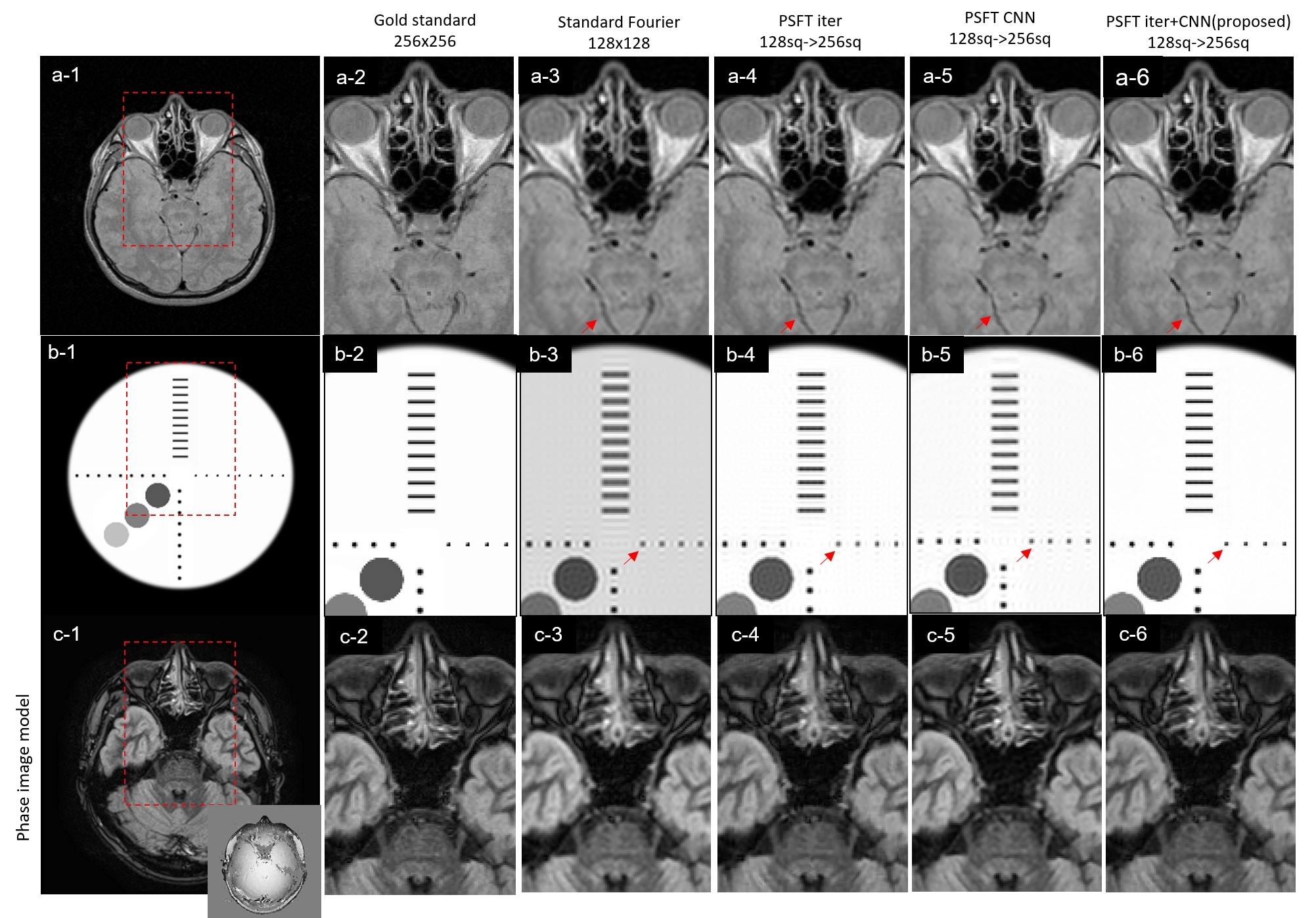
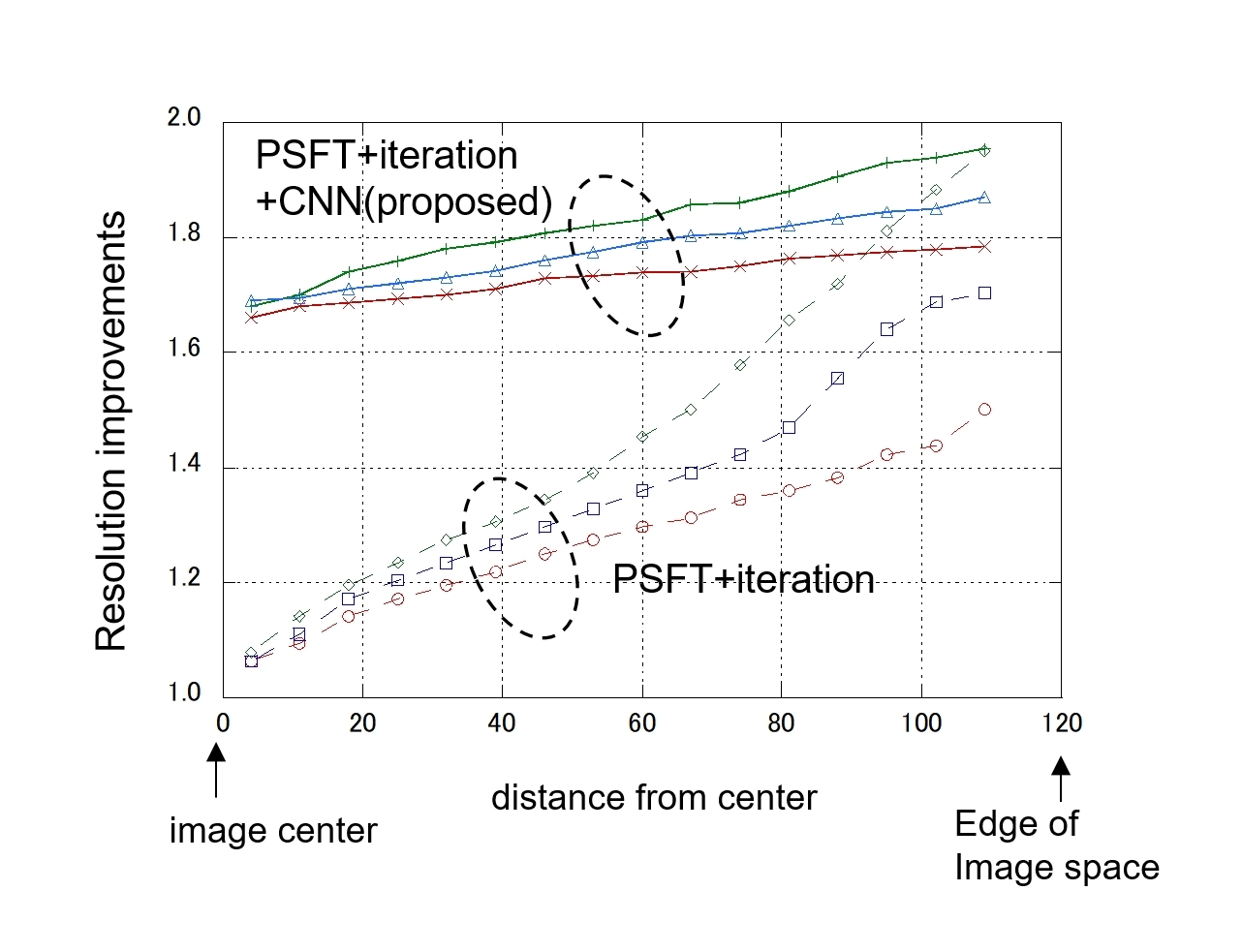
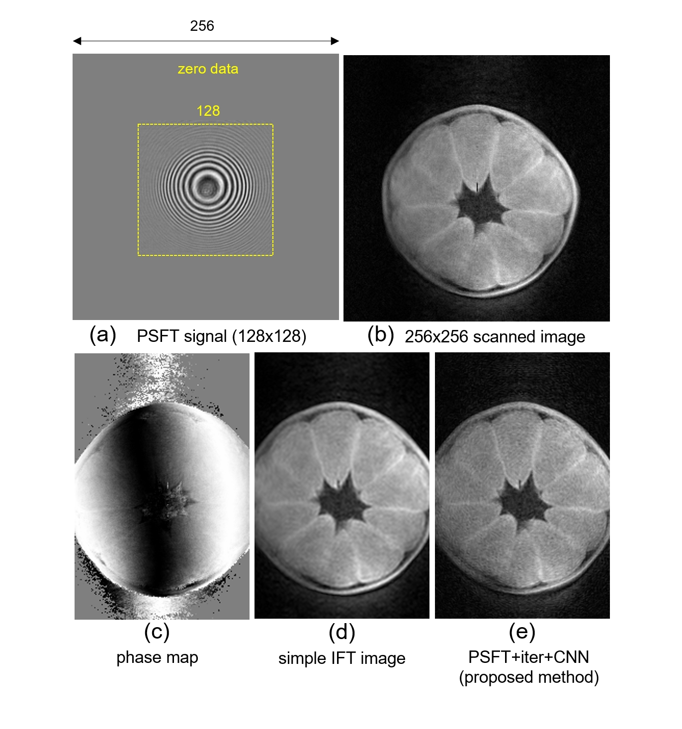
PSFT signals used in the simulation experiments were synthesized according to the Eq. (1) using the MR volunteer image. PSFT signals with 128x128 matrix size were calculated and then extrapolated to be 256x256 by filling the zero data outside the acquired signal. Figure 3 shows the results for $$$m/N=1.0$$$ (corresponding to $$$a_1$$$ or $$$a_2$$$ in Eq.(2)). Figures (a-1 to 6) and (b-1 to 6) are fully scanned (256x256) images, close-up images of (a-1) , zero filled images (256x256), images after iterative resolution improvement (10 times iteration; PSFT+iter)[2], images after CNN (PSFT+CNN)[4] and CNN output images in proposed method (PSFT+iter+CNN), respectively for real-value images. Figure 3(c-1 to 6) show the results using phase varied image. It is shown that high resolution is obtained in the center of image space as shown in (a-6) and (b-6). The results of resolution improvements which is measured using the numerical phantom like (b-1) is shown in Fig.4. Spatial resolution is drastically improved in the central area of image space. The resolution improvements introduced by CNN may depend on the type of image and high resolution improvements are likely to be given when using a numerical phantom with sharp edges like Fig.3 (b-1). However, similar results is obtained in MR image with phase variation as shown in (a,b,c-6). Figure 5 shows the results of application to experimentally obtained PSFT signal. PSFT imaging was realized by using a quadratic field gradient with 0.2 T hand-made MRI. Imaging conditions and matrix size are same as Fig.3 (a-1). Figure 5(a) - (e) are band-limited PSFT signal, fully scanned, phase map, simple IFT image, PSFT+iter+CNN images, respectively. Sharpened image compared to image (d) was obtained in image (e). Even though proposed method require quadratic phase shifting, it does not require sub-sampling and resulting aliasing artifacts do not appear on the image. Stable image quality will be expected.Conclusion
A new MR fast imaging using dual resolution improvement methods is proposed. It was demonstrated that high resolution is obtained in all image space.Acknowledgements
This study was supported in part by JSPS KAKENHI(19K04423). We would like to thank Canon Medical Systems.References
1. Maudsley AA., Dynamic Range Improvement in NMR Imaging Using Phase Scrambling. J Magn Reson 1988; 76, 287-305
2. Ito S, Liu. N, Yamada Y, Improving Super-resolution by adopting Phase-scrambling Fourier Imaging. ISMRM2007, 1907, Berlin, Germany
3. Ito S, Liu. N, Yamada, Improvement of Spatial Resolution in Magnetic Resonance Imaging Using Quadratic Phase Modulation. IEEE International Conference on Image Processing 2009; 2497-2500, Cairo, Egypt
4. Ito S, Super-resolution based on the signal extrapolation in Phase scrambling Fourier Transform Imaging using Deep Convolutional Neural Network, ISMRM2019, 4576, Montreal, Canada
5. Zhang K,Zuo W,Chen Y et al: Beyond a Gaussian Denoiser: Residual Learning of Deep CNN for Image Denoising. IEEE Tran Image Proc 2017; 26, 3142-3155
Figures