3724
Rapid whole-brain imaging with sub-mm resolution using sampling on tilted hexagonal grids (t-Hex)1Institute for Biomedical Engineering, ETH Zurich and University of Zurich, Zurich, Switzerland, 2Skope Magnetic Resonance Technologies, Zurich, Switzerland
Synopsis
In this work, we show high-resolution stacks of spirals on a tilted hexagonal grid. The new scheme provides flexibility in balancing readout and scan time, thereby allowing for high-quality images in a temporal resolution regime suitable for fMRI. 0.6 mm whole-brain coverage is achieved in below 5.
Introduction
Rapid MR scanning of 3D volumes is aimed at in applications like fMRI with high spatial and temporal resolution (1). 3D Fourier encoding lends itself to this task: it provides high SNR due to data acquisition from the entire volume, while being amenable to undersampling by optimal parallel imaging acceleration in all three dimensions.The most widespread and obvious encoding strategies used for this purpose include stacks of EPIs and spirals(2). Each k-space plane is covered by one shot of such a 2D trajectory. However, just like in the 2D case, when aiming for high spatial resolutions, it is not praticable to cover the entire k-space plane within a single shot. For such a long readout train, local field inhomogeneity would introduce severe distortions and blurring in the image. Additionally, the broadening of the PSF due to T2* decay renders the resolution gain more and more inefficient(3).
This challenge is typically met by covering each k-space plane by multiple interleaved shots. However, the overhead for other sequence modules such as RF excitation and spoiling has to be repeated for every shot and makes multiplication in the number of shots unattractive since it entails an increase in the total scan time. This effect is highly undesirable for time-resolved applications such as fMRI.
In this work, we propose to balance readout length and scan-time more flexibly than possible so far, relying on a tilted hexagonal grid (t-Hex) for fast k-space sampling (4,5). We show 0.6mm resolved in-vivo whole-brain images using t-Hex spirals.
Methods
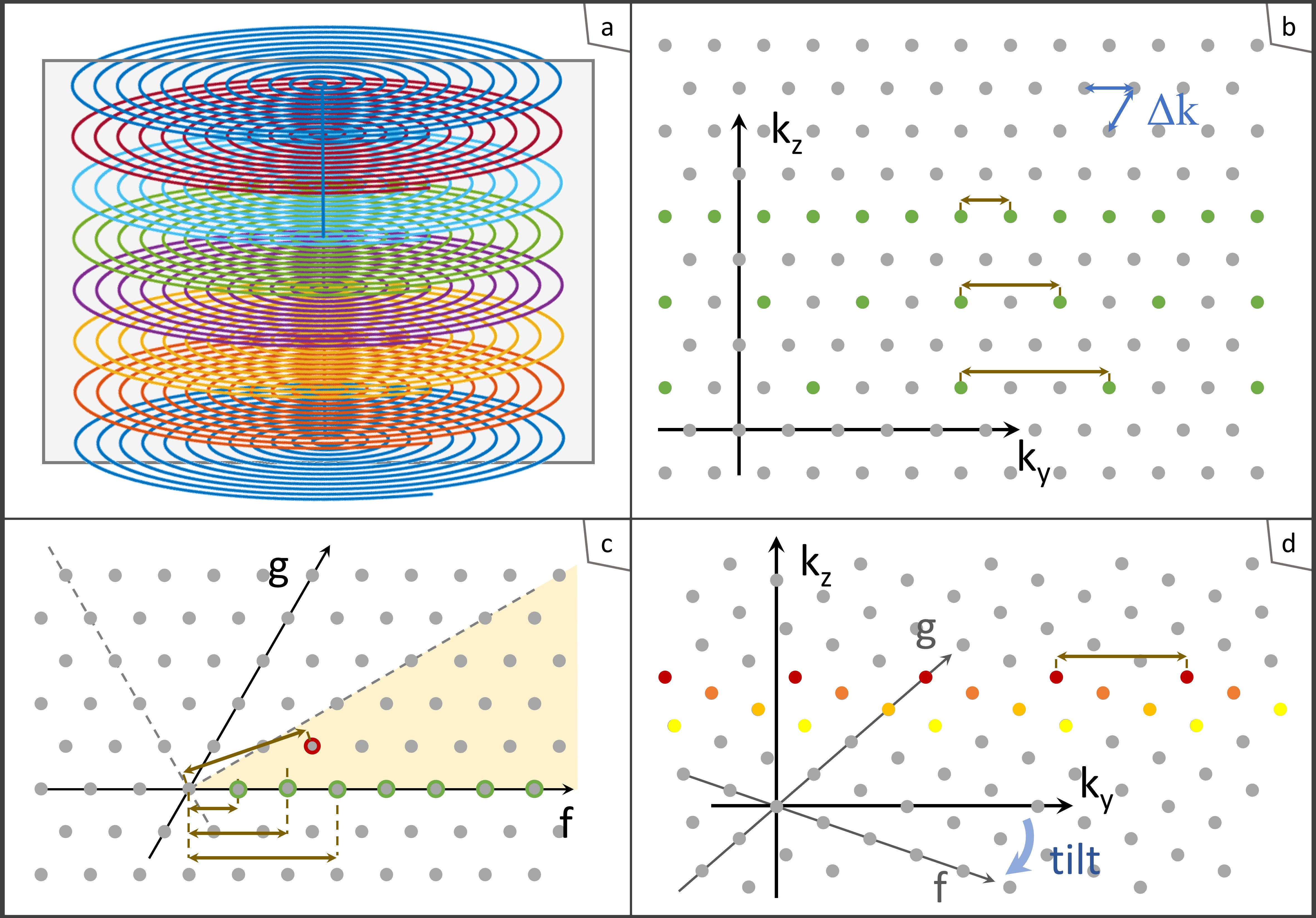
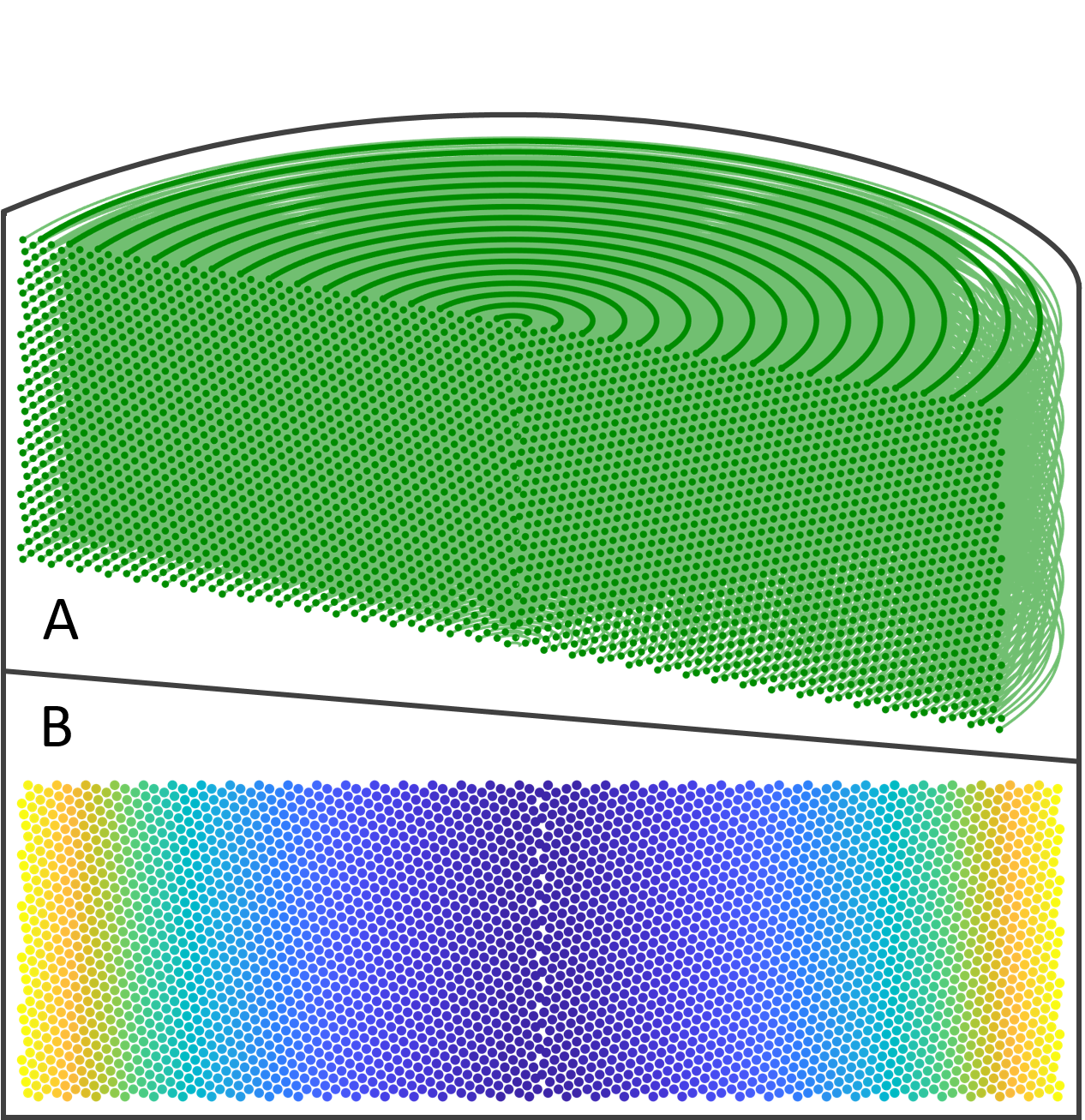
T-Hex: The tilting of the hexagonal grid (6,7) underlying a stack of spirals is shown in Figure 1. Each point with coordinates [f,g] in the oblique coordinate system corresponds to one possibility of tilting the grid. Its distance to the origin determines the TAQ per shot.Hardware:
- Philips 7T Achieva system
- 32 channel head array (Nova Medical)
- Field camera consisting of an array of 16 1H-based NMR field probes and a dedicated MR acquisition system (8)
- TE = 15 ms, TR = 56 ms, Total scan time = 4.8s
- TE = 25 ms, TR = 66 ms, Total scan time = 5.7s
Results
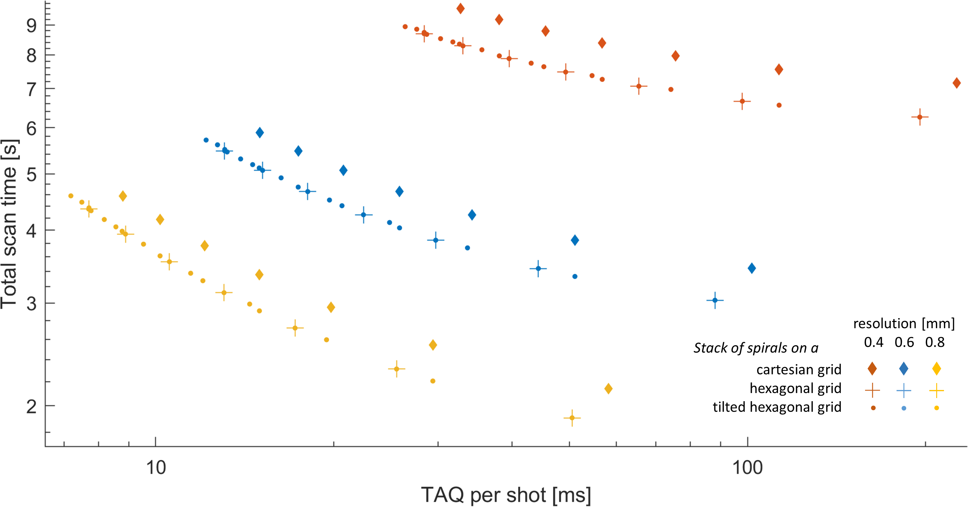
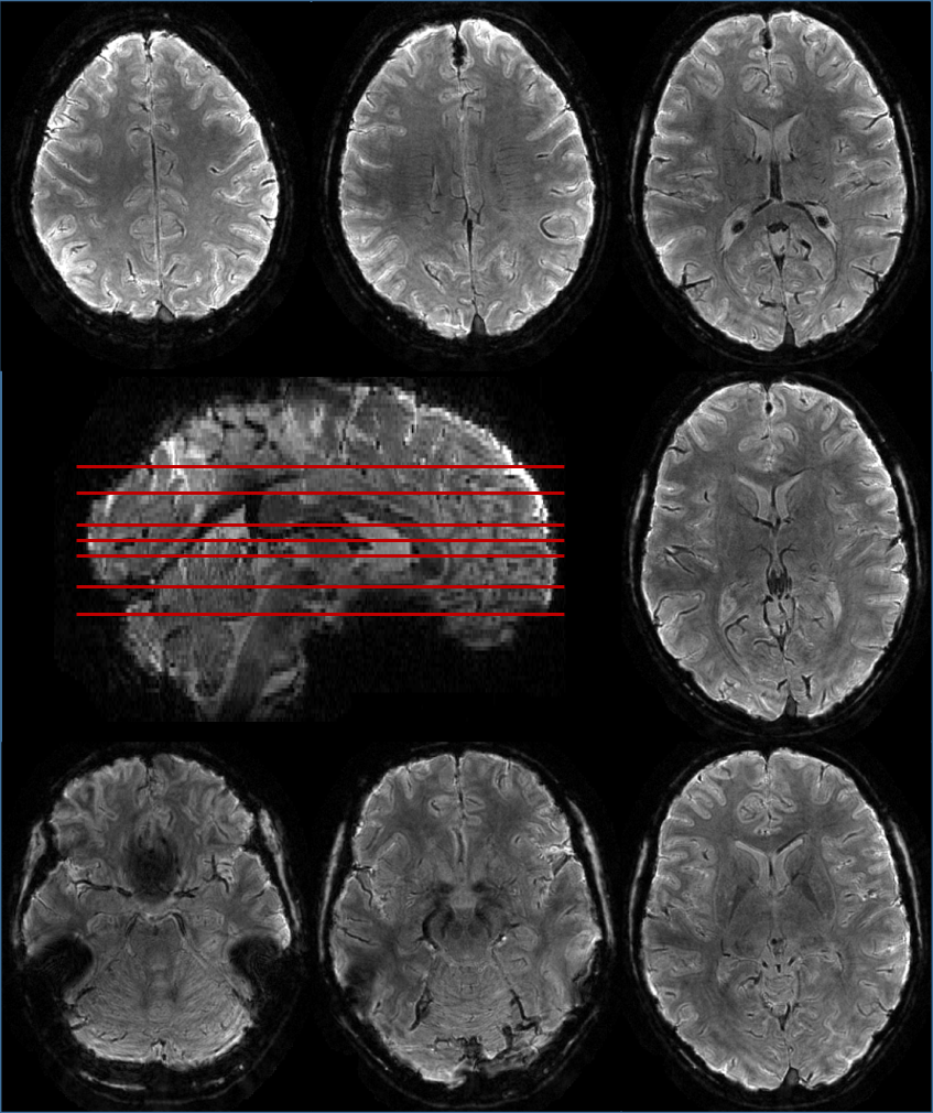
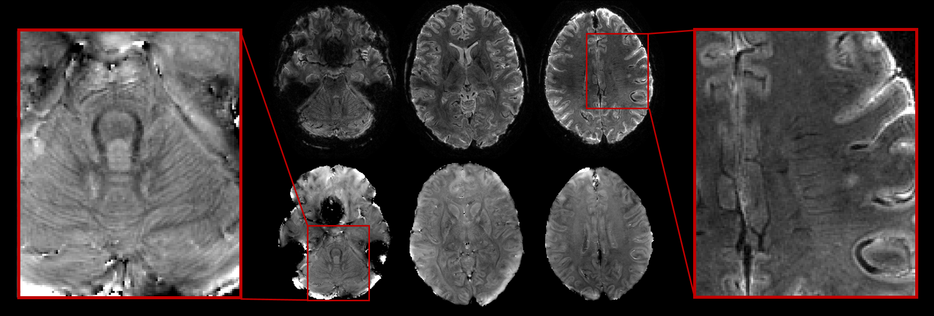
In Figure 3, scan time is plotted against TAQ per shot. Ideally one would aim for moving towards the lower left corner. Spirals stacked such that forming a hexagonal grid in the cross-section outperform in all resolution regimes conventional stacks (Cartesian grid). T-Hex provides additional flexibility in balancing total scan time and TAQ per shot. Figure 4 and 5 show in-vivo results.Discussion
The T-Hex spiral for rapid 3D imaging introduced in this work is tailored to sampling less k-space volume than one k-space plane per shot. Just like its kindred t-Hex trajectories (7,4,5) which sample more k-space volume than one k-space plane per shot, it comprises a uniform undersampling, smooth T2* weighting and high average k-space speed. However unlike them, it does not need time-consuming blips and is attractive for the contiguous high-resolution domain. Its main benefit is to gain flexibility, when balancing readout and scan duration. This is especially true for a regime, where few shots per plane are suitable. Beyond that, an integer number of shots per plane is increasingly flexible on its ownTo achieve appealing image quality, the reconstruction was based on the expanded signal model described above. Only dephasing in the ear canals and lower brain regions compromises those partially. This effect is mitigated for the shorter TE of 15ms.
The proposed t-Hex trajectory is equally applicable to different sampling schemes such as EPI or spiral-in. It may furthermore prove useful for multiband imaging.
Acknowledgements
No acknowledgement found.References
1. Feinberg DA, Moeller S, Smith SM, et al. Multiplexed Echo Planar Imaging for Sub-Second Whole Brain FMRI and Fast Diffusion Imaging Valdes-Sosa PA, editor. PLoS ONE 2010;5:e15710 doi: 10.1371/journal.pone.0015710.
2. Poser BA, Koopmans PJ, Witzel T, Wald LL, Barth M. Three dimensional echo-planar imaging at 7 Tesla. NeuroImage 2010;51:261–266 doi: 10.1016/j.neuroimage.2010.01.108.
3. Engel M, Kasper L, Barmet C, et al. Single-shot spiral imaging at 7 T. Magn. Reson. Med. 2018;80:1836–1846 doi: 10.1002/mrm.27176.
4. Engel M, Kasper L, Barmet C, Schmid T, Pruessmann KP. A rapid 3D spiral readout with uniform sampling density and smooth T2* weighting. In: Proceedings of the ISMRM 2018.
5. Engel M, Kasper L, Wilm B, Dietrich BE, Vionnet L, Pruessmann KP. T-Hex: Spiral sampling on a tilted hexagonal grid. In: Proceedings of the ISMRM 2019. ; 2019.
6. Mersereau RM. The Processing of Hexagonally Sampled Two-Dimensional Signals. Proc. IEEE 1979;67:930–949.
7. Engel M, Kasper L, Pruessmann KP. Rapid 3D imaging with multiplanar spirals. Proc. ISMRM 2017.
8. Dietrich BE, Brunner DO, Wilm BJ, et al. A field camera for MR sequence monitoring and system analysis. Magn. Reson. Med. 2016;75:1831–1840 doi: 10.1002/mrm.25770.
9. Pruessmann KP, Weiger M, Börnert P, Boesiger P. Advances in sensitivity encoding with arbitrary k-space trajectories. Magn. Reson. Med. 2001;46:638–651 doi: 10.1002/mrm.1241.
10. Man L-C, Pauly JM, Macovski A. Multifrequency interpolation for fast off-resonance correction. Magn. Reson. Med. 1997;37:785–792 doi: 10.1002/mrm.1910370523.
11. Barmet C, Tsao J, Pruessmann KP. Sensitivity encoding and B0 inhomogeneity – A simultaneous reconstruction approach. In: Proceedings of the ISMRM. ; 2005. p. 682.
12. Barmet C, Wilm BJ, Pavan M, et al. Concurrent higher-order field monitoring for routine head MRI: an integrated heteronuclear setup. In: Proceedings of the 18th Annual Meeting of ISMRM, Stockholm, Sweden. ; 2010. p. 216
Figures