3711
Using dielectric bags to increase SNR in dynamic speech imaging1Bioengineering, University of Illinois at Urbana-Champaign, Urbana, IL, United States, 2Beckman Institute, University of Illinois at Urbana-Champaign, Urbana, IL, United States, 3Carle-Illinois College of Medicine, University of Illinois at Urbana-Champaign, Urbana, IL, United States, 4Carle Foundation Hospital, Urbana, IL, United States, 5Leiden University, Leiden, Netherlands
Synopsis
Dynamic speech MRI has seen increasing use in the clinic for cleft palate and oral cancer and in research for examining the articulatory patterns of speech. Although potential frame rates have increased, in order to optimize acquisitions, special RF coils may be required to match the anatomy of the population of interest. In this work, we examine the potential of low-cost dielectric pads to significantly improve the SNR of acquisitions using standard head/neck coils. We show a 20% improvement in SNR in speech articulator regions with two dielectric bags placed on either side of the face.
INTRODUCTION
Dynamic speech imaging has seen increasing use in both research and clinical settings to study the dynamic implications of pathologies such as oral cancer and cleft palate and for studying articulatory variations over languages in linguistics. MRI pulse sequences have enabled high frame rates and good image quality, yielding frame rates from 10’s to 166 frames per second and spatial resolutions on the order of 2 mm [1-3]. However, to get the best signal-to-noise ratio (SNR) for these fast acquisitions, it is critical to have an RF coil that is well suited to the speech task [1]. Such coils are not readily available and must be custom made, potentially requiring variable sizes for imaging adults and children, and usually carrying significant costs. Recently, dielectric pads have been constructed from high permittivity materials and used to improve transmit and receive sensitivity locally, reducing SAR and improving image quality for a variety of MRI applications from cardiac imaging to ultra high field imaging [4, 5]. High permittivity pads are a low-cost solution that can potentially provide significant gains for dynamic speech imaging.METHODS
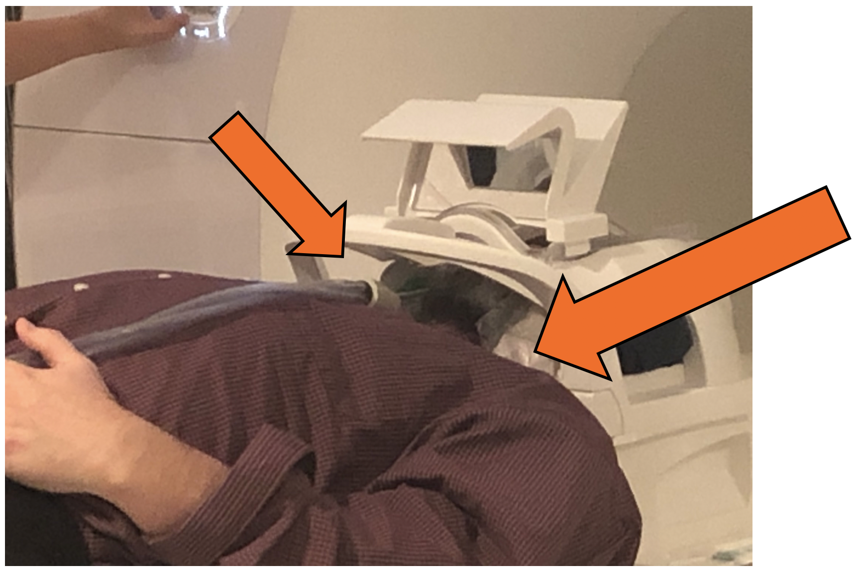
Two dielectric bags were constructed, each using 500 g of calcium titanate (Alfa Aesar, Thermo Fisher Scientific) and distilled water to make a gel of ~600 mL per bag. Total cost for construction of the two bags was under $300. The gel was placed in plastic bags and placed on either side of the sample (left/right) for imaging. The bags were aligned with the bottom of the maxilla and placed just posterior to the mouth opening, as shown in Figure 1. A dynamic GRE pulse sequence was used to measure the SNR on a water bottle phantom and in a volunteer on a Siemens 3 T Prisma. Sequence parameters were: TE=1.1 ms, TR=2.8 ms, 128 Matrix size, 24 cm field of view, 6 mm single slice thickness, and a 30° flip angle, 200 time points were acquired. The Siemens 20 channel head/neck coil was used, which has neck elements that are relevant for imaging speech, but do not sit close to the anatomy (Figure 1). SNR was measured in a water bottle phantom with 1 dielectric bag placed to the left side of the phantom and 2 bags placed one on either side (left/right). Human volunteer data was acquired with the bag placed on both the left and right side, while the subject remained stationary in a resting position. SNR was measured as the mean of the signal over the temporal standard deviation. In addition, a 2D dynamic speech experiment was performed with the subject counting with no bags and two dielectric bags in place. This counting experiment used a PS-model based imaging and reconstruction approach [3].RESULTS
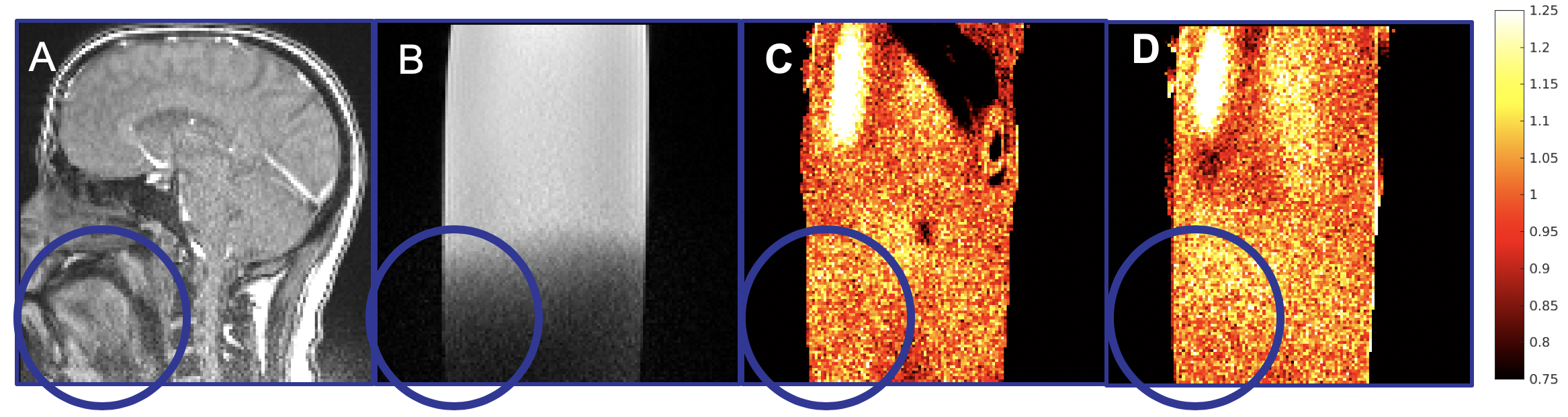
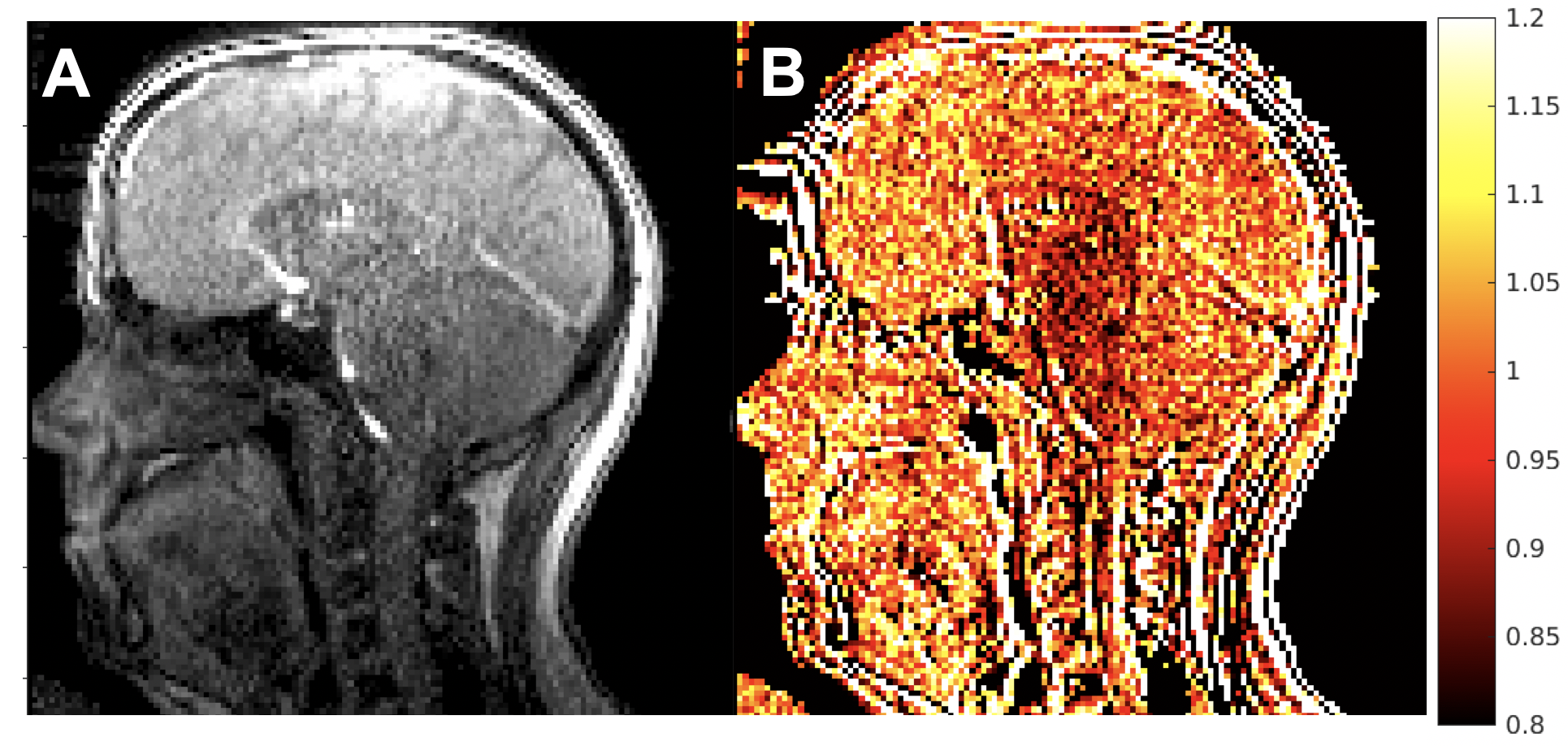
Figure 2 shows the phantom SNR measures results. In Figure 2A, a reference image of a head in the coil imaged in a similar location as the phantom is shown for reference. You can see the fall-off of the SNR that starts at the base of the maxilla bone and results in lower SNR for the speech muscles. Figure 2C shows the ratio of the SNR of the single bag GRE scan to the SNR of the GRE scan with no dielectric bag, showing significant changes in the SNR. Figure 2D shows the ratio of the SNR from two dielectric bags with the SNR of no dielectric bag, indicating gains of about 20% in the speech region and showing more uniform gains in the speech area.Figure 3 shows the results on a human volunteer scanned with the same GRE sequence, with and without two dielectric bags. The SNR ratio of 2 dielectric bags to no dielectric bags shows that the gains of about 20% in SNR are replicated in vivo. This is in-line with reported gains in SNR with the use of similar pads at 3 T of about 30% [4].
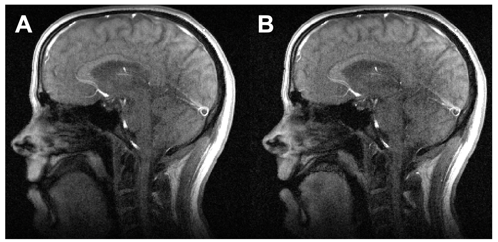
Figure 4 shows a frame from each of the dynamic speech scans with the subject counting. Figure 4A shows the result without any dielectric bags and Figure 4B shows the much improved SNR with two dielectric bags.
DISCUSSION
Fast, dynamic speech MRI suffers from low SNR in regions that are critical for imaging speech due to the poor coverage of this anatomy with existing head and neck coils. However, obtaining customized coils for imaging this region may be costly and require specialized coils for different ages and populations. The use of high permittivity materials can provide significant benefits in optimizing SNR even with variable anatomy across populations. In this work, we have shown that a gain of about 20% SNR is achievable with a pair of simple dielectric bags arranged on either side of the anatomy of interest.CONCLUSION
Dielectric pads consisting of high permittivity materials can be used to improve receiver coil sensitivity when the anatomy of interest is not a good match to the coils available. We have shown a 20% improvement in SNR for dynamic GRE imaging of speech with the use of two cost-effective dielectric pads arranged on the left and right side of the head.Acknowledgements
Research reported in this publication was supported by the National Institute Of Dental & Craniofacial Research of the National Institutes of Health under Award Number R01DE027989. The content is solely the responsibility of the authors and does not necessarily represent the official views of the National Institutes of Health.References
1. Lingala, S.G., B.P. Sutton, M.E. Miquel, and K.S. Nayak, Recommendations for real-time speech MRI. J Magn Reson Imaging, 2016. 43(1): p. 28-44. 2.
2. Fu, M., B. Zhao, C. Carignan, R.K. Shosted, J.L. Perry, D.P. Kuehn, Z.P. Liang, and B.P. Sutton, High-resolution dynamic speech imaging with joint low-rank and sparsity constraints. Magn Reson Med, 2015. 73(5): p. 1820-32. 42610623.
3. Fu, M., M.S. Barlaz, J.L. Holtrop, J.L. Perry, D.P. Kuehn, R.K. Shosted, Z.P. Liang, and B.P. Sutton, High-frame-rate full-vocal-tract 3D dynamic speech imaging. Magn Reson Med, 2017. 77(4): p. 1619-1629. 4.
4. Brink, W.M. and A.G. Webb, High permittivity pads reduce specific absorption rate, improve B1 homogeneity, and increase contrast-to-noise ratio for functional cardiac MRI at 3 T. Magn Reson Med, 2014. 71(4): p. 1632-40. 5.
5. Koolstra, K., P. Bornert, W. Brink, and A. Webb, Improved image quality and reduced power deposition in the spine at 3 T using extremely high permittivity materials. Magn Reson Med, 2018. 79(2): p. 1192-1199. PMC5811912
Figures