3710
A flexible linear reorder scheme for improved fat saturation in 3D VIBE imaging
Qiong Zhang1, Yong Xiao Zhang2, and Yulong Liu3
1DL, Siemens Shenzhen Magnetic Resonance Ltd, Shenzhen, China, 2MR Collaborations, Siemens Healthcare Ltd., Shenzhen, China, 3Beijing Institute of Technology, BeiJing, China
1DL, Siemens Shenzhen Magnetic Resonance Ltd, Shenzhen, China, 2MR Collaborations, Siemens Healthcare Ltd., Shenzhen, China, 3Beijing Institute of Technology, BeiJing, China
Synopsis
The fast (‘Quick’) fat saturation technique (Q-fat sat) [1], which acquires several k-space lines after each Fat-Sat module, is widely used for suppression of signals from fat in MR imaging. In this work, we developed and evaluated a linear flexible reorder scheme for improved fat saturation VIBE imaging. In vivo experiments indicated that this technique markedly improves suppression of fat saturation signals compared with conventional Q-fat saturation imaging. Studies are underway to validate the clinical value of the technique.
Introduction
The fast (‘Quick’) fat saturation technique (Q-fat sat) [1], which acquires several k-space lines after each Fat-Sat module, is widely used for suppression of signals from fat in MR imaging.This technique is based on the principle that the central low frequency data in k-space controls the image contrast—preferred with complete fat saturation—while collecting the edge data during fat signal recovery. The Q-fat technique also balances issues of scan time and fat saturation effects by controlling the number of echoes following each fat saturation pulse.In this work, we used a fat signal evolution model for 3D VIBE imaging (within Q-fat saturation). This model was based on the Bloch Equation. The flip angle of fat saturation pulses and echo numbers, followed by each fat-saturation module, are flexibly modified to improve fat saturation effectiveness.Methods
Sequence: Figure 1 presents a diagram of the Q-fat saturation VIBE sequence. Because all transversal magnetizations are spoiled before the next radio frequency (RF) pulse, the magnetization of fat in all tissues experiences a steady state incoherent process [2, 3]. Fat tissue related magnetization experiences periodical notations and precessions, and each period contains a “β” pulse for fat-selective excitation, as well as a series of “θ” pulses for normal “fat and water” excitation. Without losing generality, assuming a periodic n, the longitudinal magnetization of fat tissue starts from initialized magnetization Mo and evolves according to Formulas 1 - 4 shown in Figure 2. Magnetizations in the fat saturation train (Figure 1) (followed by each fat saturation module) can be calculated using Formulas 5 and 6(Figure 2). It should be noted that the longitudinal magnetization at the end of period n is the initial value of period n+1 (Formulas 7 and 8). Principally, the calculation procedures start with initial condition (n=0), and repeat formula from (1) to formula (7) in each fat saturation train, and then the longitudinal magnetizations evolution curve can be achieved, which is a direct reflection of the fat signal. Figure 3 (A and B) shows a virtual 3D imaging with 60 phase-encoding lines collected after each fat saturation module. It should be noted that the fat signal oscillates in only a few trains at the start of the sequence, and then goes to smooth and periodic variation. By neglecting this oscillation, the steady state cross section curve can be used to trace the fat signal evolution, which can be modulated by changing the angle of β pulse or the number of followed echoes. Figure 3 (C and D) shows that the fat signal evolution curve can be modulated by changing the flip angles of the fat saturation pulses, where the vertex of amplitude curve shifts left by decreasing β angle, and shifts right by increasing β angle. In real Q-fat saturation implementation, assuming all the partition encoding lines were acquired after each fat saturation module, the angle of fat saturation pulse (β) is modulated to make the vertex point shifts to the partition center. Fat saturation train length may also be split for achieving a stronger saturation effect (Figure 4). Thus, the purpose of the proposed “lineal flexible” method is twofold: one is related to the fat saturation angle, and the other is related to the train length.Experiments:The IRB-approved experiments were performed on 8 healthy volunteers using a 3T system (MAGNETOM Spectra, Siemens Healthcare, Erlangen, Germany) with a 16-channel Head/Neck coil, and were implemented on a modified prototypical 3D VIBE sequence with the following protocol parameters: TR/TE = 6.42/2.47 ms, BW=250 Hz/pixel, FOV=173x190 mm2, Imaging Matrix =230x288, slices per slab=56, slice over sampling factor =28.6%, voxel size=0.7x0.7x1.0 mm3, Q-fat saturation lines per shot =60, FA=9o. The conventional Q-fat saturation VIBE sequence with the same parameters was also conducted for comparison.
Results
A representative result from one subject is shown in Figure 4. It is clear that the images acquired with the proposed linear flexible reordering achieve better fat saturation effects when compared to the conventional Q-fat VIBE technique, which uses an empirical value for β angle (indicated by white arrows in Figure 5). The fat saturation effect is also enhanced by splitting fat saturation trains in our proposed implemented VIBE sequences, without adding much scan time (Figure 5 C).Conclusions
In this work, we developed and evaluated a linear flexible reorder scheme for improved fat saturation VIBE imaging. We validated this technique and showed that it produces a better and more controllable fat saturation effect compared with conventional Q-fat saturation imaging .It should be noted that only longitudinal magnetizations are considered in our calculation process, so the method is only practical for steady state incoherent (SSI) sequences, and are not applicable to other steady state coherent (SSC) or spin echo sequences. Studies are underway to validate the clinical value of the technique.Acknowledgements
References
[1] Haase A,et,al. 1H NMR chemical shift selective (CHESS) imaging. Phys Med Biol. 1985;30(4):341–344[2] Elster AD. Gradient echo imaging: techniques and acronyms. Radiology 1993; 186:1-8.
[3] Haase A, et,al. FLASH imaging: rapid NMR imaging using low flip angle pulses. J Magn Reson 1986: 67: 258-266.
Figures
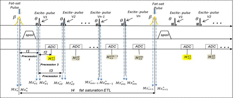
Fig1. The sequence diagram of VIBE with fast fat saturation pulses. β represents fat selective pulses. θ represents normal excitation pulse

Fig2. Calculation formulas used in each Fat
saturation trains
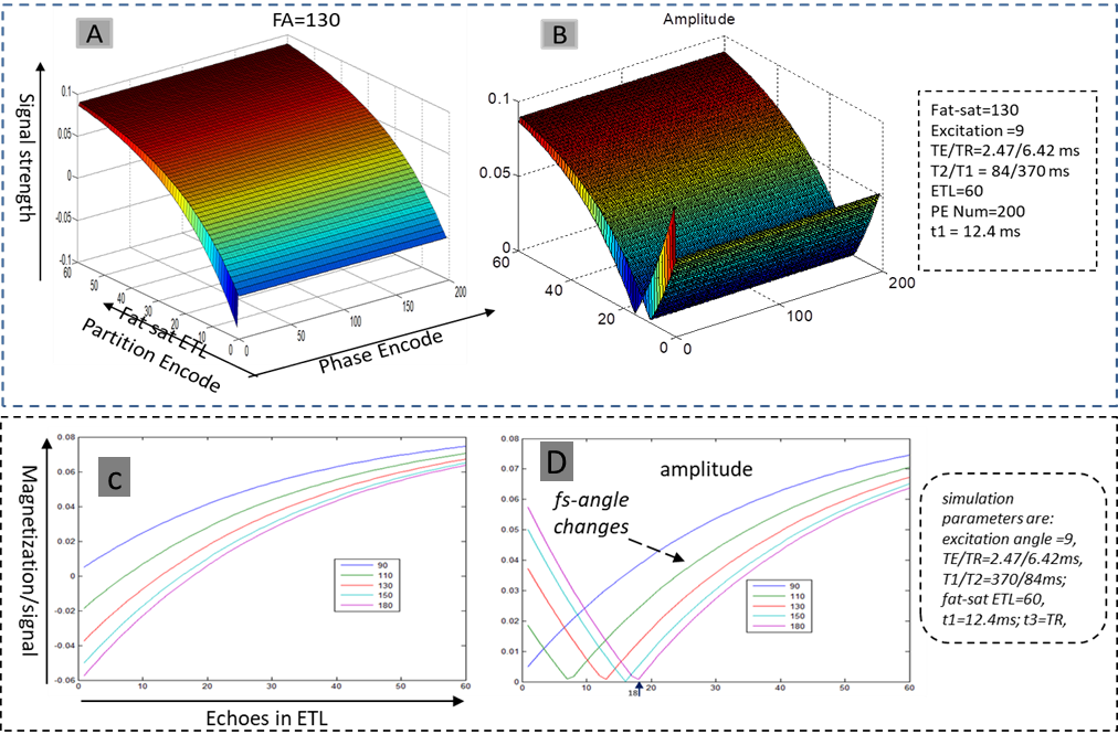
Fig3. Fat signal evolution in fast fat saturation Vibe protocol; (A) fat signal evolution curve simulation, fat saturation angle is 130 degree, 60 phase encode lines are acquired after each fat saturation module; (B) is the amplitude of (A); (C) is a simulation the cross section curve modulated by changing fat saturation angle; (D) is the amplitude of (C)
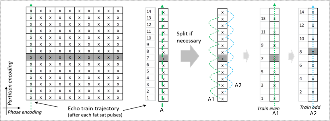
Fig4. The lineal reorder acquisition trajectory in a 3D VIBE Q-fat imaging with “Partition in Line” acquisition loop (A) All the partition encode lines are acquired after each fat saturation module, the center line were acquired with lowest fat signal. The fat saturation train A could be split to A1 and A2 for better fat saturation purpose.
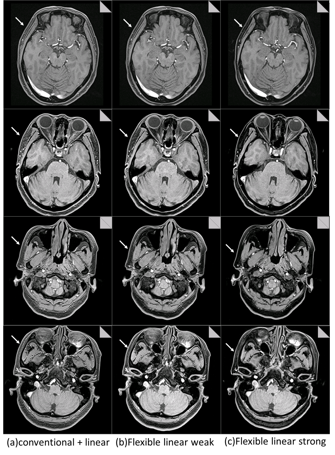
Fig5 Representative Results from one subject using convention Q-fat VIBE and the proposed implemented VIBE techniques. (a) Images acquired with conventional Q-fat VIBE sequence; (b) images acquired with the proposed implemented VIBE sequence. It is clear that fat saturation effects are improved with the new VIBE sequence (white arrows); (c) images acquired with the proposed implemented VIBE, in which fat saturation trains are reduced for enhancing the saturation effects, with scan time increased by 10s.