3703
Rapid pre-saturated TFL transmit field mapping with an optimized 3D centric single-shot readout
Dario Bosch1, Jonas Bause1, Philipp Ehses2, Moritz Zaiss1,3, and Klaus Scheffler1,4
1High Field Magnetic Resonance Center, Max Planck Institute for Biological Cybernetics, Tuebingen, Germany, 2German Center for Neurodegenerative Diseases, Bonn, Germany, 3Department of Neuroradiology, University Hospital Erlangen, Erlangen, Germany, 4Institute for Biomedical Magnetic Resonance, University Hospital Tuebingen, Tuebingen, Germany
1High Field Magnetic Resonance Center, Max Planck Institute for Biological Cybernetics, Tuebingen, Germany, 2German Center for Neurodegenerative Diseases, Bonn, Germany, 3Department of Neuroradiology, University Hospital Erlangen, Erlangen, Germany, 4Institute for Biomedical Magnetic Resonance, University Hospital Tuebingen, Tuebingen, Germany
Synopsis
Robust and fast measurements of the transmit field strength is of great importance particularly in parallel transmit applications at ultra-high field. In this work, a pre-saturation TurboFLASH B1+ mapping sequence was optimized for 3-dimensional single-shot acquisition with spiral-centric reordering. Improved SNR and reduced artifacts were achieved by using a variable flip-angle readout. The ability of the proposed sampling scheme to perform fast whole-brain B1+ mapping with low SAR was demonstrated in phantom and in-vivo experiments at 9.4 T. The obtained B1+ maps were comparable to those obtained with the conventional 2D-SatTFL approach but required significantly less scan time.
Introduction
Fast and accurate mapping of transmit-B1 (B1+) is of great importance for Ultra-High Field (UHF) MRI. The required time and deposited energy for creating B1+ maps becomes even more relevant when B1+ maps need to be recorded for several transmit channels.The saturated TurboFLASH method1 (SatTFL) allows for accurate B1+ mapping with a high dynamic range and low SAR2. It has, however, only been described with a 2D readout (2D-SatTFL), which is unsuitable for whole-brain applications due to its poor temporal efficiency. This work investigated the feasibility of single-shot SatTFL with a 3D spiral-centric reordering (3D-SatTFL).
Methods
In SatTFL two small flip-angle (FA, α) spoiled GRE images are acquired. The initial longitudinal magnetization (MZ) in one of the images is prepared with a high FA (β) presaturation pulse which reduces the detectable signal by a factor of cos(β). The ratio between the saturated (sigsat) and unsaturated (sigref) image intensity permits the calculation of the effective presaturation FA β:$$β=acos(\frac{sig_{sat}}{sig_{ref}})$$
However, this technique is limited by signal decay as well as the number and FA of the excitations in the image acquisition module. These effects drive both signals (sigsat, sigref) towards a common steady state without any dependency on β.
Due to the low spatial frequency of B1+ inhomogeneities and the fact that the largest signal difference exists at the beginning of the readout modules, a 3D-SatTFL sequence with spiral-centric readout order was implemented (Fig.1). All measurements were performed on a 9.4T whole-body MRI scanner (Siemens Healthineers, Germany), 8 independent transmit channels (pTx step 2.3), using a custom built head coil (8 transceiver, 8 receive-only elements)3.
Simulations:
Bloch Simulations to determine the maximum readout duration and optimal readout FA were performed on a Shepp-Logan-Phantom (T1=2.0s; matrix=64x64x64, spiral-centric single-shot readout). In order to reduce signal blurring compared to a readout with constant FA (3D-SatTFLconst), a variable FA readout module (3D-SatTFLvar) was investigated.
Phantom scans:
Phantom scans were performed in a head-shaped agar phantom (T1=2.6s) using both 3D-SatTFL variants. The resulting FA maps were compared against the conventional 2D-SatTFL. The B1+ linearity was evaluated by performing measurement with varying the saturation pulse voltage using the 3D-SatTFLconst readout.
In vivo scans:
Whole-brain maps were obtained from two healthy volunteers. B1+ maps were recorded using 2D-SatTFL, 3D-SatTFLconst, and 3D-SatTFLvar. As in the phantom scans, the B1+ linearity of the 3D-SatTFLconst was verified. In addition, individual channel maps (magnitude and phase) were recorded using B1+ interferometry4.
Sequence parameters:
- 3D-SatTFL: TR=2.72ms; 3.2 mm isotropic, matrix=64x64x64; readout-FA=constant (4°) or variable (4°-0°); GRAPPA5 2x2; elliptical scanning; k-space lines/volume=795; TA=22s/volume.
- 2 D-SatTFL: TR=4.7ms; FOV=205x205mm; 3.2x3.2x3.5 mm³, matrix=64x64, readout-FA=4°; k-space lines/slice=64; TA=18s/slice.
-
Common parameters: non-selective saturation-FA=90°; recovery time
7.5s; acquisition in circular polarized mode (45° phase
shift/channel).
Results
Simulations:The simulations of the signal and estimated FA for the single-shot 3D-SatTFL illustrate that the majority of presaturation decays over the course of the readout scan (Fig.2). This effect can be partially reduced by using a variable readout FA. A numeric estimation of the point spread function yielded a blurring of approximately 30% when using the 3D-SatTFLvar.
Phantom Scans:
The 3D-SatTFL data shows a B1+ pattern similar to the reference 2D-SatTFL (Fig.3). However, there are differences in areas with very high and very low preparation FA. Compared to the 3D-SatTFLconst data, the 3D-TFLvar scan results in a lower deviation from the 2D map, as well as reduced ringing. Linearity for B1+ is only given for 30°<preparation FA<150°.
In vivo Scans:
The in vivo scans confirm the similarity between 2D and 3D maps as well as the linear relation between pulse voltage and measured presaturation FA (Fig.4a). The individual channel maps, as they would be required for pTX pulse-calculation, are depicted in Fig.4b.
Discussion
While the variable readout FA reduced artifacts, it also introduced image blurring and resulted in a lower effective resolution of the B1+ maps. It may therefore be further improved by optimizing the sequence with attention to the readout point spread function. We also observed slight differences between transmit field maps obtained with the 2D and 3D-SatTFL which likely sourced from poor slice profiles or inaccuracies in the alignment of the different data sets. Further research will therefore include comparisons with other 3D B1+ mapping techniques such as AFI6 and DREAM7 as well as experiments with a multi-shot 3D-SatTFL in order to investigate the impact of readout blurring further. One also has to keep in mind that a presaturation FA in range of 30°-150° is required in order to obtain reliable results. However, this is even possible in case of strong B1+ inhomogeneities at 9.4T if the pre-saturation pulse voltage is chosen carefully.Conclusion
This work shows that spiral-centric reordered 3D-SatTFL could be used for efficient B1+ mapping at 9.4 T despite the decay of presaturation magnetization during the readout train. The implementation of a variable FA readout allowed for a minimization of imaging artifacts at the cost of slightly increased image blurring. Nevertheless, the presented approach has the capability to perform fast and robust B1+ mapping with low SAR which is essential for pTx pulse calculation, RF coil testing and retrospective B1+ correction at UHF.Acknowledgements
References
- Chung, Sohae, et al. "Rapid B1+ mapping using a preconditioning RF pulse with TurboFLASH readout." Magnetic Resonance in Medicine 64.2 (2010): 439-446.
- Pohmann, Rolf, and Scheffler, Klaus. "A theoretical and experimental comparison of different techniques for B1 mapping at very high fields." NMR in Biomedicine 26.3 (2013): 265-275.
- Avdievich, Nikolai I., et al. "Combination of surface and ‘vertical’ loop elements improves receive performance of a human head transceiver array at 9.4 T." NMR in Biomedicine 31.2 (2018): e3878.
- Brunner, David O., and Pruessmann, Klaas P. "B1+ interferometry for the calibration of RF transmitter arrays." Magnetic Resonance in Medicine 61.6 (2009): 1480-1488.
- Griswold, Mark A., et al. "Generalized autocalibrating partially parallel acquisitions (GRAPPA)." Magnetic Resonance in Medicine 47.6 (2002): 1202-1210.
- Yarnykh, Vasily L. "Actual flip‐angle imaging in the pulsed steady state: a method for rapid three‐dimensional mapping of the transmitted radiofrequency field." Magnetic Resonance in Medicine 57.1 (2007): 192-200.
- Nehrke, Kay, and Börnert, Peter. "DREAM—a novel approach for robust, ultrafast, multislice B1 mapping." Magnetic Resonance in Medicine 68.5 (2012): 1517-1526.
Figures
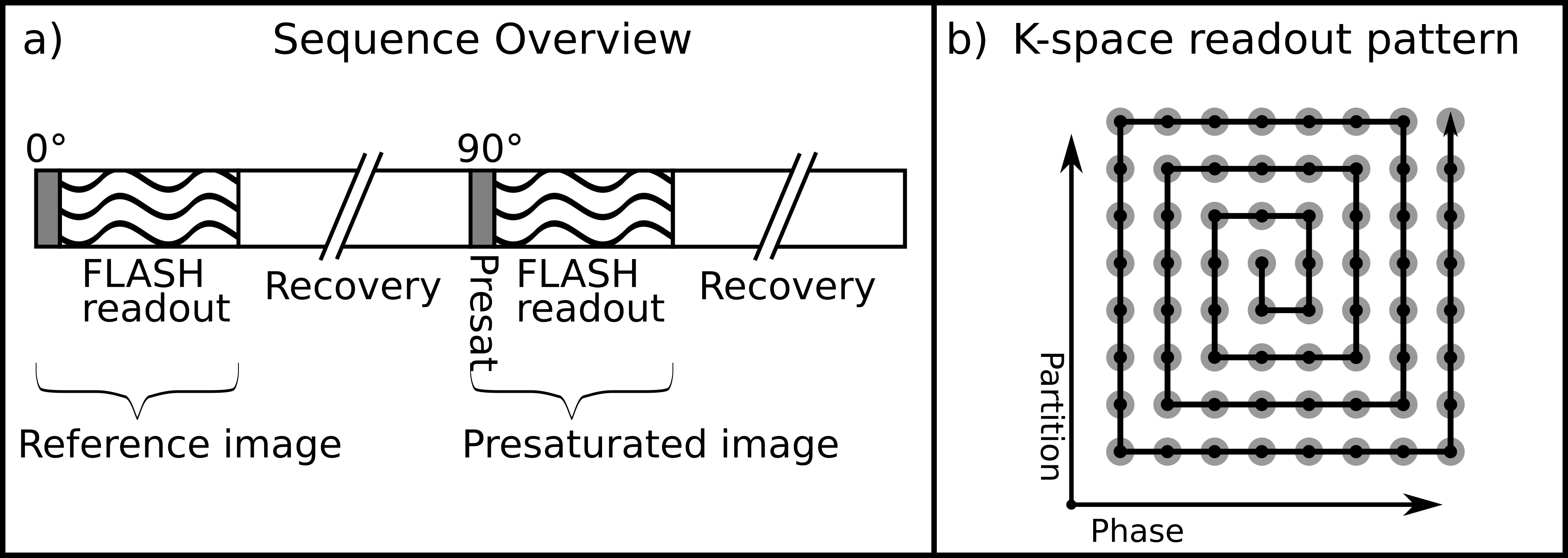
Figure 1: a) Scheme of a SatTFL sequence b) Illustration of the spiral-centric readout order.
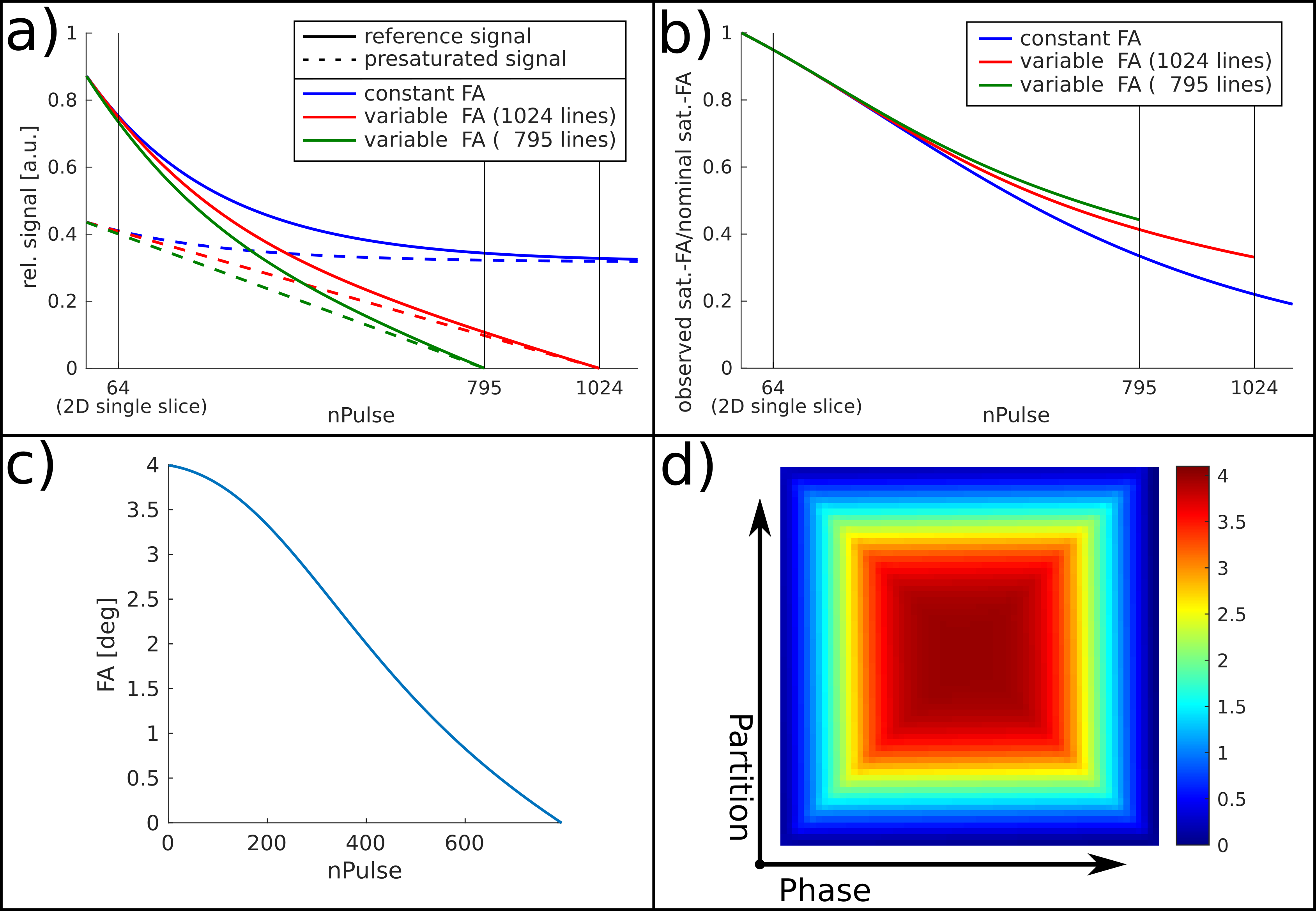
Figure 2:
Signal evolution of sigref and sigsat (a). A
constant flip-angle during the readout is compared to a readout with
variable FA which was optimized for 1024 and 795 k-space lines,
respectively. b) Shows the ratio of the observed FA and the actual FA
for readouts with a constant and variable FAs, respectively. c)
Depicts the variable FA calculated for a spiral-centric single-shot
readout with a total of 795 k-space lines and (d) the corresponding
FA distribution in the k-space trajectory.
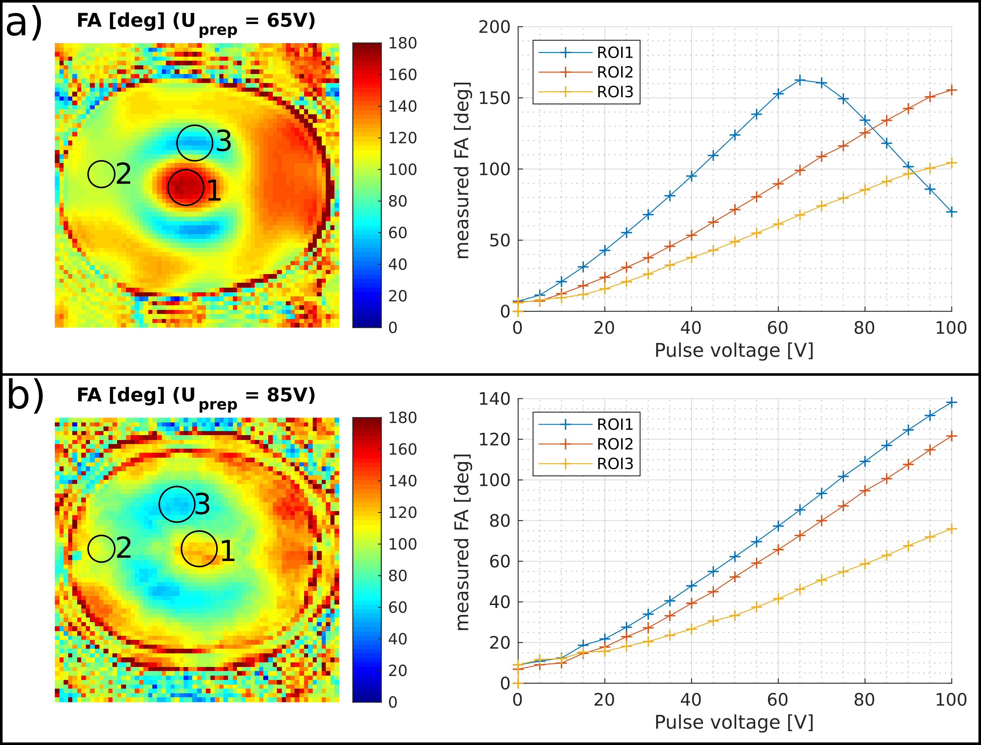
Figure 3: Preparation flip-angle measured with the 3D-SatTFLconst sequence as a function of preparation pulse voltage, in an agar phantom (a) and in vivo (b). Linearity below 30° and above 150° preparation flip-angle is poor. Preparation flip-angles above 180° cannot be measured and will be wrapped.
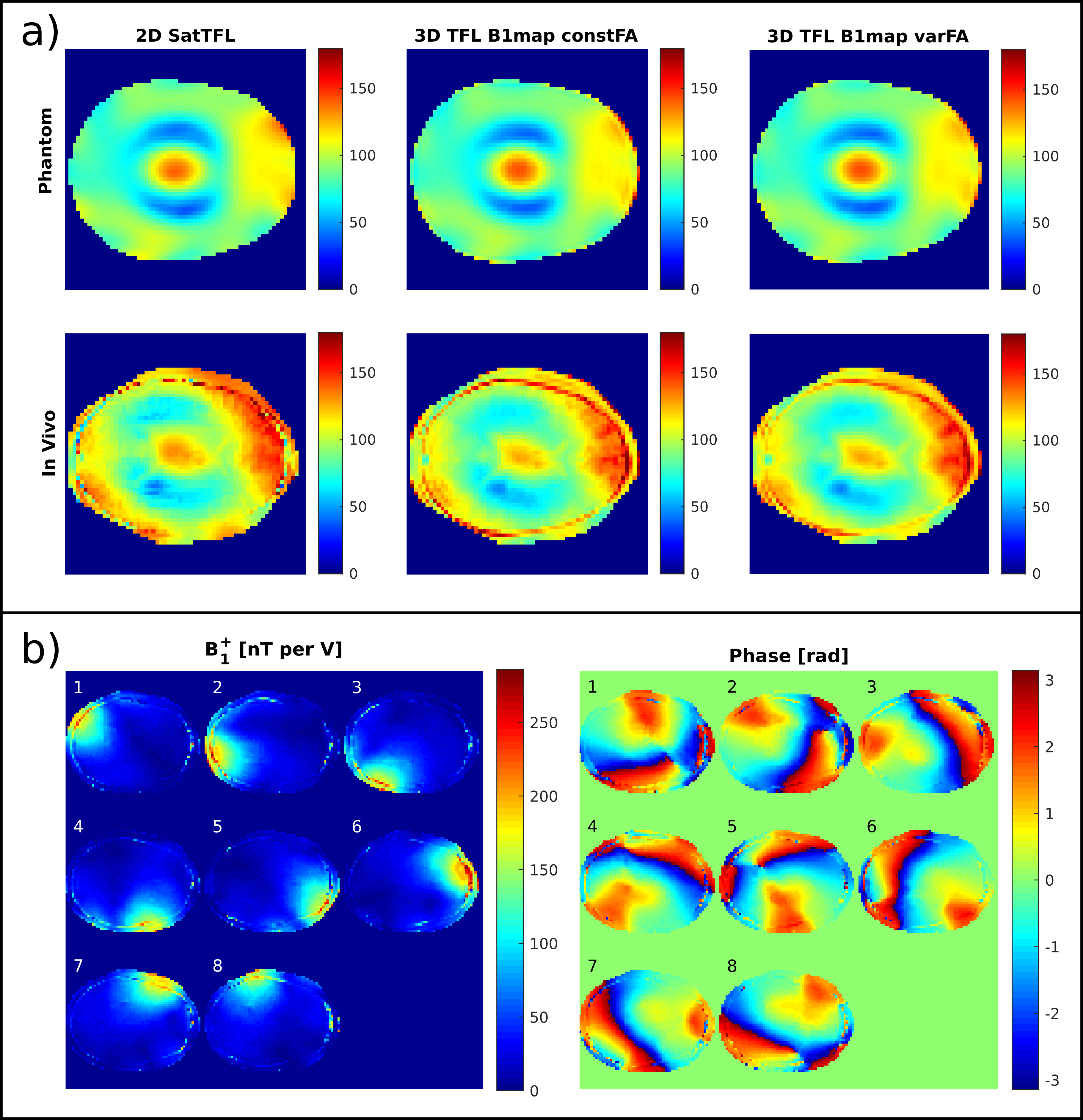
Figure
4: 2D-SatTFL compared to 3D-SatTFLconst and 3D-SatTFLvar,
in a head-shaped phantom (a, top) and in vivo (a, bottom). The
3D-SatTFLconst map shows ringing in the frontal section of
the phantom, which is reduced in the 3D-SatTFLvar map.
Deviations in the measured flip-angle from the reference map obtained
with the 2D-SatTFL mostly occur in regions with very low or very high
presat-FA.
Individual-channel
in-vivo B1+ maps recorded using the
3D-SatTFLvar are plotted in (b).