3688
MRI with Sub-millisecond Temporal Resolution over a Reduced Field of View1Bioengineering, University of Illinois at Chicago, Chicago, IL, United States, 2CMRR, University of Illinois at Chicago, Chicago, IL, United States, 3Radiology, University of Illinois at Chicago, Chicago, IL, United States, 4Neurosurgery, University of Illinois at Chicago, Chicago, IL, United States
Synopsis
Sub-millisecond Periodic Event Encoded Dynamic Imaging or SPEEDI (also known as SMILE) has been reported to be capable of achieving sub-millisecond temporal resolution. However, the total scan times of this technique is typically very long. In this study, we have incorporated two techniques – reduced field of view (rFOV) and echo-train acquisition – into a SPEEDI sequence to substantially reduce the total scan times. This sequence, which we call re-SPEEDI, has been demonstrated in phantom experiments for capturing rapidly changing currents in a wire loop with a temporal resolution of 0.6ms-0.8ms.
Introduction
A recently reported technique – Sub-millisecond Periodic Event Encoded Dynamic Imaging or SPEEDI (also known as SMILE1,2) – has demonstrated sub-millisecond temporal resolution in MRI using an FID or spin-echo acquisition. However, the total scan time is long, which prevents SPEEDI from being practically used in a wide range of potential applications. Decreasing the matrix size can reduce the scan times, but compromises the spatial resolution. One possible approach is to simultaneously reduce the field-of-view (FOV) by focusing on a region of interest3–5 so that the spatial resolution can be maintained. In addition, the scan times of SPEEDI can be further shortened by replacing FID-based or spin-echo-based acquisition with echo-train-based acquisition. In this study, we report a variation of the SPEEDI sequence, which we call re-SPEEDI, by incorporating the two aforementioned strategies – reduced FOV and echo-train acquisition.Methods
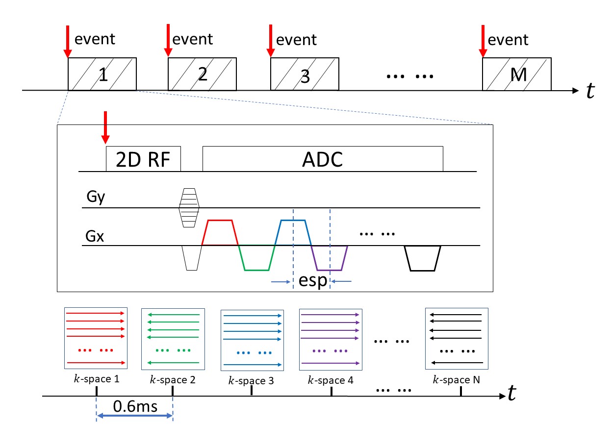
re-SPEEDI:To reduce the FOV, re-SPEEDI employed a 2D RF excitation pulse, which was designed by tilting 2D excitation k-space3,4. To incorporate echo-train acquisition so that frequency-encoding can be applied along one spatial direction to reduce the scan time, an FID signal in the original SPEEDI sequence was replaced with a non-phase-encoded gradient-echo train (Fig. 1). Each echo in the echo train was positioned in an individual k-space raster, and all echoes in the echo train were spread across a series of time-resolved k-space rasters (k-space 1, k-space 2, … k-space n), as shown in Fig. 1. This process was repeated with different phase-encoding values until all 2D k-space rasters were adequately sampled. After a 2D Fourier transform, a collection of time-resolved images was obtained with a temporal resolution determined by echo spacing (esp). Compared to SPEEDI, re-SPEEDI considerably reduces the total scan times because of a smaller matrix size and the use of frequency-encoded echo train signals.
Data Acquisition:
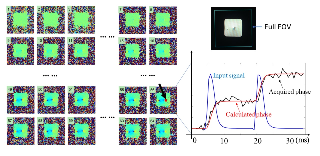
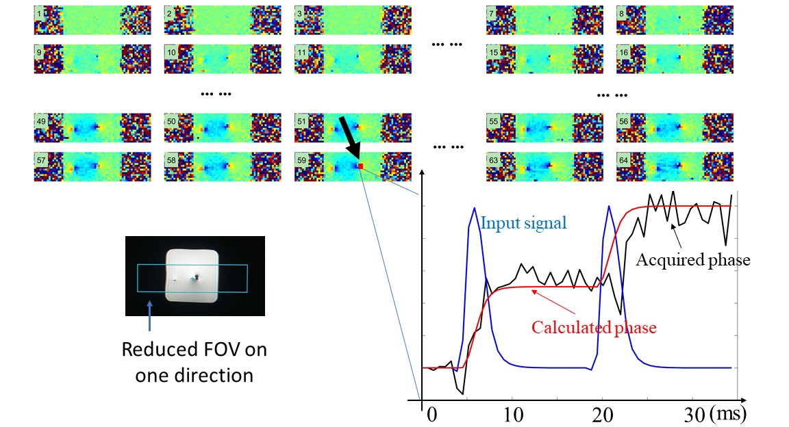
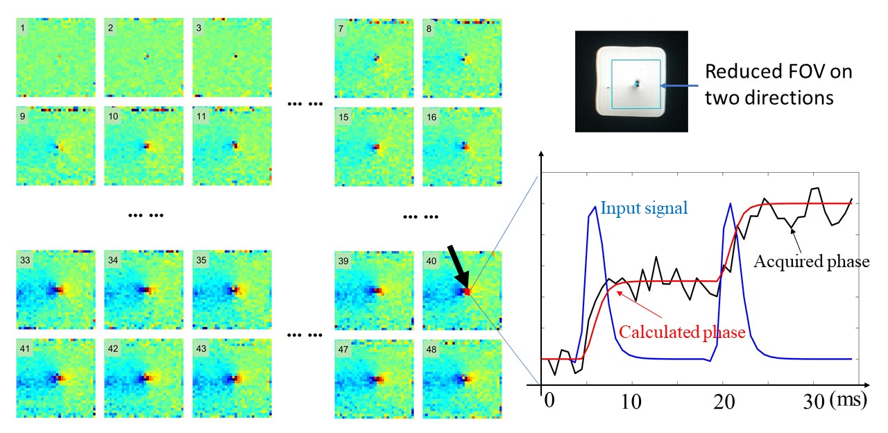
A re-SPEEDI sequence (Fig. 1) was implemented on a 3T GE MR750 scanner. Experimental demonstration was carried out using a phantom that contained a rectangular wire loop submerged in water. A pulse generator was used to supply a rapidly time-varying current to the wire loop, which was synchronized with the RF unblanking signal from the scanner. The same signal was also used to trigger the re-SPEEDI sequence in order to capture the dynamics of the phase change produced by the time-varying current. The experiments were performed three times, each with a different FOV: (a) full FOV that covered the entire phantom (FOV=12cm×12cm, matrix=64×64, scan time=2:08, esp=0.6ms), (b) a reduced FOV in the phase-encoding direction only (FOV=12cm×3cm, matrix=64×16, scan time=32s, esp=0.6ms), and (c) reduced FOV in both frequency-encoding and phase-encoding directions (FOV=4cm×4cm, matrix=32×32, scan time=1:04, esp=0.8ms). For each experiment, a scan was first performed without current in the wire loop to establish a reference, followed by another scan after applying a 3mA peak current with a duration of 35ms and a shape that imitated an action potential. The other acquisition parameters were: TR=2000ms, TE=2.8ms, and slice thickness=5mm.
Image Analysis:
After acquisition of time-resolved images using re-SPEEDI, the phase maps of these images were produced at each time point. The phase evolution from a specific point in the water phantom was extracted and compared with the simulated phase change calculated using the following equation: $$\phi(t)=\gamma\int_{0}^{t} B(t^{'})dt^{'}, [1]$$ where B(t) is the magnetic field produced by the time-varying current in the wire loop.
Results
The dipole pattern produced by the current can be clearly seen on the phase maps from all three experiments, as shown in Figs. 2-4, respectively. The fast-changing phase evolution was captured with a 0.6ms or 0.8ms temporal resolution, and agreed well with the simulation results using Eq. [1]. Compared to conventional SPEEDI, the scan times were shortened by a factor of 4, 16, and 8 in the three experiments, respectively. In addition, the approach with reduced FOV in both in-plane directions improved the spatial resolution from 1.8×1.8mm2 in full FOV to 1.2×1.2mm2.Discussion and Conclusion
We have demonstrated the ability of re-SPEEDI for substantially reducing the overall scan times while offering sub-millisecond temporal resolution for capturing the dynamic current change in a wire loop. The rFOV approach, which has been used to decrease the acquisition times in previous studies6–8, was successfully incorporated into SPEEDI. Imaging with an rFOV can also improve the spatial resolution without suffering from aliasing artifacts. The echo-train acquisition method was a variable alternative to the FID-based acquisition and balanced the conflicting requirements between the total scan time and the temporal resolution. By combining rFOV and echo-train acquisition, re-SPEEDI is expected to be an important contributor to broadening the applications based on SPEEDI-type sequences.Acknowledgements
This work was supported in part by NIH 1S10RR028898.References
1. Zhong, Z., Karaman, M. M., Claiborne, T. and Zhou, X. J. Capturing Time-Dependent Electric Currents Using MRI with A Sub-Millisecond Temporal Resolution. in ISMRM2019 P4574.
2. Zhong, Z., Karaman, M. M. and Zhou, X. J. MRI with Sub-Millisecond Temporal Resolution: An Example Employing Spatially Resolved Eddy Current Characterization. in ISMRM2019 P0247.
3. Sui, Y., Damen, F., Xie, K. and Zhou, X. J. Image Domain Segmented Diffusion Imaging Using A 2D Excitation RF Pulse for Distortion Reduction. in ISMRM 22 (p. 0608) (2014).
4. Zhou XJ, Sui Y. Image Domain Segmented Echo Planar Magnetic Resonance Imaging Using a 2D Excitation Radiofrequency Pulse. U.S. Patent No 9797970 B2. 2017.
5. Finsterbusch, J. Improving the performance of diffusion-weighted inner field-of-view echo-planar imaging based on 2D-Selective radiofrequency excitations by tilting the excitation plane. J. Magn. Reson. Imaging 35, 984–992 (2012).
6. Zhao, L., Madore, B. and Panych, L. P. Reduced field-of-view MRI with two-dimensional spatially-selective RF excitation and UNFOLD. Magn. Reson. Med. 53, 1118–1125 (2005).
7. Alizadeh, M. et al. Reduced FOV diffusion tensor MR imaging and fiber tractography of pediatric cervical spinal cord injury. Spinal Cord 55, 314–320 (2017).
8. Saritas, E. U., Cunningham, C. H., Lee, J. H., Han, E. T. and Nishimura, D. G. DWI of the spinal cord with reduced FOV single-shot EPI. Magn. Reson. Med. 60, 468–473 (2008).
Figures