3685
A Combined Approach of Variable Flip Angle, Keyhole and Averaging (CAVKA) for Accelerating the Acquisition of a low SNR Image Series
Hen Amit Morik1, Patrick Schuencke1, and Leif Schröder1
1Leibniz-Forschungsinstitut für Molekulare Pharmakologie (FMP), Berlin, Germany
1Leibniz-Forschungsinstitut für Molekulare Pharmakologie (FMP), Berlin, Germany
Synopsis
We propose a method that is a Combined Approach of Variable Flip Angle (VFA), Keyhole undersampling and Averaging (CAVKA). It is designed to optimize the use of the limited magnetization and to accelerate the acquisition in MRI series that suffer from low SNR and thus require averaging. The method is applied to the acquisition of a CEST (chemical exchange saturation transfer) image series, where the sensed nucleus is hyperpolarized 129Xe. There it provides ~4-fold SNR increase compared to conventional imaging without averaging or 7-fold acceleration with the same SNR compared to imaging with averaging.
Introduction
X-nuclei based imaging (e.g. 13C, 23Na, 129Xe) usually suffers from low signal relative to 1H MRI. Increasing the SNR by means of conventional averaging is very time-consuming especially when a series of images is required. Such image series are for example acquired in chemical exchange saturation transfer (CEST), where each image is encoded after applying a saturation pulse with a different frequency. In this study, we present a new approach to increase the SNR and/or accelerate the acquisition of CEST measurements with hyperpolarized 129Xe.Methods
Our proposed CAVKA method combines variable flip angle (VFA) excitations1 with the “keyhole” technique and averaging. Keyhole is a view-sharing approach that includes the acquisition of two components: a fully sampled reference image and a series of undersampled images2. Undersampling is achieved by encoding only a square in the center of k-space (called keyhole). Missing data in the periphery of the undersampled images is retrieved computationally from the reference image. In the proposed CAVKA approach, this reference image is acquired using averaging and the keyhole series is encoded with VFA. Merging the k-space data of the keyhole and reference images and the subsequent image reconstruction are done in self-written software in Python. All measurements were performed on a 9.4T pre-clinical MRI scanner (Bruker, Germany). Xenon hyperpolarization is achieved through spin exchange optical pumping3 using a home-built polarizer. All results shown come from a two-compartment phantom experiment, where the outer compartment is filled with H2O and the inner one with a CEST agent solution (10 μM Cryptophane-A in H2O + 0.2% DMSO). Hyperpolarized 129Xe gas was bubbled directly into the solutions.Results
Our approach increases SNR since VFA divides the available magnetization according to the chosen number of phase encoding steps. The lower this number, as in an undersampled image, the higher the magnetization per readout line. Additional de-noising is achieved, for the entire CEST image series, by the averaged reference image. Fig.1 shows three images: a conventional (non-hybrid) image (A), a hybrid image with VFA-enhanced center and non-averaged periphery (B) and the CAVKA image with VFA-enhanced center and averaged periphery (C). The transition from A to B shows the signal intensity increase due to VFA encoding and the transition from B to C shows the noise reduction due to averaging of the periphery. Importantly, the combination of both enables a ~4-fold SNR increase from A to C. Quantification of the SNR in the images that were composited according to our method is presented in Fig.2. The plot shows the SNR as a function of the number of averages of the reference image for three different undersampling factors of R = 1.33 (green), R = 2 (orange) and R = 4 (blue). In addition, a linear fit is included (dashed gray line) to show the theoretical SNR values when both, the periphery and the keyhole region, were averaged. For a fixed undersampling factor, the SNR increases for increasing number of averages. More important, the SNR further increases with increasing undersampling factors and almost reaches the level of the reference line for CAVKA-4 (i.e., R = 4). The outcome of applying the method to an image series is presented in Fig. 3. It shows the comparison between two z-spectra: one based on the proposed CAVKA method (blue) and the other based on fully sampled and averaged images (orange). It displays a very good agreement between the two spectra by their almost complete overlap. In particular, this match applies to the CEST contrast at -132 ppm. However, using CAVKA leads to a significant reduction (7-fold) in the acquisition time compared to the fully sampled and averaged series.Discussion
The two components of CAVKA optimize the utilization of the available magnetization according to the number of phase encoding steps. In addition, the accelerated acquisition of this method is achieved by the reuse of averaged data from the reference image for the entire undersampled series. The CAVKA method is particularly useful to accelerate image acquisitions for setups that suffer from low SNR and demands for averaging. Some limitations apply for capturing dynamic changes of small areas along the image series where an excessive undersampling factor corresponds to a small keyhole and might not capture such dynamic changes that manifest in the periphery. However, in the presented CEST case, a keyhole size of just 4x4 was still enough to capture the CEST contrast between the two different compartments.Conclusion
In this work, we have introduced a method that deals with low SNR in MRI series. Such conditions particularly occur for X-nuclei or limited magnetization from dilute hyperpolarized agents. Such scenarios often do not allow for long acquisition times. In addition, future in vivo applications of the HyperCEST method face reduced SNR compared to in vitro experiments where a large potential of this method has been demonstrated.Acknowledgements
No acknowledgement found.References
- Zhao L, Mulkern R, Tseng CH, et al. Gradient-echo imaging considerations for hyperpolarized 129Xe MR. J Magn Reson B. 1996;113:179-183.
- Varma G, Lenkinski RE, Vinogradov E. Keyhole Chemical Exchange Saturation Transfer. Magn Reson Med Off J Soc Magn Reson Med Soc Magn Reson Med. 2012;68(4):1228-1233. doi:10.1002/mrm.23310
- Walker TG, Happer W. Spin-exchange optical pumping of noble-gas nuclei. Rev Mod Phys. 1997;69(2):629-642. doi:10.1103/RevModPhys.69.629
Figures
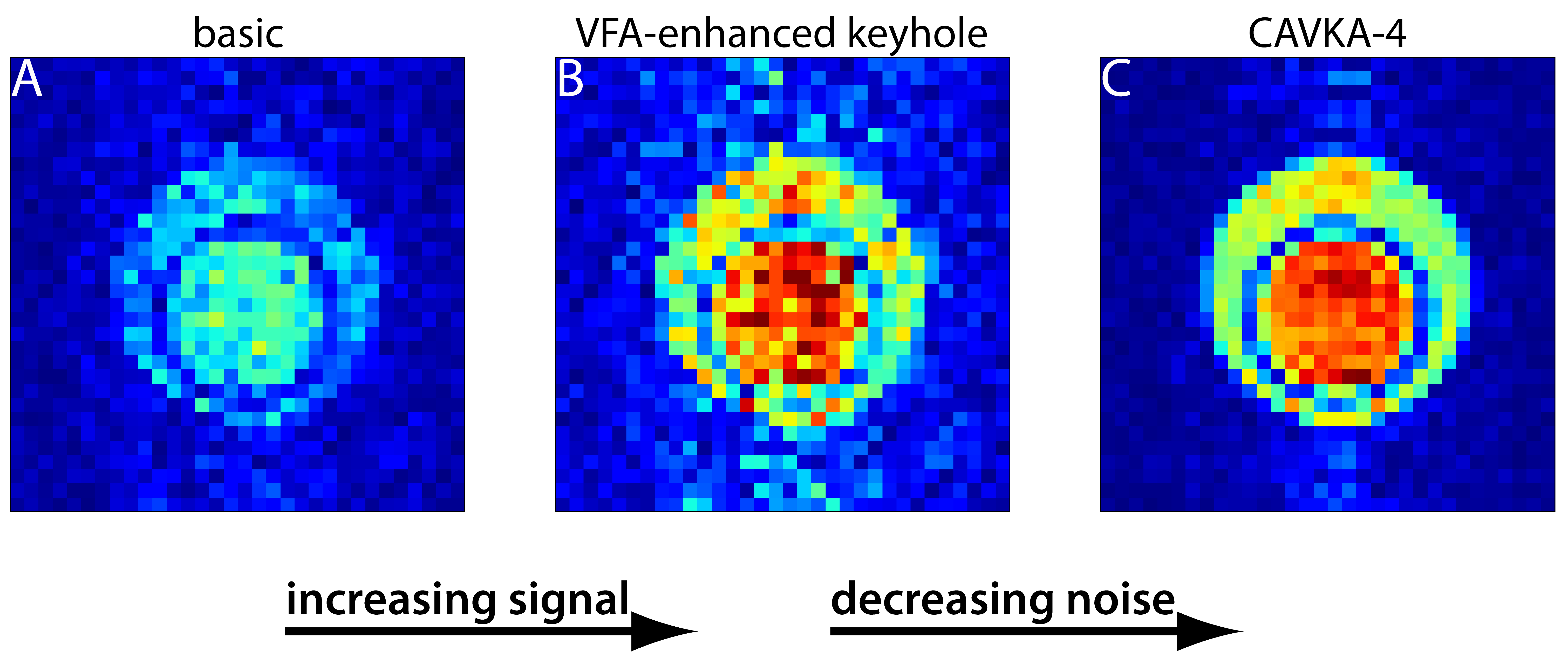
Figure 1: The two-step CAVKA image composition. Results from a phantom
experiment. Left to right: Non-hybrid image with matrix size of 32x32, VFA
encoded (A). Hybrid image with 8x8 keyhole, VFA-encoded and non-averaged reference image (B). Hybrid image with 8x8 keyhole, VFA-encoded and 16-times averaged reference image (C). The final image illustrates the increase of signal
intensity due to VFA enhancement (A to B) and the reduction of noise due to
averaging of the periphery (B to C).
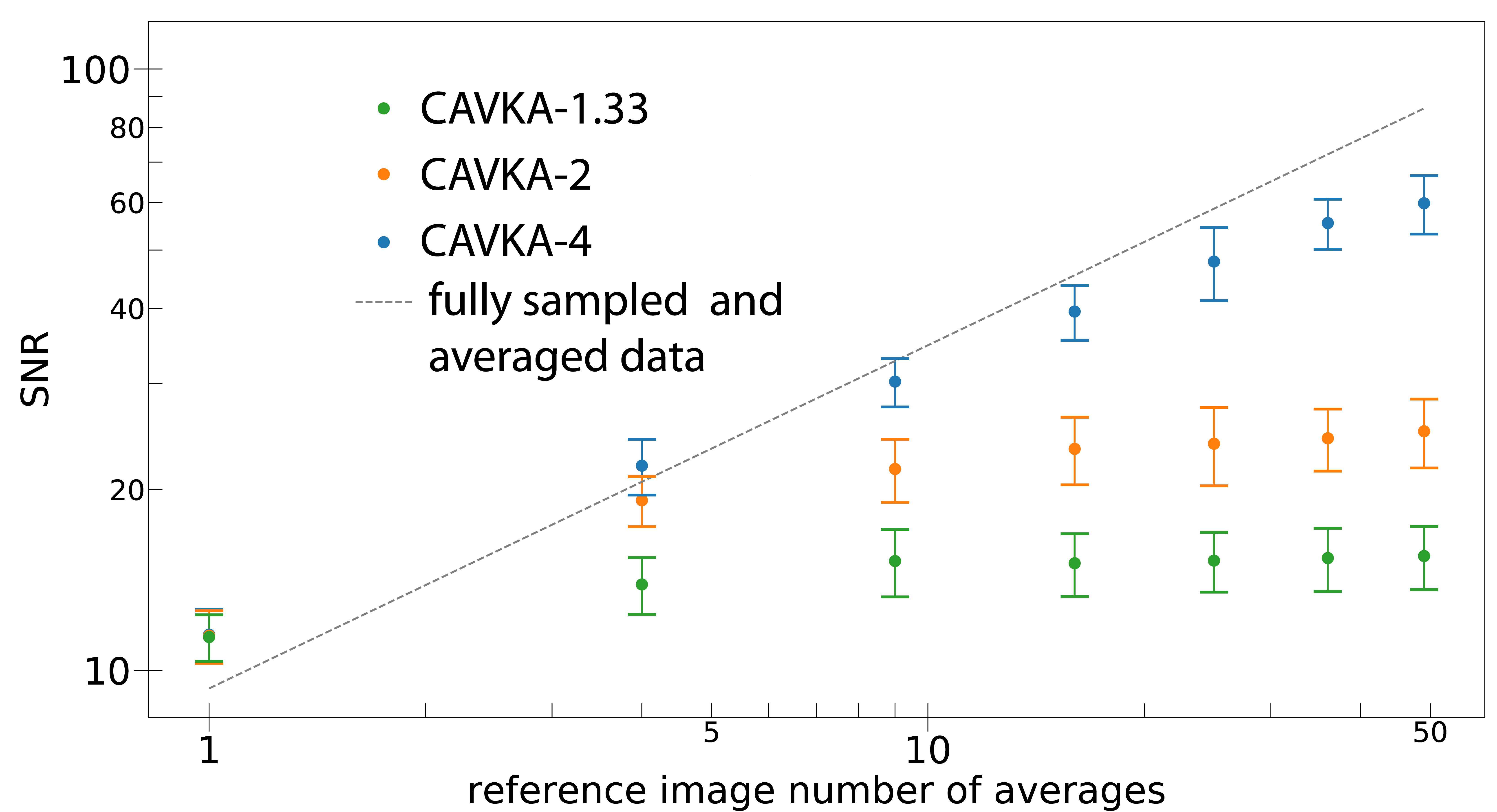
Figure 2: SNR in CAVKA images. SNR vs. the number of averages of the
reference image for three undersampling factors of 1.33 (green), 2 (orange) and
4 (blue). A comparison (fitted) line shows SNR vs. number of averages when both, the periphery and the keyhole region,
were averaged (dashed, gray). Values are mean ± 1 SD of 10 independent replicates
and plotted with a double logarithmic scale.
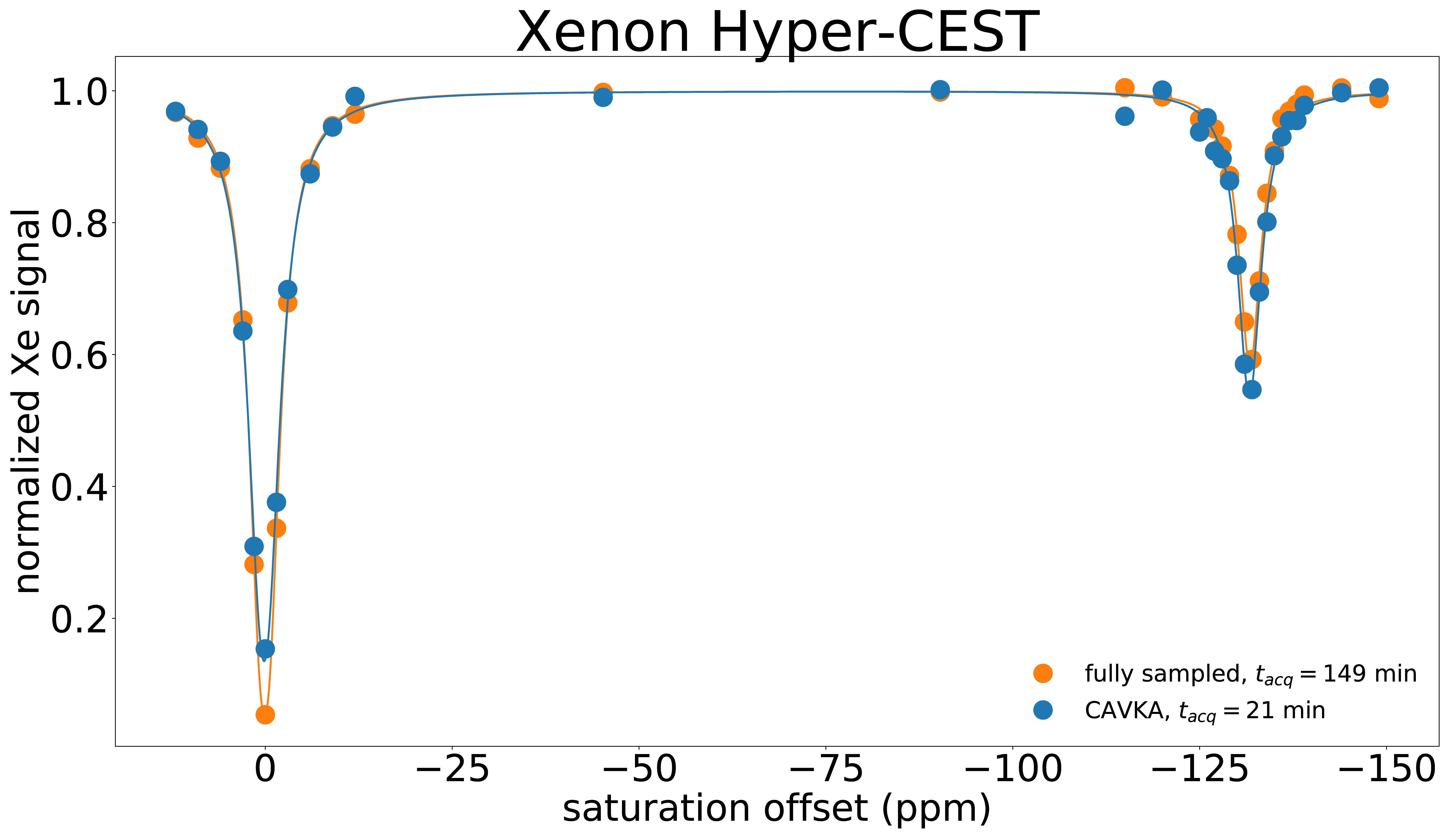
Figure 3: Applying CAVKA to CEST image series of hyperpolarized 129Xe.
Z-spectrum from non-hybrid and individually 9-times averaged images (orange)
and from CAVKA-4 with 9-times averaged reference image (blue). Data points
represent ROI-averaged and normalized signal intensities. Image acquisition
times are 21 min and 149 min for the CAVKA based and the fully sampled z-spectrum,
respectively.