3684
Joint Compressed Sensing and Sensitivity Estimation for Free-Breathing Whole Heart CINE MRI Without ECG Gating1UIH America, Inc., Houston, TX, United States, 2Beijing LuHe Hospital, Capital Medical University, Beijing, China, 3United Imaging Healthcare, Shanghai, China
Synopsis
This abstract presents a new approach to compressed sensing cardiac MRI, which enables free-breathing whole heart coverage cine imaging within 30 seconds. Using a phased array coil, data was acquired continuously along Cartesian sampling trajectories using a lookup table without ECG gating. Each slice was continuously sampled for a fixed period of time, before the slice-selective RF excitation pulse switch to the next slice. In reconstruction, the approach jointly updates coil sensitivity maps and images, integrated with compressed sensing. In post-processing, virtual ECG is calculated based on unsupervised machine learning.
Introduction
To date, compressed sensing magnetic resonance imaging (CSMRI) using multichannel receiver coils has emerged as an effective tool to reconstruct images from highly accelerated scan in various applications. [1-2] However, the issue of accurate estimation of coil sensitivities in cardiac cine imaging combined with respiration motion has not been fully addressed, which limits the level of speed enhancement achievable with the technology.The self-calibrating (SC) technique for sensitivity extraction has been well accepted, especially for dynamic imaging, and complements the common calibration technique that uses a separate scan. However, the existing method to extract the sensitivity information from the SC data is not accurate enough in cardiovascular MRI, and thus erroneous sensitivities affect the reconstruction quality when they are directly applied to the reconstruction equation.
This abstract presents a new approach to compressed sensing cardiac cine imaging. Using a phased array coil, data was acquired continuously along Cartesian sampling trajectories using a lookup table (pre-calculated based on Latin Hyper Sampling [4]) without ECG gating. Each slice was continuously sampled for a fixed period of time (roughly 2 seconds) to cover the whole cardiac cycle, before the slice-selective RF excitation pulse switch to the next slice. In reconstruction, the approach jointly updates coil sensitivity maps and images, [4, 5] integrated with compressed sensing. In post-processing, the approach uses unsupervised k-median method to detect end-diastolic phases without ECG. The whole cardiac cycle images were processed slice-by-slice.
Method
Model: The main goal is to design and implement a sampling and reconstruction strategy that enables full heart coverage from highly accelerated free breathing real-time acquisitions, with a relatively high spatial resolution (2.5 × 2.5 mm2) and temporal resolution (40 ms). We adopt the VALAS [4] sampling pattern, and multi-channel blind deconvolution model [5] to update the coil sensitivities. Because respiration motion is involved, we assume each time frame / phase has its own coil sensitivity map.Problem formulation: Incorporating the above models, the data consistency model can be formulated as $$d_{t,j}=\Omega_t FS_{t,j} \mathbf x_t$$ where $$$d_{t,j}$$$, $$$S_{t,j}$$$ represent the undersampled k-space data and coil sensitivity profile from the j-th coil at the t-th time frame, respectively; $$$F$$$ is the 2-dimensional fast Fourier transform, $$$\Omega_t$$$ represents the undersampling operator.
Initialization: An initial coil sensitivity map is estimated from the temporal average k-space data.
Alternating optimization: The images and coil sensitivity maps are calculated and updated by the following optimization function alternatively: $$$\mathbf x_t=\underset{\mathbf x_t}{\arg\min} \frac{1}{2} \sum_{j} ||\Omega_t FS_{t,j} \mathbf x_t-d_{t,j}||^2+\lambda_1||TV_s(\mathbf x_t)||_1+\lambda_2||TV_t(\mathbf x_t)||_1$$$, $$$S_{t,j}=\underset{\mathbf S_{t,j}}{\arg\min}||\Omega_t FS_{t,j} \mathbf x_t-d_{t,j}||^2+\lambda||S_{t,j}F_t||_1$$$. $$$F_t$$$ represents temporal FFT.
Virtual ECG: After the cine images were acquired, unsupervised k-median method [6] is used to cluster the cine images to three groups. k-median clustering, originally from signal processing, that is popular for cluster analysis in data mining. The median image was used as reference image to calculate the correlation curve for all $$$\mathbf x_t$$$. Virtual ECG is calculated based on the correlation curve and temporal resolution.
Results
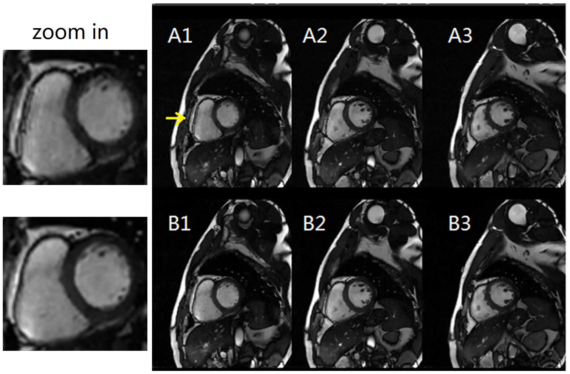
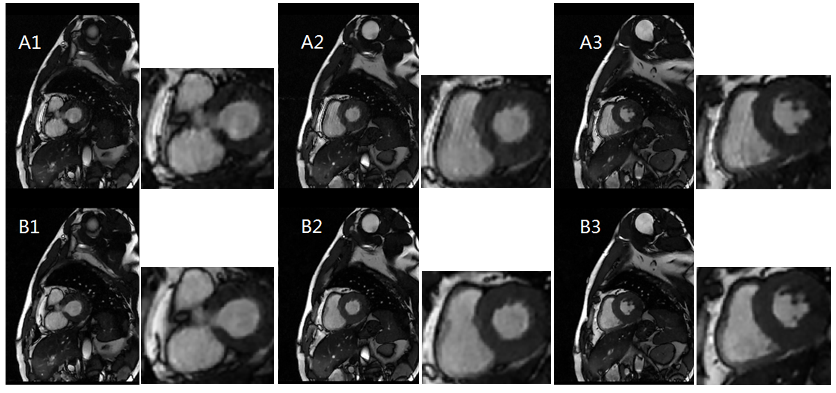
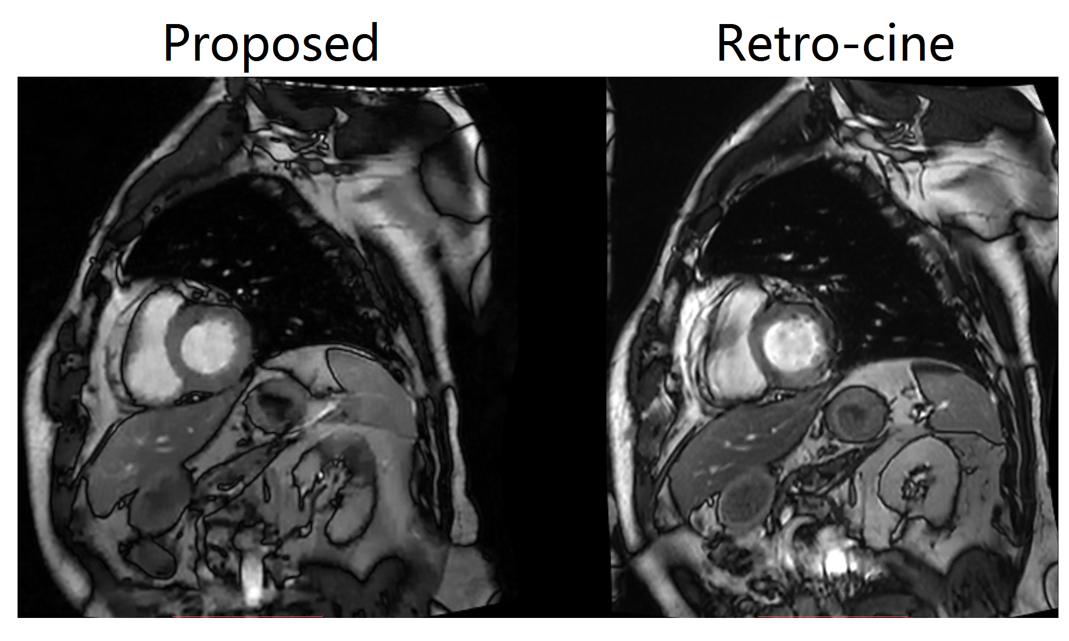
The proposed method was implemented with a bSSFP sequence on a clinical 3T scanner (uMR 780 United Imaging Healthcare, Shanghai, China) with the approval of local IRB. Imaging parameters were: imaging matrix: 192(readout) x 165(PEs) x 36(phases) x11(slices), TR/TE = 2.81/1.40 ms, slice thickness = 8 mm, flip angle =80°, bandwidth = 1300Hz/pixel, spatial resolution = 1.82x1.82 mm2, 15 PEs/phase, 36 phases/slice, Scan time: 1.7s/slice. Fig. 1 shows all image frames from one slice; Fig.2 shows end-diastolic phase images from the proposed method, compared with traditional compressed sensing without jointly reconstruction; Fig. 3 shows reconstructed end-systolic images from both methods; Fig.4 shows the spatial-temporal profiles of the myocardium. The ventricular morphology is better preserved from the proposed reconstruction method, without any wall motion artifacts and/or bSSFP banding artifacts.For comparison, whole heart coverage retro cine was also implemented. Imaging parameters were: TR/TE=3.05/1.4ms, bandwidth=1000Hz/pixel, Scan time: 14.6s/slice. 11 breath-hold for the whole heart retro-cine scan. Fig. 5 compares the proposed method with retro-cine. Motion artifacts were found in retro-cine, when the volunteer cannot hold the breath well; whereas the reconstructed image from proposed method is in a real-time fashion, without motion artifacts.
Conclusion
We have proposed a novel approach to real-time cardiac cine without breath hold and ECG gating. The approach effectively combines compressed sensing, multichannel blind deconvolution, auto phase detection in the same framework. The proposed method has shown success to recover whole heart cine images (11 slices) without ECG from highly accelerated free breathing acquisitions.Acknowledgements
No acknowledgement found.References
[1] Uecker, Martin et al., ESPIRiT--an eigenvalue approach to autocalibrating parallel MRI: where SENSE meets GRAPPA. Magnetic resonance in medicine, vol. 71,3: 990-1001, doi:10.1002/mrm.24751, 2014.
[2] Feng L, et al., Highly accelerated real‐time cardiac cine MRI using k–t SPARSE‐SENSE. Magnetic resonance in medicine, vol. 70.1: 64-74, 2013.
[3] Sheng J., Liu B., Ying L., Improved self-calibrated spiral parallel imaging using JSENSE, Medical Engineering and Physics, vol. 31, No. 5, pp. 510-514, June 2009.
[4] Lyu J., Ding Y., Zhong J., Zhang Z., Zhao L., Xu J., Liu Q., Peng R., and Zhang W., Toward single breath-hold whole-heart coverage compressed sensing MRI using VAriable spatial-temporal LAtin hypercube and echo-Sharing (VALAS), no.4752, ISMRM, 2019.
[5] Lyu J., Nakarmi U., Zhou Y., Zhang C., Ying L., Calibration-free Parallel Imaging Using Randomly Undersampled Multichannel Blind Deconvolution (MALBEC), no. 3232, ISMRM, 2016.
[6] Jain A. K. and Dubes R. C., Algorithms for Clustering Data. Prentice-Hall, 1988.
Figures