3667
Optimizing a preprocessing pipeline for structural 7T MR analyses in FreeSurfer
Giske Opheim1,2, Oula Puonti3,4, Jan Ole Pedersen5, Vincent O. Boer3, Ane Kloster1,2, Martin Prener1,2, Helle Juhl Simonsen6, Olaf B. Paulson1,2, Lars H. Pinborg1,2, and Melanie Ganz1,7
1Neurobiology Research Unit, Rigshospitalet, Copenhagen University Hospital, Copenhagen, Denmark, 2Faculty of Health and Medical Sciences, University of Copenhagen, Copenhagen, Denmark, 3Danish Research Centre for Magnetic Resonance, Funktions- og Billeddiagnostisk Enhed, Copenhagen University Hospital Hvidovre, Copenhagen, Denmark, 4Dept. of Health Technology, Technical University of Denmark, Kongens Lyngby, Denmark, 5Philips Healthcare, Copenhagen, Denmark, 6Functional Imaging Unit, Department of Clinical Physiology, Nuclear Medicine and PET, Rigshospitalet Glostrup, Copenhagen, Denmark, 7Dept. of Computer Science, University of Copenhagen, Copenhagen, Denmark
1Neurobiology Research Unit, Rigshospitalet, Copenhagen University Hospital, Copenhagen, Denmark, 2Faculty of Health and Medical Sciences, University of Copenhagen, Copenhagen, Denmark, 3Danish Research Centre for Magnetic Resonance, Funktions- og Billeddiagnostisk Enhed, Copenhagen University Hospital Hvidovre, Copenhagen, Denmark, 4Dept. of Health Technology, Technical University of Denmark, Kongens Lyngby, Denmark, 5Philips Healthcare, Copenhagen, Denmark, 6Functional Imaging Unit, Department of Clinical Physiology, Nuclear Medicine and PET, Rigshospitalet Glostrup, Copenhagen, Denmark, 7Dept. of Computer Science, University of Copenhagen, Copenhagen, Denmark
Synopsis
Automated cortical segmentations benefit from higher SNR and spatial resolutions on 7T MR images, but are also challenged by B1 inhomogeneities, causing faulty surface inflations primarily in the temporal lobes. We investigated how FreeSurfer outputs were affected by applying eight different preprocessing schemes prior to reconstructions of submillimeter 7T MPRAGE images. The highest segmentation robustness across subjects was obtained by setting bias-field correction FWHM to 60mm and adding light regularization, and additionally performing intensity normalization.
Introduction
Computational analysis of structural MRI scans provides valuable insights into anatomical changes in different brain diseases. Ultra-high field (UHF) scanners provide unprecedented spatial resolution and SNR1 with potentially higher sensitivity to detect minute anatomical changes aiding the discovery of novel biomarkers2-3. However, non-uniform intensity distributions and inhomogeneous B1 transmit and receive fields at UHF MRI lead to undesired variations in image intensity and hamper the automated segmentation of structural images using software such as FreeSurfer (FS)4. These issues could lead to selection bias where only the subjects in which the automated analysis is successful can be included. On the FS wiki-page5, bias-field correction with 18mm FWHM and sampling distance = 2 is recommended prior to running recon-all on 7T data, together with tuned expert options adapted to the temporal lobe intensities in your images. These settings somewhat improve recon-all output, but still entail consistent failures in several regions in our datasets. We therefore investigated how different preprocessings affect the robustness of FS segmentations based on a submillimeter 3D MPRAGE sequence acquired using a the most widely used commercial head-coil for 7T.Methods
3D MPRAGE images (0.7mm isotropic) were acquired on a 7T MR system (Philips, Achieva, Best, The Netherlands), with 19x19cm dielectric pads on both sides, with a quadrature 32/2 Rx/Tx coil (Nova Medical, Wilmington, MA). Six subjects were included in the study, informed consent was obtained according to the local ethical guidelines. The data were processed using FS’s recon-all stream (conf2hires) with eight different preprocessing schemes: 1) No bias-field correction (BFcorr), 2) BFcorr with low (30mm) FWHM and no regularization, 3) BFcorr with high (60mm) FWHM with liberal regularization, and 4) BFcorr with high FWHM with tight regularization - all performed with and without intensity normalization by a spatially adaptive non-local means filter. An overview of the different preprocessing steps can be seen in Figure 1. BFcorr was performed with a downsampling factor of 3, and done in SPM12. Intensity normalization was done in SPM12 with functions provided in the CAT12 toolbox. All 48 segmentation outputs were visually quality controlled by the same observer. Variances in average thicknesses across six typically problematic cortical regions and across all six patients were calculated for all eight preprocessing combinations.Results
Visual inspection of segmentations revealed improved quality after applying scheme 2, 3 and 4, compared to scheme 1. Scheme 1, 2 and 3 further improved quality after combining it with intensity normalization, whereas scheme 4 did not. Scheme 2 seemed to yield the lowest variances (see Figure 2), but visual quality control revealed consistent failures in all subjects. Scheme 3 with intensity normalization gave the highest robustness based on qualitative assessment of segmentations (see Figure 3), and on the variances (Figure 2).Discussion
The results above highlight that both visual inspection and quantitative validations are important when assessing output from FreeSurfer. One should also note that even though we significantly improve the segmentation performance after preprocessing, manual edits might still be necessary. Furthermore, our results show that it is better to do bias-field correction and intensity normalization than not to, but also that surface quality control failures occur more often and consistently in the least and most flexible schemes (“fwhm60_tightreg” and “fwhm30_noreg”). The reason for these failures in the former case is likely that the bias correction is not flexible enough to correct for all the intensity inhomogeneities, whereas in the latter case it might be too flexible and start fading out contrast differences between tissues.Conclusion
Based on our findings, we recommend performing bias-field correction with FWHM set to around 60mm along with intensity normalization before automatic whole-brain segmentation on 7T MPRAGE images from a classical 7T MR scan set-up.Acknowledgements
The project is supported by the Independent Research Fund Denmark. The 7T scanner was donated by the Danish Agency for Science, Technology and Innovation grant no. 0601-01370B, and The John and Birthe Meyer Foundation.References
- Trattnig, S., Springer, E., Bogner, W., Hangel, G., Strasser, B., Dymerska, B., ... & Robinson, S. D. (2018). Key clinical benefits of neuroimaging at 7 T. Neuroimage, 168, 477-489.
- Fujimoto, K., Polimeni, J. R., Van Der Kouwe, A. J., Reuter, M., Kober, T., Benner, T., ... & Wald, L. L. (2014). Quantitative comparison of cortical surface reconstructions from MP2RAGE and multi-echo MPRAGE data at 3 and 7 T. Neuroimage, 90, 60-73.
- Zaretskaya, N., Fischl, B., Reuter, M., Renvall, V., & Polimeni, J. R. (2018). Advantages of cortical surface reconstruction using submillimeter 7 T MEMPRAGE. Neuroimage, 165, 11-26.
- Fischl, B. (2012). FreeSurfer. Neuroimage, 62(2), 774-781.
- https://surfer.nmr.mgh.harvard.edu/fswiki/HighFieldRecon
Figures
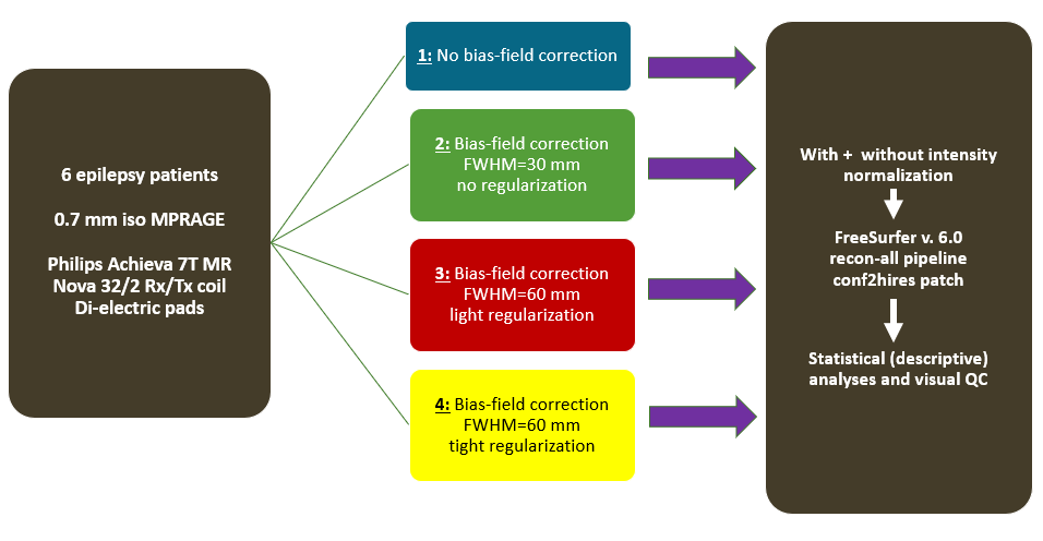
Figure 1: Schematic overview of the examined preprocessing work-flow.
Six subjects were processed using FreeSurfer’s recon-all stream (conf2hires)
with eight different pre-processing schemes:
1) No bias-field correction (BFcorr), 2) BFcorr with low (30mm) FWHM and
no regularization, 3) BFcorr with high
(60mm) FWHM with liberal regularisation, and 4) BFcorr with high FWHM with
tight regularization - all performed with and without intensity normalization
by a spatially adaptive non-local means filter (scheme 5-8).
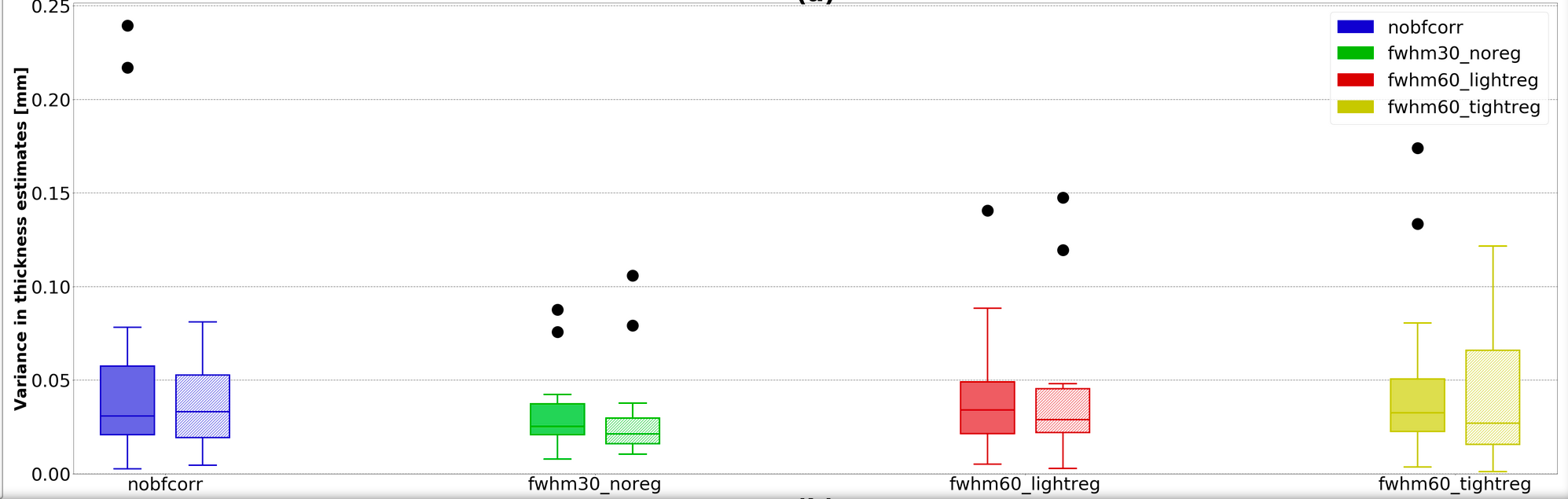
Figure 2: Boxplot showing the variance in thickness estimates for the different preprocessing pipelines . Colors correspond to different preprocessing
schemes, depicted in the Figure 1. Full color represent the schemes without
intensity normalization, and dashed represent the schemes with intensity
normalization. Variances
in six problematic regions for all patients with the eight preprocessing
combinations. “fwhm30_noreg” seems more stable across patients. However, visual
inspections of the segments revealed consistent segmentation errors in these
outputs.
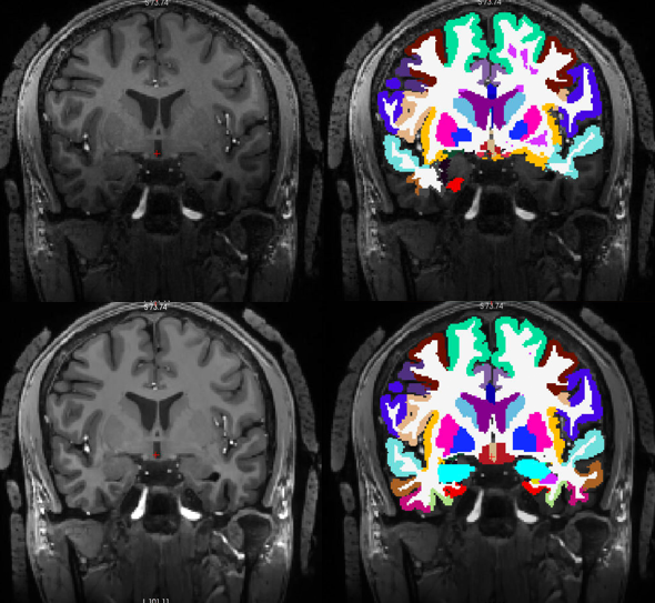
Figure 3: Example showing the
effect of preprocessing vs. no preprocessing on a single coronal slice. Top: Raw image without any preprocessing (left) overlayed
with FreeSurfer (FS) segmentations (right). Bottom: Bias-field corrected
(FWHM=60 mm, light regularisation) and intensity normalized image (left) and corresponding FS
segmentations (right). Visual inspections of these outputs revealed consistently
higher quality in this scheme.