3579
Rapid phase unwrapping with deep learning for shimming applications1Corporate Research Institute, United Imaging Healthcare, Shanghai, China
Synopsis
The implementation of shimming relies on phase unwrapping techniques to measure the underlying field inhomogeneity. This work presents a simple and effective deep learning based phase unwrapping method to accelerate the field mapping stage for shimming applications. The method significantly reduces the compulational time and shows similar performance compared to the conventional path-following method for shimming applications.
Purpose
This work presents a rapid deep learning based phase unwrapping method to accelerate the field mapping stage for shimming applications. Moreover, the comparison of this method with a conventional path-following algorithm demonstrates its superiority in routine shimming tasks.Introduction
In magnetic resonance imaging, shimming is used to homogenise the magnetic field. To measure the field inhomogeneity for compensation, field mapping techniques are implemented which rely on calculating the phase change of MR signal over a certain duration. However, the phase of signal only takes vales in 2$$$π$$$ range. In order to recover the underlying phase signal, phase unwrapping algorithms are conventionally adopted.Conventional phase unwrapping algorithms take an iterative way to minimize the gap between a pixel/region and its neighbors, e.g., the quality-guided path-following method and minimum-norm method.1,2 However, the computational time scales with the number of pixels/regions in image volumes. Previously it has been shown that the convolutional networks, U-Net, could be used for phase unwrapping.3,4 In this work, we extend the scope and provide a simple and effective U-Net based phase unwrapping method. Moreover, the performance of the U-Net based method is evaluated on realistic field maps for shimming applications.
Methods
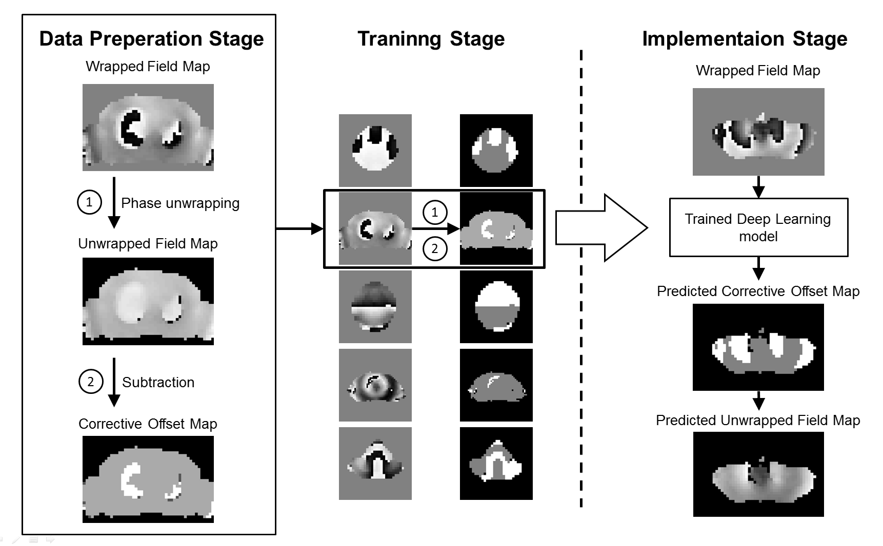
The experiments were conducted on a simultaneous PET/MR scanner (Unites Imaging, Shanghai, China) in Zhongshan Hospital, Shanghai, China. A total of 245 low-resolution field maps of different body parts (FOV=10 mm*10mm*10 mm, Slices=45) were acquired from different subjects as part of clinical studies for shimming applications.The procedures of the deep learning based phase unwrapping method are illustrated in Figure 1:
- Data preparation stage: The field maps in the database were unwrapped using a quality-guide path-following method. For a given voxel $$$i$$$ in the field map, the ground truth unwrapped phase $$$θ$$$ is given by $$θ_{i}=Φ_{i}·N_{i},$$ where $$$Φ$$$ denotes the measured phase, N = {1, 2, 3 …} denotes the corrective offset parameter of the voxel. Then, the corresponding ground truth corrective offset maps were obtained by subtracting the measured field maps from the unwrapped field maps.
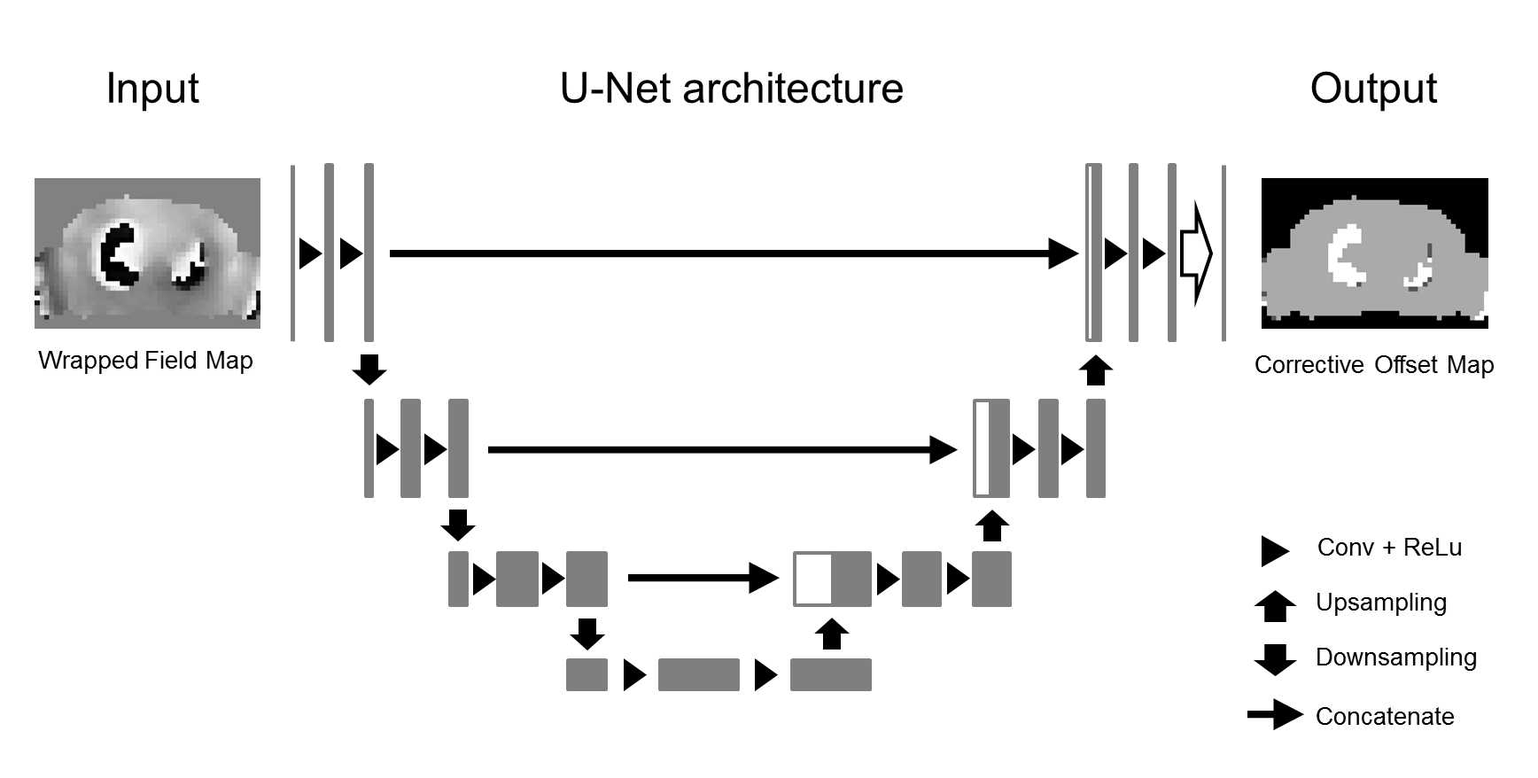
- Training stage: The dataset was split into training and validation set. An U-Net model was adopted and trained to predict the corrective offset maps on a slice-by-slice basis by taking measured field maps as input with the Adam optimization algorithm. The U-Net architecture is shown in Figure 2.
- Implementation stage: For a wrapped field map, the corresponding corrective offset map was predicted using the trained U-Net model slice by slice. Then, the slice-wise unwrapped field map was calculated using the equation above and concatenated together.
Results
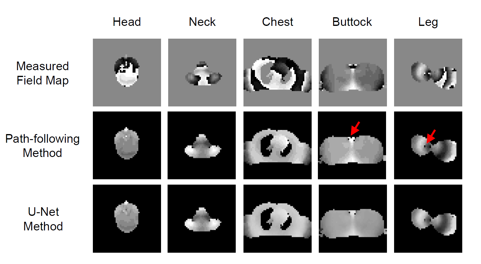
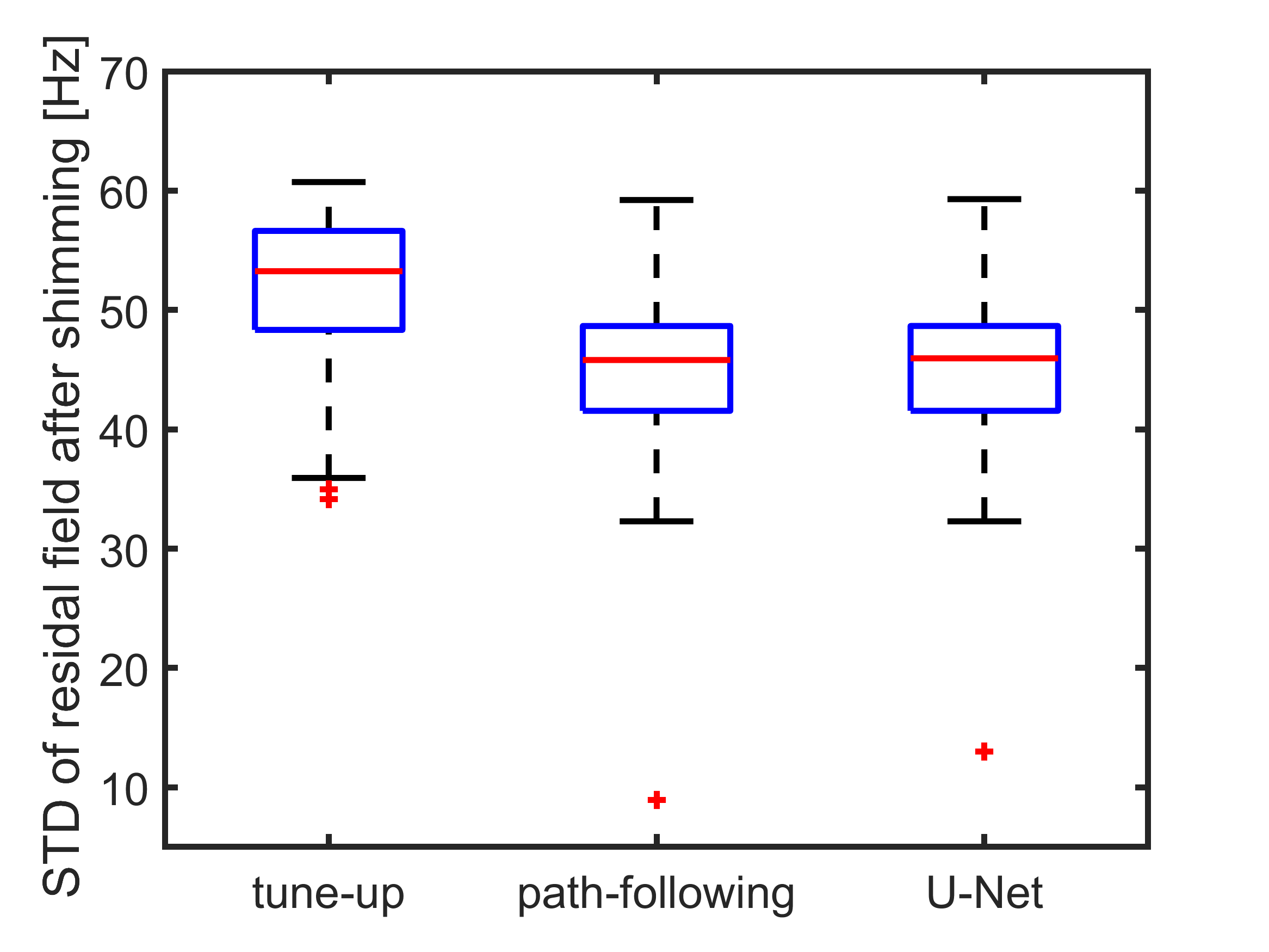
Figure 3 shows measured field maps of different body parts with no phase unwrapping, the path-following method and the U-Net based methods, respectively. As can be seen from the figure, the path-following method unwrapped phase in high-quality regions and discarded ambiguous voxels (red arrow). The U-Net based method shows similar performance.Figure 4 quantitatively compares the residual field standard deviation after shimming with the two methods, compared to the tune-up shim. The U-Net based method yields similar results compared to the path-following method.
In terms of computational time, the U-Net based method takes ~200ms per image volume. However, the path-following method could take up to 2 seconds in torso regions as a result of involving more voxels in calculation.
Disscussion
The reasons for the good performance of the U-Net based method are twofold. First, the U-Net convolution neural network comprises of encoder and decoder stages, which is able to extract both high-level (local field gradients and edges) and low-level (rough field distribution pattern) field map features for identifying corrective phase offsets. Second, the field maps used for shimming usually have fewer wraps and phase wrapping patterns are fairly consistent with different body parts.Whilst the path-following method yields good phase unwrapping results, this comes at a cost of computational time. Additionally, the field map used for shimming is low-resolution and noisy, it doesn't necessarily translate to better shimming results. The U-Net based method trades some slight phase unwrapping accuracy for much-improved speed, which is considered more practical for shimming applications. For futher research, the accuracy and speed of U-Net based method can be improved by using 3D convolution kernel and the pruning operation to accelerate the inference.
Conclusions
The U-Net based method has demonstrated its superior performance in identifying corrective offset maps for phase unwrapping in a short time. Meanwhile, it shows similar performance compared the path-following phase unwrapping results. The use of this phase unwrapping method could effectively accelerate the field mapping stage for shimming applications.Acknowledgements
No acknowledgement found.References
1. Wang H, Weaver JB, Perreard II, et al. A three-dimensional quality-guided phase unwrapping method for MR Elastography. Phys Med Biol. 2011;56(13): 3935–3952
2. Jenkinson M. Fast, automated, N‐dimensional phase‐unwrapping algorithm. Magn Reson Med. 2003; 49: 193–197.
3. Ronneberger O, Fischer P, and Brox T. U-net: Convolutional networks for biomedical image segmentation. International Conference on Medical image computing and computer-assisted intervention. Springer, 2015;234–241.
4. Spoorthi GE, Gorthi S, Gorthi RKSS. Phasenet: A deep convolutional neural network for two-dimensional phase unwrapping. IEEE Signal Process. Lett. 2019;26: 54–58
Figures