3518
Anisotropic Deep Learning Multi-planar Automatic Prostate Segmentation
Tabea Riepe1, Matin Hosseinzadeh1, Patrick Brand1, and Henkjan Huisman1
1Diagnostic Image Analysis Group, Radboudumc, RadboudUMC, Nijmegen, Netherlands
1Diagnostic Image Analysis Group, Radboudumc, RadboudUMC, Nijmegen, Netherlands
Synopsis
Optimized acquisition of prostate MRI for detection of clinically significant prostate cancer requires automatic prostate segmentation. State of the art automatic prostate segmentation is performed with convolution neural networks (CNNs). Exention of our previously developed anisotropic single plane CNN to handle multi-planar input is expected to decrease segmentation problems caused by the low inter-plane resolution of t2-weighted images. Data preprocessing includes volume alignment, intensity clipping and normalization. Comparing the performance to a similar axial network, the multi-stream model shows a visually relevant improvement in prostate segmentation.
Introduction
Prostate cancer (PCa) is the most common cancer among men with 174,650 new cases and 31,650 deaths in the US expected in 20191. Prostate MRI was recently established as a good modality for diagnosing clinically significant cancer (csPCa)2. Automatic segmentation of the prostate is an important part of current research in computer aided diagnosis (CAD) and optimized acquisition of MRI. State-of-the art segmentation uses standard convolution neural networks (CNN) using only axial t2-weighted MRI. The anisotropy caused by the much lower inter-plane MRI resolution results in segmentation errors in the apex and base region. Meyer et al.3 recently showed that adding the sagittal and coronal t2-weighted series to a conventional isotropic CNN improves segmentation performance. We propose an MRI specific CNN that is designed to handle anisotropic, multi-planar acquisitions and reduce annotation and computational effort by extending our previously developed anisotropic single plane CNN4.Methods
The dataset is a subset of 40 from 340 of our institutional data (imaging and lesion annotations) that was exposed in the ProstateX challenge. MRI data was acquired on Siemens 3TMR scanners (MAGNETOM Trio and Skyra). T2-weighted turbospin echo sequences had around 0.5mm in-plane and 3.6mm interplane resolution5. Prostate segmentation annotations for this subset are publicly available from Meyer et al.3 Preprocessing included resampling the inplane resolution to 0.9x0.9, and gray value rescal-ing/clipping the 1st and 99th percentiles to [0,1]. The proposed CNN architecture is based on a modified 3D U-Net for anisotropic MR images proposed by Mooij et al.4 Three input streams, one for each t2-weighted imaging plane, are used instead of only one axial input stream (see figure 1). The multi-resolution filterbanks down sample to isotropic volumes and prior to concatenation of these the coronal and sagittal volume are rotated to match the axial view. Skip connections are added only for the axial stream as the other input streams do not match the size ofthe upsampled volume. The network is trained with 5×5-fold nested cross-validation. The baseline model has a similar architecture with only one axial input stream. Segmentation performance was compared quantitatively with Dice overlap score using paired t-testing and visually by inspection.Results
Mean Dice scores of the 5×5-fold nested cross-validation are shown in table 1. The multi-planar model shows a significant performance gain of 2.3% in Dice score (p < 1e-5). Visual analysis (see example in figure 2) reveals better overall prediction of the prostate boundary compared to the axial model.Discussion
Our proposed solution provides a small but significant and visually relevant segmentation improvement of the prostate by extending an MRI compatible anisotropic CNN to handle multi-planar t2-weighted images. Our improvement by extending the CNN to multi planar imaging is similar to 3, but we can use annotations on the raw images instead of high resolution resampled isotropic images. Our smaller CNN model will also be faster and likely generalize better. During our research we note that prostate segmentation is especially challenging because of lack of general consensus on the exact prostate outline. This ambiguity confuses the network and significantly affects performance. An additional difficulty when using multiple separately acquired series are anatomic deformation processes. Some cases required manual registration to compensate significant patient movement, bowel movement or other artifacts that occur during imaging. Preliminary results using a similar network to segment both transition and peripheral zone also show beneficial effects on the performance of the network when combining multiple imaging planes.Conclusion
The extension of the MRI compatible anisotropic CNN to handle multi-planar t2-weighted images increases performance resulting in better prostate segmentation results.Acknowledgements
No acknowledgement found.References
- Siegel R, Miller, K , Jemal A. Cancer statistics, 2019. CA Cancer J Clin. 2019;69(1):7–34.
- van der Leest M, Cornel E, Israël B, et al. Head-to-head comparisonof transrectal ultrasound-guided prostate biopsy versus multiparametric prostate resonance imaging with subsequent magnetic resonance-guided biopsy inbiopsy-naïve men with elevated prostate-specific antigen: a large prospective multicenter clinical study. European Urology. 2019;75(4):570–578.
- Meyer A, Mehrtash A, Rak M, et al. Automatic high resolution segmentation of the prostate from multi-planar MRI. 2018 IEEE 15th International Symposium on Biomedical Imaging (ISBI2018). 2018:177–181.
- Mooij G, Bagulho I, Huisman H. Automatic segmentation of prostate zones. 2018:ArXiv:1806.07146.
- Armato S, Samuel G, Huisman H, et al. PROSTATEx Challengesfor computerized classification of prostate lesions from multipara-metric magnetic resonance images. Journal of Medical Imaging. 2018;5(4):044501.
Figures
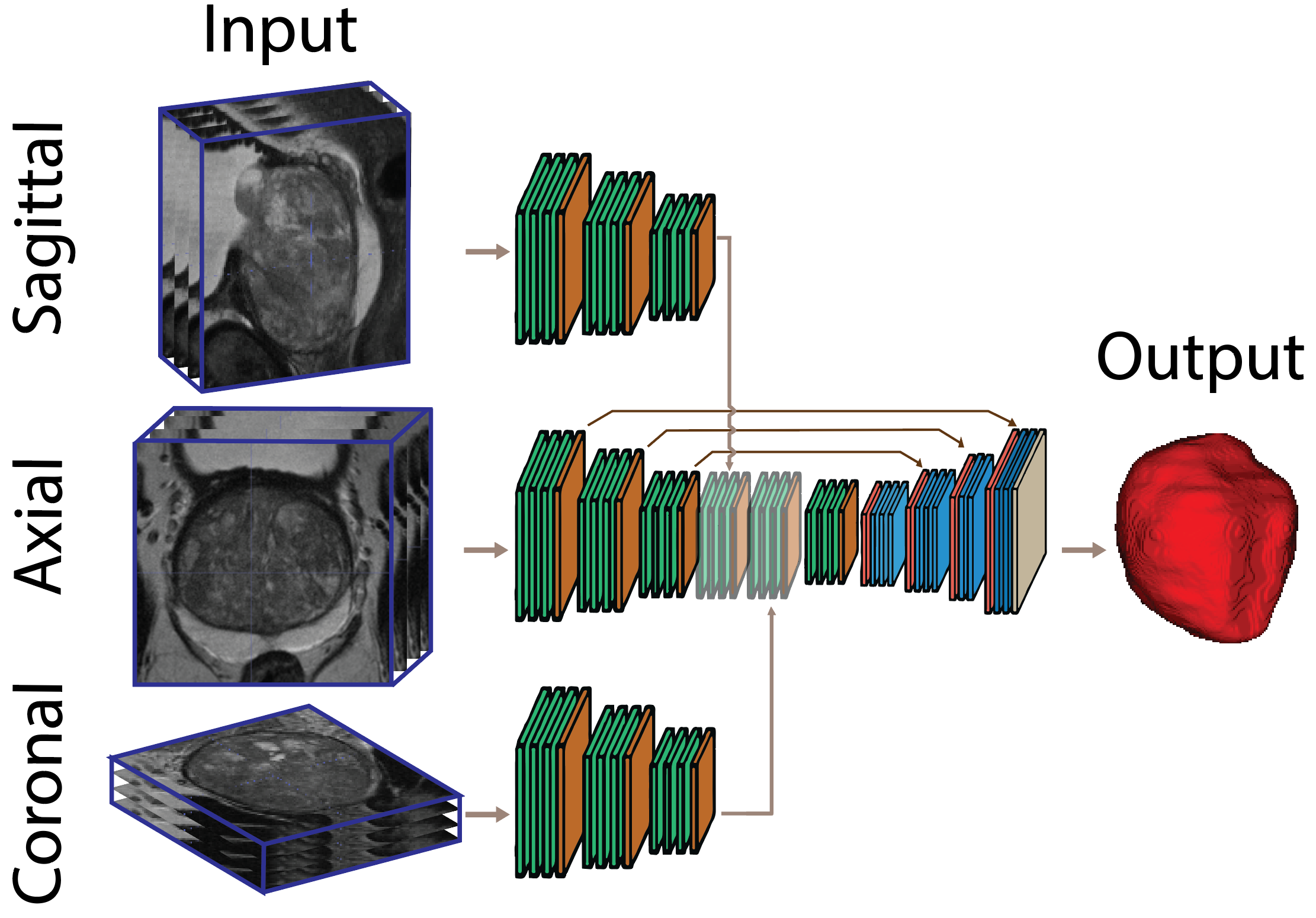
Figure1: An illustration of the multi-stream architecture.
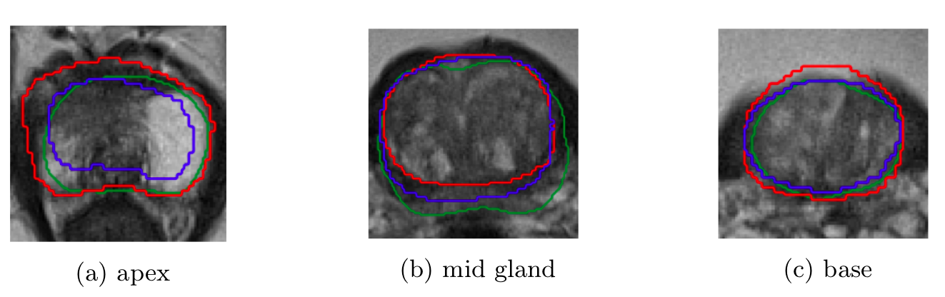
Figure 2: Prediction of the axial model (red), multi-planar model (blue) and ground truth (green) shown in overlay on the axial t2-weighted image slices through the apex, mid and base.
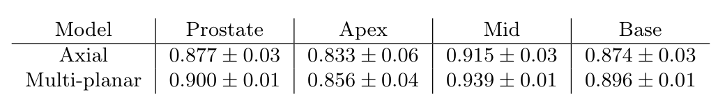
Table 1: Comparison of segmentation performance between axial only and multi-planar anisotropic CNN.