3508
A Cascaded 3D CNN Approach for Thalamic Nuclei Segmentation1Department of Electrical and Computer Engineering, University of Arizona, Tucson, AZ, United States, 2Department of Medical Imaging, University of Arizona, Tucson, AZ, United States, 3Siemens Healthcare USA, Tucson, AZ, United States, 4Department of Psychiatry & Behavioral Sciences, Stanford University, Menlo Park, CA, United States, 5Department of Biomedical Engineering, University of Arizona, Tucson, AZ, United States
Synopsis
We propose the use of conventional MPRAGE images to synthesize WMn-MPRAGE images for thalamic nuclei segmentation. We compare thalamic nuclei segmentation performance on the synthesized WMn images to those directly on CSFn-MPRAGE. We also validate the clinical utility of our method by analyzing differences in thalamic nuclei volumes between patients with alcohol use disorder (AUD) and age-matched healthy controls using conventional MPRAGE data.
Purpose
White-matter-nulled (WMn) Magnetization-prepared Rapid Gradient Echo (MPRAGE) has been proposed for better visualization of the thalamus and thalamic nuclei in the brain1,2 compared to conventional MPRAGE images (CSFn). WMn-MPRAGE data has been successfully used with a multi-atlas segmentation algorithm3 to automatically segment and quantify thalamic nuclei volumes. However, many clinical studies acquire only conventional MPRAGE to minimize total scan time and numerous online repositories like ADNI have only standard MPRAGE. While a probabilistic atlas-based method that is part of Freesurfer4 has been proposed for thalamic nuclei segmentation in MPRAGE, it is computationally burdensome and has not been rigorously validated against manual segmentation. In this work, we propose the use of conventional MPRAGE images to synthesize WMn-MPRAGE images for thalamic nuclei segmentation. We compare thalamic nuclei segmentation performance on the synthesized WMn images to those directly on CSFn-MPRAGE. We also validate the clinical utility of our method by analyzing differences in thalamic nuclei volumes between patients with alcohol use disorder (AUD) and age-matched healthy controls using conventional MPRAGE data.Methods
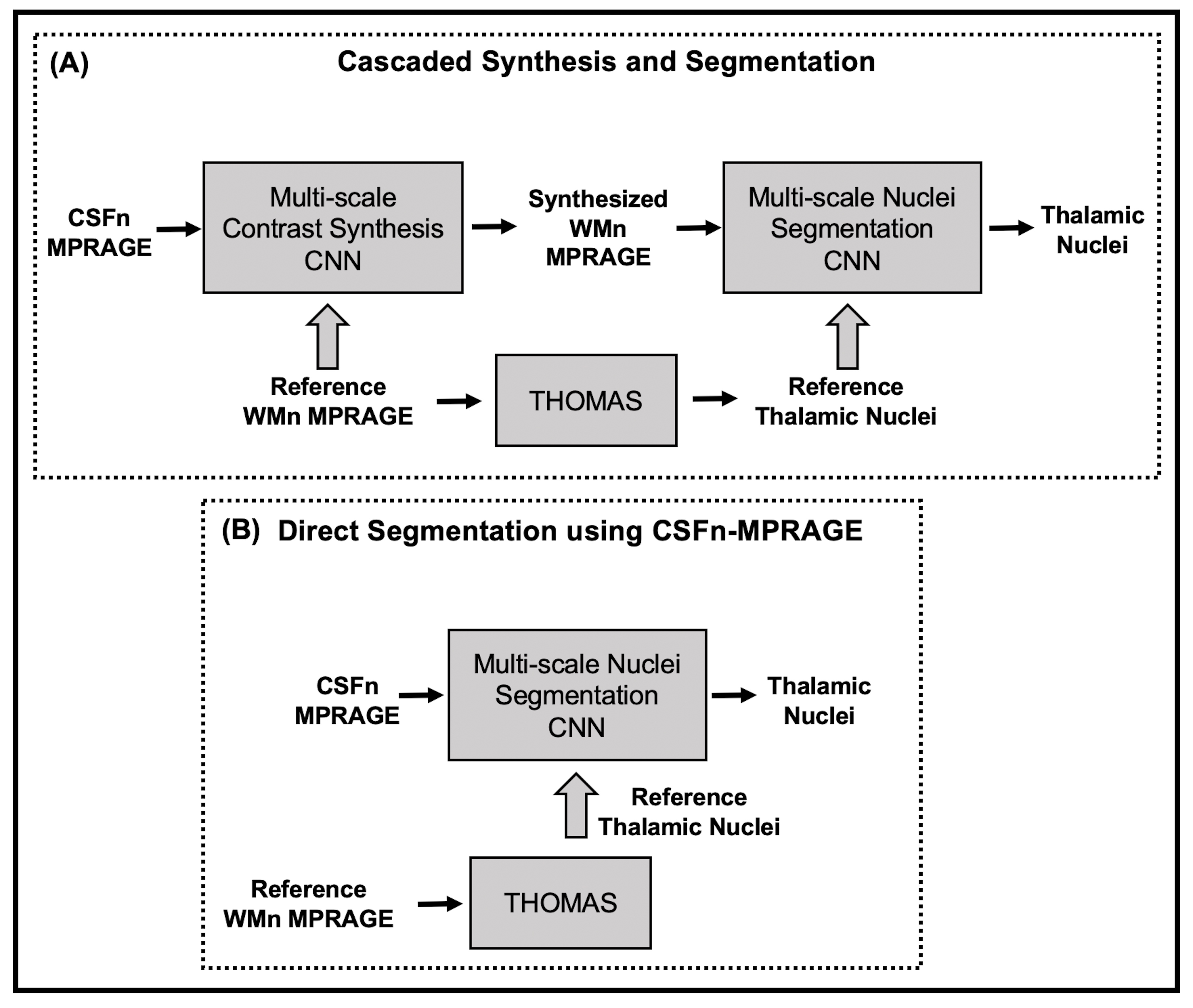
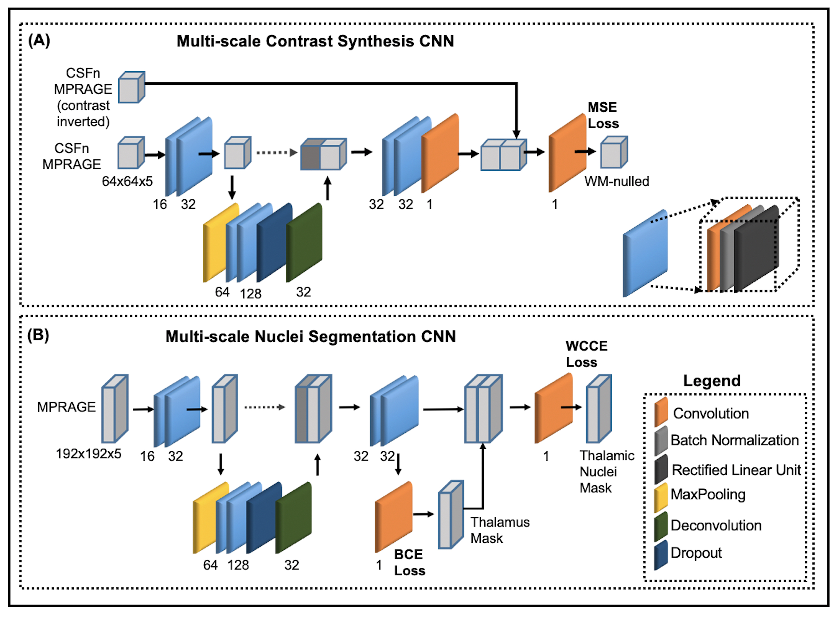
An overview of our proposed architecture is shown in Figure 1A. This cascade consists of a multi-scale 3D contrast synthesis Convolutional Neural Network (CNN) that synthesizes WMn-MPRAGE from CSFn-MPRAGE, followed by a 3D segmentation CNN that predicts thalamic nuclei on the synthesized images. Figure 1B shows the segmentation model being used to directly segment thalamic nuclei on CSFn-MPRAGE images. In the synthesis CNN (Figure 2A), feature maps extracted from the CSFn image are combined with the contrast inverted CSFn image to generate the final synthesized image. The segmentation CNN (Figure 2B) first predicts the thalamic mask from feature maps, which is then concatenated to the feature maps to predict individual thalamic nuclei.With informed consent, 47 subjects were scanned on a 3T scanner (GE Healthcare, Milwaukee, WI) using a 32-channel head array coil. MRI data acquired on all subjects included CSFn and WMn-MPRAGE. As part of pre-processing, the images were brain extracted, bias corrected, and intensity normalized. The study cohort was split into training (36), validation (1), and testing (9 subjects). Training patches (64x64x5) were extracted from the co-registered CSFn and WMn images. For training the segmentation CNN, we used 2.5D cropped image blocks (192x192x5) extracted from WMn images using a sliding window. The reference labels for the left thalamic nuclei were generated using THOMAS3 algorithm run on WMn-MPRAGE images. The CNNs in the cascade were trained independently with the following loss functions- Synthesis CNN loss: mean squared error (MSE), Segmentation CNN: binary cross entropy for thalamus, multi-class weighted cross entropy for thalamic nuclei.
The synthesized WMn image quality was evaluated using MSE, structural similarity (SSIM) and peak signal-to-noise (PSNR) ratio. The nuclei segmentation was evaluated using dice similarity. Data analysis was performed on 33 subjects (18 AUD, 15 controls) from a cohort previously used to study AUD effects on thalamic nuclei volumes using WMn-MPRAGE based segmentation5. The proposed method was used to predict right thalamic nuclei by flipping the left-right orientation of the CSFn-MPRAGE images. The estimated volumes were corrected for intra-cranial volume (svol) using the residual method6 and a t-test was performed between the two groups for the following main thalamic nuclear groups: whole thalamus, anterior-ventral (AV), ventral-lateral-posterior (VLP), ventral-posterior-lateral (VPL), pulvinar (Pul), and medial-dorsal (MD), with significance level set to 0.05/6 to account for multiple comparisons.
Results
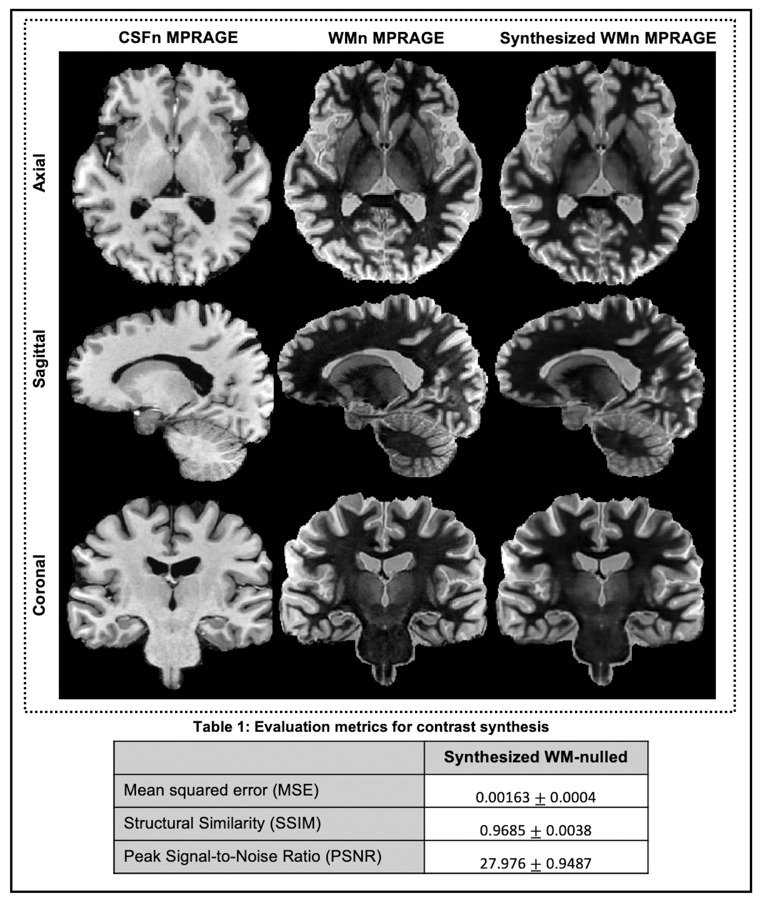
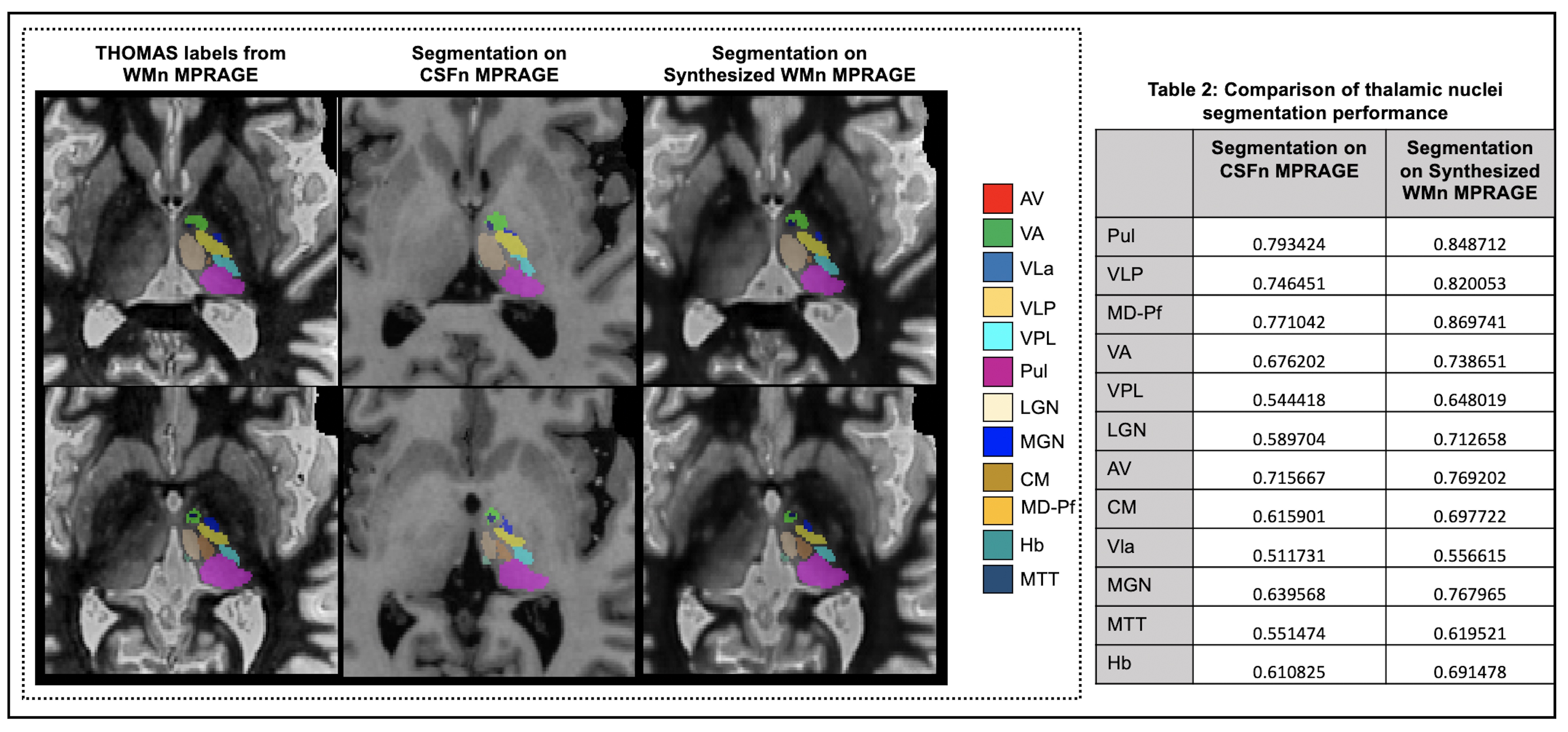
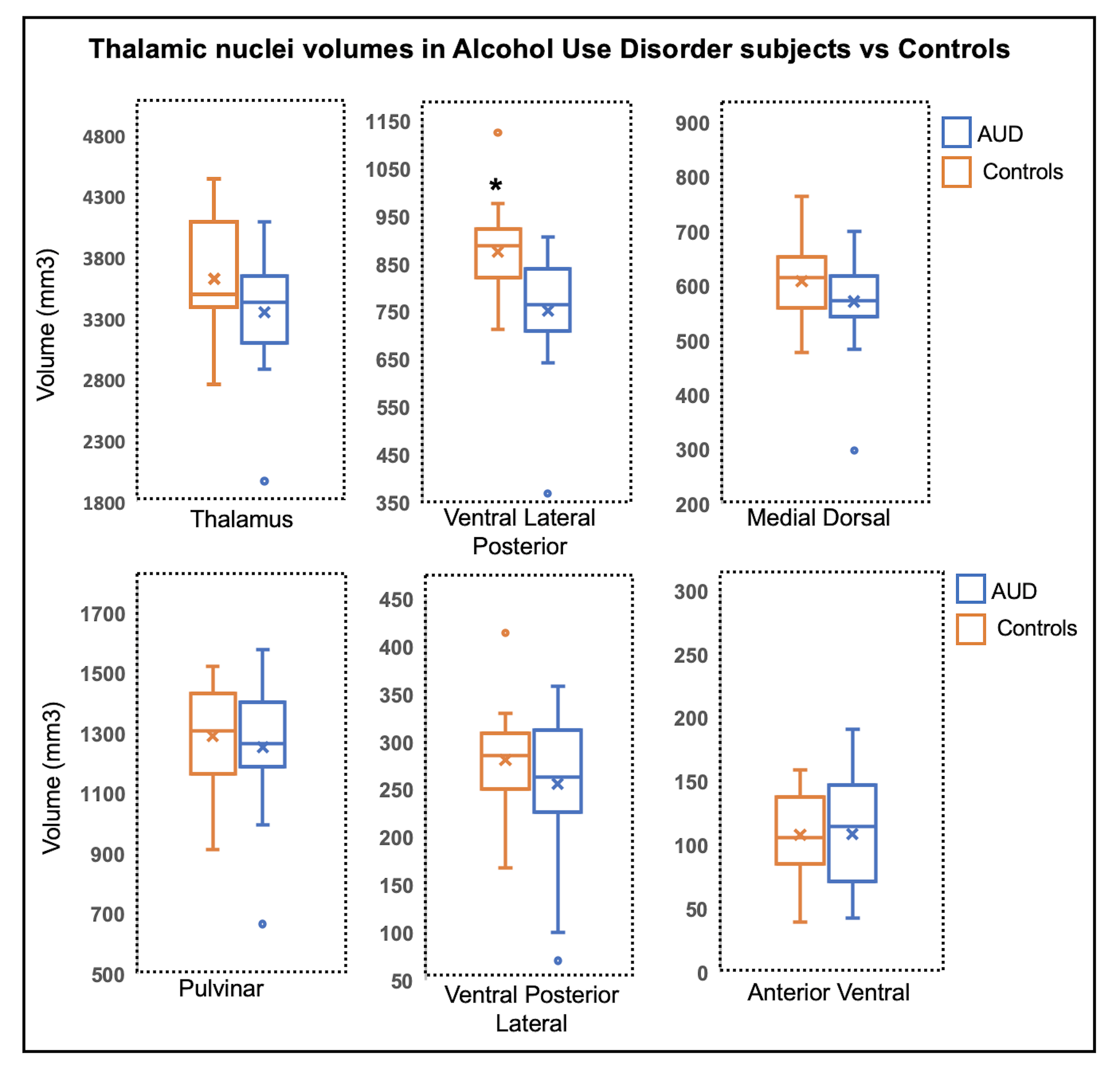
Figure 3 shows orthogonal views of WMn, CSFn and Synthesized WMn-MPRAGE from a test subject. On all test subjects, the synthesized images yielded an average SSIM of 0.9685, PSNR of 27.976 dB, and MSE of 0.00016. Figure 4 shows the thalamic nuclei masks predicted by the CNN cascade overlaid on two representative slices from a test subject. Also shown are the reference labels and nuclei masks predicted directly from CSFn-MPRAGE. For nuclei such as AV, VLP, Pul, and MD, the proposed CNN cascade achieved average dice scores of 0.7692, 0.82, 0.8487, and 0.8697, respectively. Dice scores for segmentation on CSFn-MPRAGE for the same nuclei were 0.7156, 0.7464, 0.7934, and 0.7710, respectively.The box plot in Figure 5 compares representative thalamic nuclei volumes for AUD subjects and controls. We observed a significant volume reduction in the VLP nucleus in AUD compared to controls (p = 0.0008, n=33). Logistic regression was performed using all nuclei volumes. The best model, chosen based on AIC (VLP/MD/svol), predicted diagnosis with Area Under Curve of 0.952.
Discussion
We validated a method that can provide fast (20s end-to-end prediction) and accurate thalamic nuclei segmentation using conventional MPRAGE images. While the accuracy is slightly lower than segmenting thalamic nuclei on WMn images, the Dice for most of the larger nuclei are 0.79 or higher. More significantly, our method reproduced the same reduction in VLP nucleus observed using WMn-MPRAGE with THOMAS labels5, attesting to its sensitivity and accuracy. Of the two methods proposed, segmentation on the synthesized WM-nulled images yields better performance when compared to CSF-nulled based segmentation.Conclusion
A cascade of multi-scale 3D CNNs was proposed to use traditional MPRAGE images to generate synthesized WMn-MPRAGE images and then predict thalamic nuclei masks.Acknowledgements
R21 grant from the NIH (R21AA023582)References
1. Tourdias T, Saranathan M, et al. Visualization of intra-thalamic nuclei with optimized white-matter-nulled MPRAGE at 7 T. NeuroImage. 2014. V(84): 534-545.
2. Sudhyadhom A1, Haq IU, et al. A high resolution and high contrast MRI for differentiation of subcortical structures for DBS targeting: the Fast Gray Matter Acquisition T1 Inversion Recovery (FGATIR). NeuroImage. 2009. Suppl2 T44-52.
3. Su JH, Thomas FT, et al. Thalamus Optimized Multi Atlas Segmentation (THOMAS): fast, fully automated segmentation of thalamic nuclei from structural MRI. NeuroImage. 2019. V(194): 272-282
4. Iglesias J, Insausti R, et al. A probabilistic atlas of the human thalamic nuclei combining ex vivo MRI and histology. NeuroImage 2018. V(183): 314-326.
5. Zahr N, Saranathan M. Substructural volumes of the thalamus in alcoholism. Proc ISMRM 2017.
6. In Vivo Detection and Functional Correlates of White Matter Microstructural Disruption in Chronic Alcoholism
Figures