3494
Accelerating Positive Contrast MRI Reconstruction with GPU-based Parallel Chambolle-Pock Algorithm1Shenzhen Institutes of Advanced Technology, Chinese Academy of Sciences, Shenzhen, China, 2Department of Biomedical Engineering, Guangzhou Medical University, Guangzhou, China, 3Department of Radiology, Guangzhou Panyu Central Hospital, Guangzhou, China
Synopsis
Positive contrast Magnetic Resonance Imaging (MRI) based on the susceptibility mapping requires very fast reconstruction for clinical applications. Modern graphics processing units (GPUs) are very efficient at manipulating computer graphics and image processing. And Chambolle-Pock(CP) algorithm is also an efficient algorithm to solve the minimization problem. To further reduce the reconstruction time of positive contrast MRI, a GPU-based parallel CP algorithm was proposed. The experimental results showed that the proposed GPU-based parallel CP method could achieve similar image results, and provide a faster reconstruction of up to 15 times than the conventional CPU-based CP method.
Synopsis
Positive contrast Magnetic Resonance Imaging (MRI) based on the susceptibility mapping requires very fast reconstruction for clinical applications. Modern graphics processing units (GPUs) are very efficient at manipulating computer graphics and image processing. And Chambolle-Pock(CP) algorithm is also an efficient algorithm to solve the minimization problem. To further reduce the reconstruction time of positive contrast MRI, a GPU-based parallel CP algorithm was proposed. The experimental results showed that the proposed GPU-based parallel CP method could achieve similar image results, and provide a faster reconstruction of up to 15 times than the conventional CPU-based CP method.Introduction
In the traditional MR images, interventional metallic devices become a dark hole.1 Susceptibility-based positive contrast Magnetic Resonance Imaging(MRI) is one method to image the MR-compatible metallic devices. The reconstruction of the susceptibility-based positive-contrast MRI can be considered as a minimization problem, which has been addressed by nonlinear conjugate gradient(CG) algorithm and Chambolle-Pock algorithm.2-6 Chambolle-Pock algorithm has also applied to other MRI reconstruction problem7 and shown a shorter reconstruction time than CG and other algorithms in solving this problem4. However, the CP method still need minutes to reconstruct a full 3D image. Currently, researchers has investigated using the modern competitive platforms of graphics processing unit (GPU) to accelerate MRI reconstruction.8-9 In this work, we realize a parallel CP algorithm based on GPU to accelerate the reconstruction of positive contrast MRI for applications. The current experimental results have showed that the proposed GPU-based parallel CP method could achieve very similar results and require reconstruction time of about 15 times less than the conventional CPU-based CP method. Moreover, the GPU-based parallel CP algorithm can be easily transplanted to any other similar minimization reconstruction problems with a slightly modification.Methods
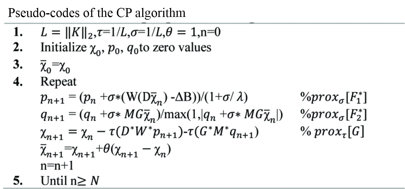
The susceptibility-based positive contrast MR technique uses an equivalent short TE by shifting the readouts gradient with Tshift during MR data acquisition4,6. Then the aquired two data sets are used to measure the local field variation $$$\Delta B $$$ induced by the metallic devices. The susceptibility mapping $$$\chi $$$ can be reconstructed as follow:$$ \chi = \mathrm{arg \displaystyle min_{\chi }} \left \| W(D\chi -\Delta B) \right \|_2^2 + \left \| MG\chi \right \|_1 \;\; ,(1)$$
where $$$D $$$ is the dipole kernel convolution operator, $$$G $$$ is the gradient operator, $$$W $$$ is the weighting matrix, $$$M $$$ is the mask matrix and λ is the regularization parameter. CP algorithm was used to solve the Eq.(1), and the iterative processes are as Figure 1.
It is well known that GPU is a specialized electronic circuit to accelerate the creation of images in a frame buffer intended for output to a display device. Current GPUs are very efficient at manipulating computer graphics and image processing8. Their highly parallel structure makes them more efficient than general-purpose central processing units (CPUs) for many computation algorithms. In order to utilize the multithreads property of the GPU and accelerate the reconstruction computations, we first realize the matrix-vector operations of the CP algorithm on GPU. These parallel execution mode operations can reduce the computation time. Furthermore, we deal with the 3D data in a parallel fashion. Therefore, the proposed GPU-based CP algorithm would be as Figure 2.
To evaluate the performance of the proposed GPU based method, two data sets were acquired. Data set 1 is the stent data, scan parameters were: FOV = 128×128 mm2, matrix size = 192×192, TR =2000 ms, TE = 18 ms, slice number = 20, in-plane resolution = 0.67×0.67 mm2, slice thickness =1.5 mm, slice gap =0.0 mm, bandwidth =134 Hz/Pixel, and Tshift =0.6 ms. Data set 2 is the seeds-phantom data, scan parameters were: TE/TR = 33/1500 ms, in-plane resolution = 0.72 × 0.72 × 2 mm3, matrix = 128 × 128 × 32, and bandwidth = 698 Hz/pixel, and Tshift =0.5 ms. CP algorithm parameters were set the same as the reference4.
Results
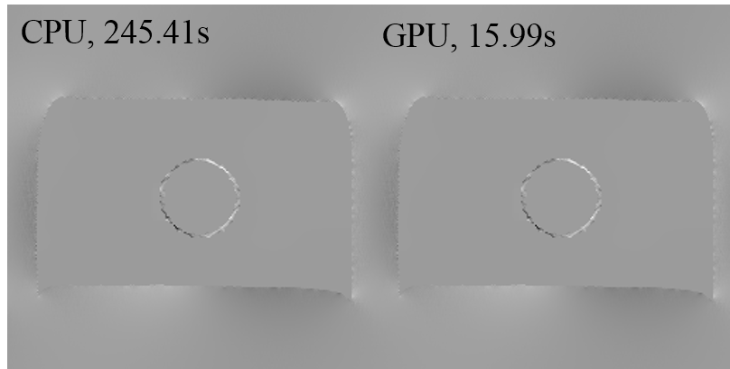
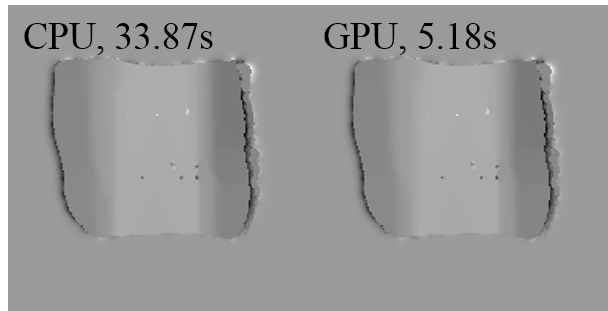
Figure 3 and Figure 4 show that the proposed GPU based CP algorithm needs 15.99s to reconstruct 25 images of stent and 5.18s to reconstruct 10 images of seeds-phantom while the traditional CPU based CP algorithm needs 245.41s and 33.87s respectively. These figures also demonstrate that the GPU-based method produce an equal image quality than CPU-based method. Apparently, the proposed GPU-based method has a higher acceleration factor on the stent data which consists of more image slices and bigger image size than the seeds-phantom data. The reason for the higher performance on the bigger data set is that the bigger data utilize more GPU threads.Conclusion
In sum, a GPU-based parallel CP algorithm has been proposed to accelerate the reconstruction of positive contrast MRI for applications. The results illustrated that the proposed scheme could be 6-15 times faster than the conventional scheme without any compromise in image quality. In the future, the proposed scheme will be seamlessly transplanted to other similar applications.Acknowledgements
Fang Cai and Caiyun Shi contributed equally to this work. This work was supported in part by the grant from the National Natural Science Foundation of China (Grant No. 81729003 and 61871373), the Strategic Priority Research Program of Chinese Academy of Sciences (Grant No. XDB25000000), the Natural Science Foundation of Guangdong Province (Grant No. 2018A0303130132), and the Shenzhen Peacock Plan Team Program (Grant No. KQTD20180413181834876).References
1. Wachowicz K, Thomas S, Fallone B. Characterization of the susceptibility artifact around a prostate brachytherapy seed in MRI. Med Phys. 2006;33:4459-4467.
2. Shi C, Xie G, Zhang Y, et al. Accelerated susceptibility-based positive contrast imaging of MR compatible metallic devices based on modified fast spin echo sequences. Phys Med Biol. 2017;62(7):2505-2520.
3. Dong Y, Chang Z, Xie G, et al. Susceptibility-based positive contrast MRI of brachytherapy seeds. Magn Reson Med. 2014;74:716-726.
4. Shi C, Cheng J, Xie G, et al. Positive-contrast susceptibility imaging based on first-order primal-dual optimization. Magn Reson Med. 2019;00:1-9.
5. Chambolle A, Pock T. A first‐order primal‐dual algorithm for convex problems with applications to imaging. J Math Imaging Vision. 2011;40:120-145.
6. Shi C, Cheng J, Su S, et al. Three Dimensional Positive Contrast Susceptibility Fast Spin Echo MR Imaging with Variable Excitation Pulses and PD Algorithm. 2019 41st Annual International Conference of the IEEE Engineering in Medicine and Biology Society (EMBC), Berlin, Germany. 2019;pp:4824-4827.
7. Wang H, Cheng J, Jia S, et al. Accelerating MR Imaging via Deep Chambolle-Pock Network. 2019 41st Annual International Conference of the IEEE Engineering in Medicine and Biology Society (EMBC), Berlin, Germany. 2019;pp:6818-6821.
8. Wang H, Peng H, Chang Y, et al. A survey of GPU-based acceleration techniques in MRI reconstructions. Quant Imaging Med Surg. 2018;8:196-208.
9. Cai F, Qiu Z, Su S, et al. [A Graphics Processing Unit-Based Modified Conjugate Gradient Method for Accelerating Wave-CAIPI Reconstruction]. Jour of Inte Tech. 2019; 8(6):1-10. (in Chinese)
Figures