3475
Accelerated Free-breathing 3D Whole-heart Black-blood TSE Imaging1Biomedical Engineering Department, School of Biomedical Engineering and Imaging Sciences, King's College London, London, United Kingdom, 2MR Research Collaborations, Siemens Healthcare Limited, Frimley, United Kingdom
Synopsis
In this study we investigate the feasibility of a 3D turbo spin-echo imaging technique with variable flip angle combined with an undersampled Cartesian variable density trajectory (3D TSE-VDCASPR) for efficient whole-heart black-blood imaging. Data from five healthy subjects are acquired with the proposed 3D TSE-VDCASPR and the conventional 3D TSE with Cartesian GRAPPA. The proposed imaging 3D TSE-VDCASPR imaging technique allows for comparable black-blood imaging, with the advantage of reduce nominal scan time (4.6±1 vs 8.1±1.6 minutes ± seconds). Future work will investigate more advanced motion compensation and image reconstruction techniques in order to achieve predictable and fast scan time.
Introduction
Black-blood imaging suppresses blood signal, allowing the visualization of the vessel walls and the morphology of the heart. Recently, 3D turbo spin-echo (TSE) imaging techniques with variable flip angle [1] have been proposed for the visualization of the aortic vessel wall and whole-heart anatomy, allowing free-breathing acquisition with high image resolution [2-3]. However, 3D TSE can be quite challenging due to prolonged scan time and image artefacts due to insufficient blood nulling. In this study, we investigate the feasibility of a 3D TSE imaging sequence with variable flip angle combined with an undersampled Cartesian variable density trajectory for efficient whole-heart black-blood imaging. The proposed imaging sequence is compared with conventional 3D TSE in five healthy subjects.Methods
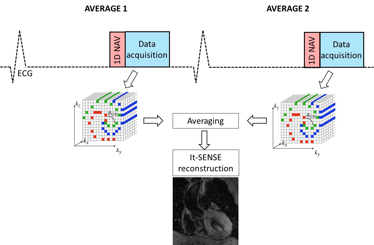
Five healthy subjects were examined on a 1.5T MR system (MAGNETOM Aera, Siemens Healthcare, Erlangen, Germany) using a 3D turbo spin-echo prototype imaging sequence with Cartesian sampling and GRAPPA reconstruction (3D TSE) and a Cartesian variable density trajectory with spiral profile order (3D TSE-VDCASPR) [4] with iterative SENSE reconstruction [ref]. The imaging parameters used for the T1 weighted 3D TSE sequence were: coronal orientation, image resolution of 1x1x2mm3, FOV = 320x320x110, echo train length = 35, echo spacing = 3.3 ms, echo time = 17 ms, number of averages = 2, ECG triggered with end-systolic image acquisition, fat suppression = SPIR, resulting in an acquisition window of 116 ms. The 3D TSE sequence uses a 90˚ slice-selective RF excitation followed by non-selective refocusing RF pulses with variable flip angles. A diaphragmatic navigator with a gating window of 5mm and a tracking factor of 0.6 was used for respiratory motion compensation for both sequences. To reduce free induction decay (FID) artefacts, the sampling points were acquired twice (two averages) and then averaged together prior the final image reconstruction [5]. The 3D TSE-VDCASPR data were acquired with an acceleration factor of 3.8 and reconstructed offline using iterative SENSE (It-SENSE), while the conventional 3D TSE data were acquired with Cartesian GRAPPA acceleration of 2 right-left (RL) and reconstructed directly on the scanner.Results
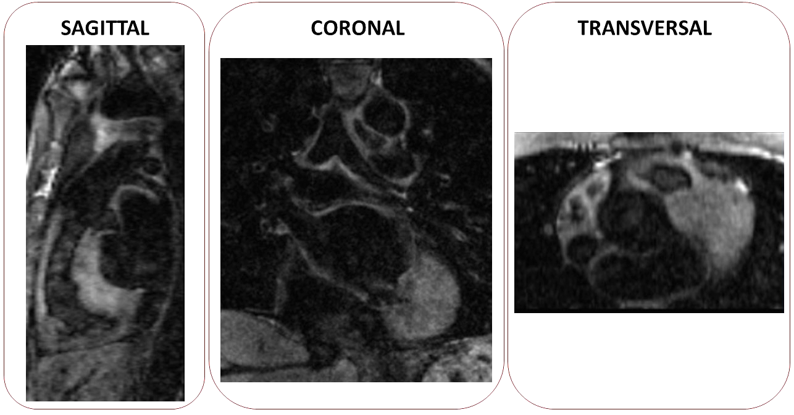
Figure 2 shows the images acquired with the conventional 3D TSE imaging sequence and the proposed 3D TSE-CASPR for two representative subjects. After It-SENSE reconstruction, the delineation of the myocardial and vessels borders is sharper in the 3D TSE-VDCASPR images. 3D TSE-VDCASPR achieves comparable image quality to the conventional 3D TSE imaging sequence. However, the nominal scan time of 3D TSE-VDCASPR was considerably lower than that of the conventional 3D TSE approach (4.6±1 vs 8.1±1.6 minutes ± seconds). Figure 3 shows the 3D TSE-CASPR images reformatted in three different orientations (sagittal, transversal and coronal) for a representative subject.Conclusions
The combination of variable flip angle 3D TSE imaging with a variable density Cartesian trajectory allows for black-blood imaging of the whole-heart in a reduced scan time. Future works will investigate the use of more advance motion compensation and reconstruction techniques in order to achieve predictable and faster scan time.Acknowledgements
This work was supported by the following grants: BHF 1) PG/18/59/33955, EPSRC 2) EP/P032311/1; 3) EP/P007619/1; and the Wellcome/EPSRC Centre for Medical Engineering (WT 203148/Z/16/Z).References
[1] Mugler P. J., Optimized Three-Dimensional Fast-Spin-Echo MRI. J Magn Reson Imaging 39:745–767 (2014)
[2]Henningsson, M., Zahr, R.A., Dyer, A. et al. Feasibility of 3D black-blood variable refocusing angle fast spin echo cardiovascular magnetic resonance for visualization of the whole heart and great vessels in congenital heart disease. J Cardiovasc Magn Reson 20, 76 (2018)
[3] Eikendal AL, Blomberg BA, Haaring C, Saam T, van der Geest RJ, Visser F, Bots ML, den Ruijter HM, Hoefer IE, Leiner T. 3D black blood VISTA vessel wall cardiovascular magnetic resonance of the thoracic aorta wall in young, healthy adults: reproducibility and implications for efficacy trial sample sizes: a cross-sectional study. J Cardiovasc Magn Reson. 2016;18:20.
[4] Prieto C, Doneva M, Usman M, et al. Highly efficient respiratory motion compensated free‐breathing coronary MRA using golden‐step Cartesian acquisition. J Magn Reson Imaging. 2015;41:738‐746
[5] [5] Magland JF, Rajapakse CS, Wright AC, Acciavatti R, Wehrli FW. 3D fast spin echo with out-of-slab cancellation: a technique for high-resolution structural imaging of trabecular bone at 7 tesla. Magn Reson Med. 2010;63(3):719–27.
Figures