3469
Near bias-free robust MP2RAGE reconstruction using a signal-dependent regularization function1MR Collaboration, Siemens Healthcare Ltd., Shanghai, China, 2Institute of Science and Technology for Brain-Inspired Intelligence, Fudan University, Shanghai, China, 3Human Phenome Institute, Fudan University, Shanghai, China
Synopsis
MP2RAGE is a B1-insensitive T1-weighted imaging and T1 mapping method with strong background noise. Introducing a regularization parameter into the reconstruction can suppress the noise at the cost of lowering the signal intensity and T1 value. We propose a robust MP2RAGE using a signal-dependent regularization function. The reconstructed images are nearly bias-free with minimal artifact and reduced background noise.
INTRODUCTION
Magnetization Prepared with 2 Rapid Gradient Echoes (MP2RAGE)1 acquires two gradient-echo (GRE) images at two inversion times. MP2RAGE is a preferred choice for high-field application, because it provides T1-weighted images and T1 maps with no dependence on B1-, proton density, T2*, and the first-order characterization of B1+. However, because of the ratio taken between two GRE images, MP2RAGE images contain strong noise at regions with low signal intensity. This noise increases the variability of brain segmentation2. A regularization method for robust MP2RAGE reconstruction has been proposed at the cost of lowering the MP2RAGE signal2. The reproducibility of brain segmentation is improved, but the low MP2RAGE signal results in biased T1 maps. In this study, we propose a new robust MP2RAGE reconstruction by using a signal-dependent regularization function to not only suppress the noise but also reduce the bias in T1 quantification.METHODS
T1 weighted images were acquired using an MP2RAGE sequence on a 7T MRI system (Magnetom Terra, Siemens Healthineers, Erlangen, Germany). Image parameters were TRMP2RAGE/TRGRE/TE = 4010ms/7ms/2.29ms, image matrix = 272x272x200, resolution = 0.7x0.7x0.7 mm. The original MP2RAGE image was computed by combining two GRE images using the following equation:$$MP2RAGE_{orig}=real\left ( \frac{GRE_{TI1}^{*}\times GRE_{TI2}}{GRE_{TI1}^{*}\times GRE_{TI1}+GRE_{TI2}^{*}\times GRE_{TI2}} \right)$$
At the low signal region, the MP2RAGE image exhibited strong noise due to the small value in the denominator. A robust MP2RAGE reconstruction2 can overcome this challenge by introducing a regularization parameter:
$$MP2RAGE_{robust}=real\left ( \frac{GRE_{TI1}^{*}\times GRE_{TI2}-\frac{\lambda }{2}}{GRE_{TI1}^{*}\times GRE_{TI1}+GRE_{TI2}^{*}\times GRE_{TI2}+\lambda } \right)$$
Here, we propose a signal-dependent regularization function for noise suppression with minimal signal reduction in MP2RAGE.
$$MP2RAGE_{robust-new}=real\left(\frac{GRE_{TI1}^{*}\times GRE_{TI2}-\frac{\beta (\lambda,S^{2}) }{2}}{S^{2}+\beta (\lambda,S^{2})}\right ), S^{2}=GRE_{TI1}^{*}\times GRE_{TI1}+GRE_{TI2}^{*}\times GRE_{TI2}$$
In this study, we compared four regularization functions β(λ,S2), including the constant function used in the robust MP2RAGE reconstruction:
$$\begin{cases}\beta _{robust}\left ( \lambda ,S^{2} \right )=\lambda\\\beta _{reciprocal}\left ( \lambda ,S^{2} \right )=\frac{\lambda^{2}}{S^{2}}\\\beta _{sigmoid_{sharp}}\left ( \lambda ,S^{2} \right )=\frac{2\lambda}{1+exp\left (S^{2}-\lambda\right )}\\\beta _{sigmoid_{ blunt }}\left (\lambda ,S^{2} \right )=\frac{2\lambda}{1+exp\left (\frac {S^{2}-\lambda}{5000}\right )}\end{cases}$$
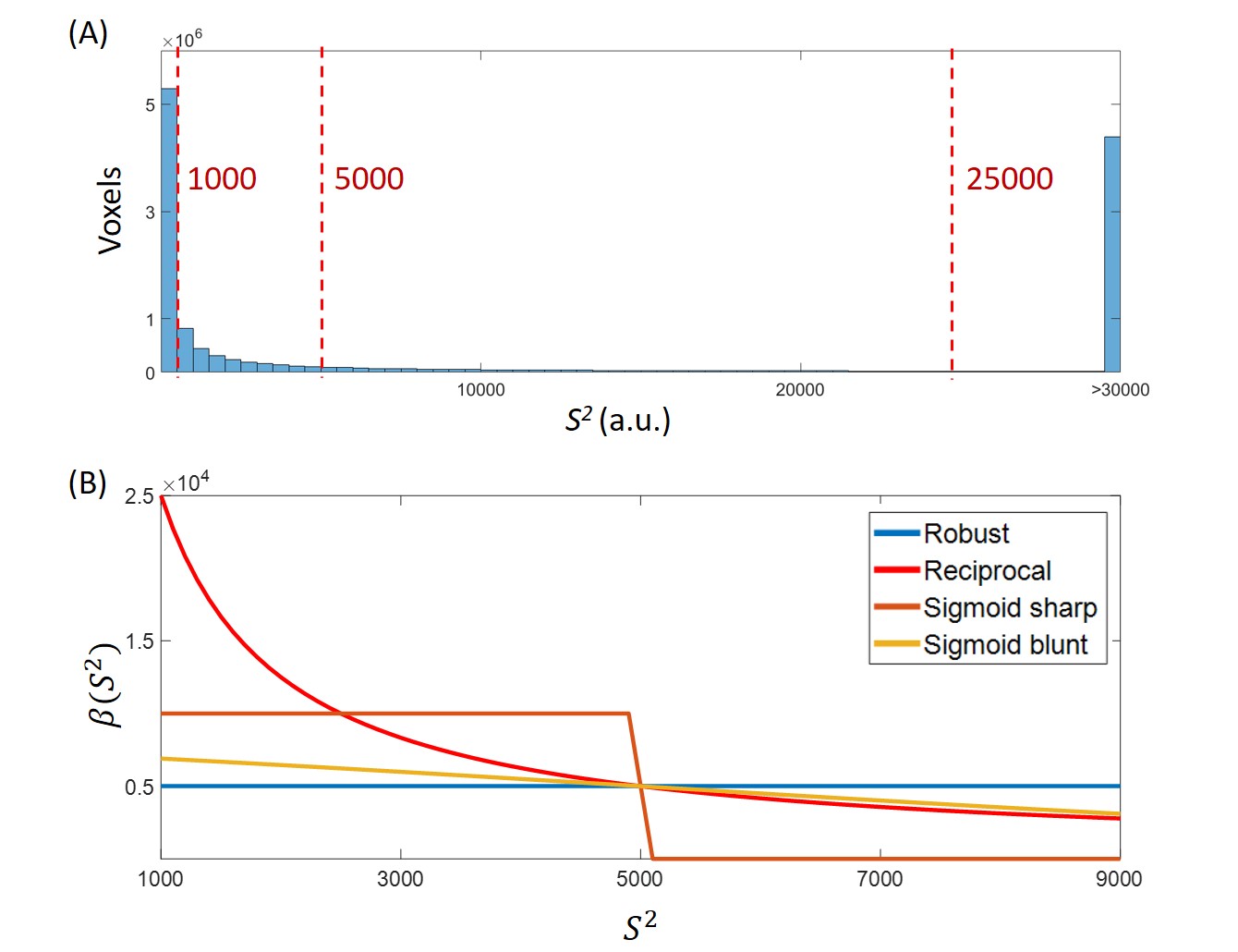
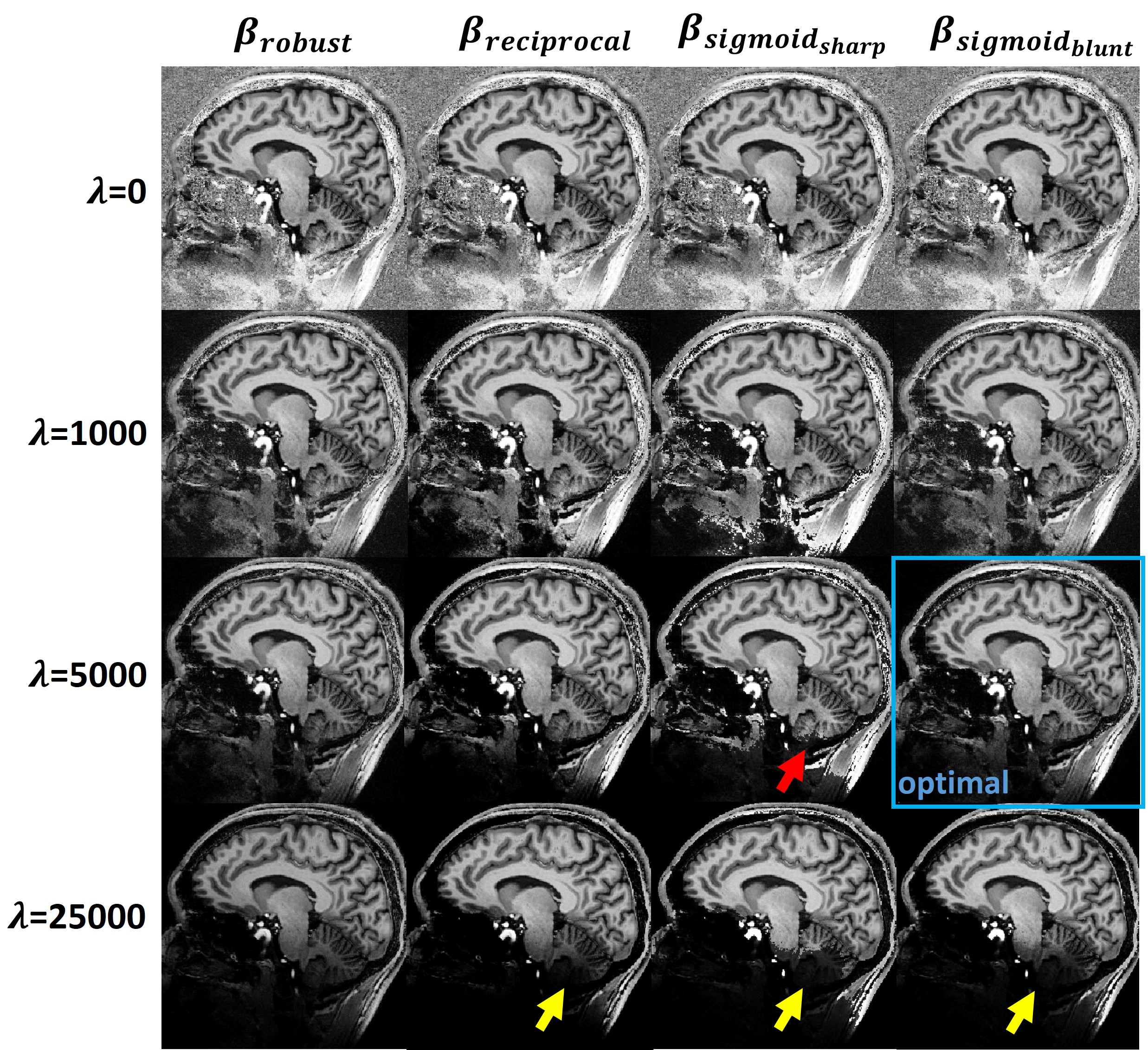
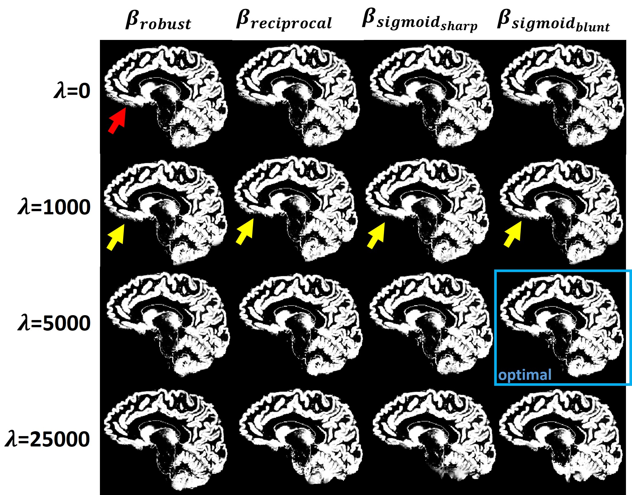
Three regularization parameters λ were used in this study: 1000, 5000, and 25000. The gray matter and white matter region were segmented using SPM12 (https://www.fil.ion.ucl.ac.uk/spm/).
RESULTS
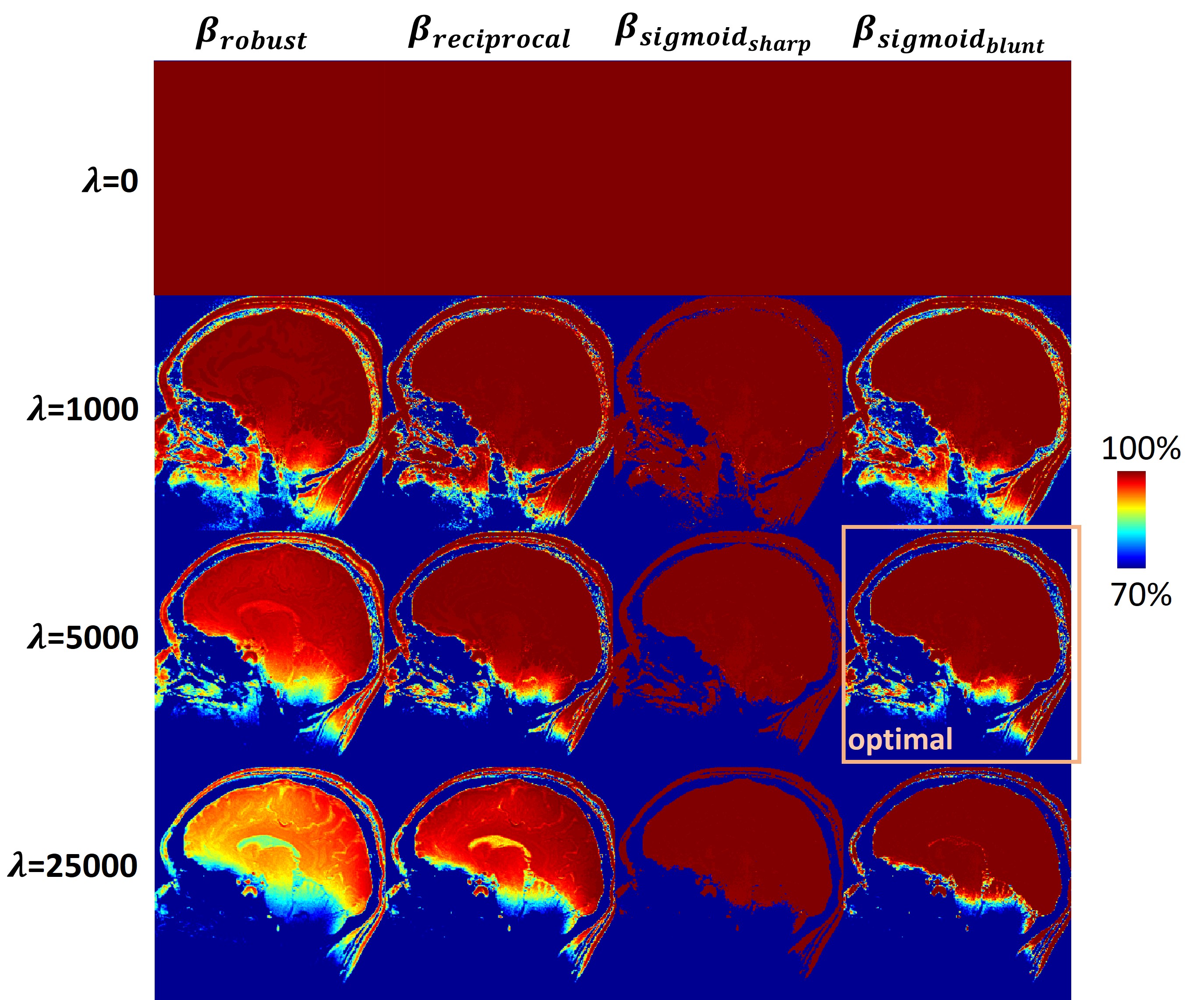
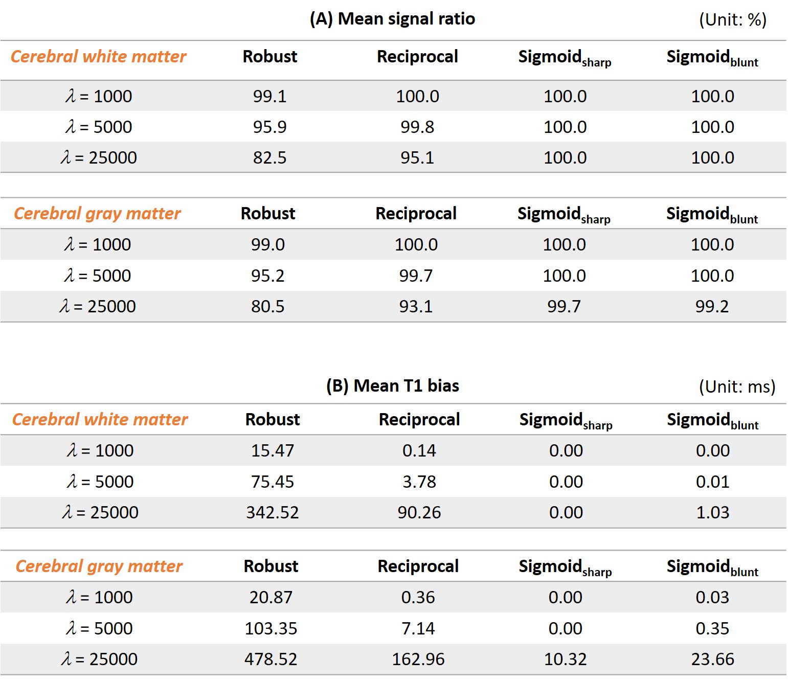
Figure 1A shows the histogram of S2. Chosen regularization parameters were marked on the S2 axis. Figure 1B shows four regularization functions used in this study with λ = 5000. All regularization functions had the same value at S2=λ. All methods suppressed more than 50% signal at S2<λ. Compared with results with a constant beta function, using a non-constant beta function suppressed more at S2<λ and less at S2>λ.Figures 2, 3, and 4 are reconstructed MP2RAGE images, segmented gray matter images, and signal ratio images, respectively, with different regularization methods and parameters. With λ=0, strong noise appeared at low signal regions (Figure 2). Image pixels in the nasal cavity were noisy and connected to the brain gray and white matter, causing the gray matter segmentation error (red arrow in Figure 3). At λ=1000, only very little noise remained in all regularized reconstructions. The gray matter segmentation error near the nasal cavity was reduced but still visible in all cases (yellow arrows in Figure 3). λ=5000 was the optimal regularization parameter. Noises were suppressed, and the segmented gray matter was accurate. Especially, the reciprocal and sigmoidblunt methods reconstructed artifact-free images and kept signal simultaneously. The robust method had reduced signals. The cerebellum was partially truncated in the sigmoidsharp image. The regularization was too strong at λ=25000, which led the signal reduction at the inferior part of the cerebellum (yellow arrows in Figure 2).
Table 1 summarizes the average signal ratio and T1 bias in the cerebral white matter and gray matter. Using the optimal regularization parameter, the robust reconstruction had 95.9% and 95.2% signal and the T1 bias was 75.45 ms and 103.35 ms for cerebral white matter and gray matter, respectively. Using a non-constant regularization method, the signal decreased by less than 1%, and the average T1 bias was less than 7.14 ms for all methods.
DISCUSSION
Based on the results, we suggested using sigmoidblunt as the regularization function with λ=5000 to obtain robust MP2RAGE reconstructions. This choice led to good noise suppression, minimal visible artifact, near 100% signal ratio, and less than 1 ms T1 bias. Although the sigmoidsharp method achieved the minimal bias in the cerebral region, the reconstructed images had a sharp intensity transition and caused truncation of the interested anatomy (red arrow in Figure 2).In this study, we only compared the average signal ratio and the average T1 bias in the cerebral region. Because of the strong B1 inhomogeneity at 7T, the inversion efficiency near the cerebellum is low. It is difficult to derive accurate T1 values even using a regularized reconstruction.
Here we neglected the B1 effect for T1 quantification. The B1 correction is important for accurate T1 mapping3, and together with our proposed method, a more accurate T1 mapping can be achieved.
CONCLUSIONS
We demonstrated that the proposed robust MP2RAGE method can suppress noise at the low signal region and keep the signal close to 100% in the cerebral region. Compared with the conventional robust reconstruction, the proposed reconstruction also reduced the bias in T1 mapping from 75.45 ms and 103.35 ms to less than 1ms at gray and white matter, respectively.Acknowledgements
No acknowledgement found.References
[1]Marques, J. P., Kober, T., Krueger, G., van der Zwaag, W., Van de Moortele, P. F., & Gruetter, R. (2010). MP2RAGE, a self bias-field corrected sequence for improved segmentation and T1-mapping at high field. Neuroimage, 49(2), 1271-1281.
[2]O'Brien, K. R., Kober, T., Hagmann, P., Maeder, P., Marques, J., Lazeyras, F., ... & Roche, A. (2014). Robust T1-weighted structural brain imaging and morphometry at 7T using MP2RAGE. PloS one, 9(6), e99676.
[3]Haast, R. A., Ivanov, D., & Uludağ, K. (2018). The impact of correction on MP2RAGE cortical T 1 and apparent cortical thickness at 7 T. Human brain mapping, 39(6), 2412-2425.
Figures