3463
Highly Accelerated Free Breathing Whole Heart Real-time Cine using AuTO-calibrated Multiband Imaging and Compressed Sensing (ATOMICS)
Yu Ding1, Lele Zhao2, Zhongqi Zhang2, Qi Liu1, Jian Xu1, and Yuan Zheng1
1UIH America, Houston, TX, United States, 2United Imaging Healthcare, Shanghai, China
1UIH America, Houston, TX, United States, 2United Imaging Healthcare, Shanghai, China
Synopsis
Conventional whole heart cine scan requires multiple breath-holds, and the prolonged scan time may limit its clinical use. We propose a highly accelerated whole heart real-time cine technique that combines AuTO-calibrated Multiband (MB) Imaging and Compressed Sensing (ATOMICS). With specially designed temporal phase modulation and in-plane undersampling pattern, single-band reference images can be extracted from the multiband data themselves, and cine images can be reconstructed using the CS framework. The proposed technique was demonstrated on a healthy volunteer with 16-fold acceleration (MB=2, undersampling factor=8), and free-breathing whole heart cine with diagnostic quality was acquired in about 12 seconds.
Introduction
Cardiac cine MRI is an important clinical tool for assessing cardiac disease1. Conventional cine only scans one or two slices per breath-hold (BH), and multiple BHs are needed for whole-heart coverage, leading to prolonged scan time which not only causes patient discomfort but is also challenging for patients having difficulties in repeated breath-holding. Multiband (MB) imaging2 and compressed sensing (CS)3 are two promising acceleration techniques to tackle this problem. MB imaging leverages coil array sensitivity variation and scans multiple slices simultaneously, and either separately acquired reference scans or auto-calibration achieved by temporal phase modulation4 is needed for image reconstruction; CS exploits signal sparsity and only acquires a fraction of the k space.In this study, we propose the ATOMICS technique that combines autocalibrated MB and CS to achieve highly accelerated whole heart real-time cine imaging. A temporal phase modulation is applied to the simultaneously excited slices, which eliminates the need for dedicated single-band reference scans. An efficient temporal interleaved pseudo-random sampling pattern is also utilized. With these techniques, 16-fold acceleration is achieved, enabling real-time free breathing whole heart cine scan in about 12 seconds.
Theory
Phase modulation through temporal encoding and pseudo random in-plane undersampling in cardiac cine imaging are demonstrated in Fig.1 using MB = 2 as an example.Taking the first slice as reference, the temporal phase modulation adjusts the phase of the second slice as follows: 1, the phase advances by π for each phase-encoding (PE) step (before undersampling); 2, the global phase offset alternates between 0 and π for adjacent frames, so that the two slices are in-phase in odd frames and out-of-phase in even frames. Such phase modulation not only produces controlled aliasing for better preserving SNR, but also allows self-calibration, i.e., extracting single-band reference images from the MB data themselves.
The pseudo random in-plane sampling pattern is designed according to the Latin Hypercube method5. The patterns are different and complementary for adjacent frames6, which is a favorable property for the temporal total variation (TV) operation in reconstruction.
Single-band reference images are extracted as follows: Even and odd frames are first averaged separately; Non-aliasing images of the two slices can then be calculated by taking the sum or difference of the even and odd averages, as shown in Fig. 2. Coil sensitivity maps are subsequently estimated from the reference images. Cine images are then reconstructed by minimizing the following cost function:
$$\underset{x_1, x_2}{\operatorname{argmin}}\frac{1}{2} \parallel{p_1}DF(s_1x_1)+p_2DF(s_2x_2)-y\parallel_2+\lambda\parallel Tx_1\parallel_1 + \lambda\parallel Tx_2\parallel_1$$
In which x1 and x2 are the two cine image series of two slices, s1 and s2 are coil sensitivity maps calculated from the aforementioned single-slice reference images, p1 and p2 are phase modulations described in Fig.2, y is the acquired multi-slice k-space data, D represents the k-space sampling operator, F represents the Fourier transform operator, and T represents the temporal TV operator which is the sparsifying transform for l1 regularization, λ is an adjustable parameter of the regularization strength.
Methods
The proposed ATOMICS technique was implemented in a bSSFP sequence, and phase modulation was achieved by adjusting the rephasing and prephasing gradient pairs7(Fig. 3). A healthy volunteer was scanned on a 3.0 T scanner (uMR 790, United Imaging Healthcare, Shanghai, China) with 40 receive channels (a 24-channel cardiac coil and a 16-channel spine coil). The volunteer was told to breath normally during the scan. Imaging parameters include: FA = 45 deg, FOV = 350×340 mm2, matrix = 128×124, bandwidth = 1200Hz/pixel, TR/TE = 2.88/1.37 ms. Sixteen-fold acceleration was achieved with MB = 2 and in-plane undersampling factor = 8 (15 lines/frame). Real-time cine of 10 short-axis slices were acquired to cover the whole heart. Three heartbeats were collected for each of the 5 multiband pairs, and the total scan time was about 12 sec. Another scan with the same k-space acquisition pattern but no multiband (CS only) was also acquired for comparison, which took twice as long.Results
ATOMICS and CS images acquired at end-diastole both well delineated the heart structures and had similar image quality, as shown in Fig. 4. The use of 2 times MB acceleration did not lead to obvious image quality reduction. Cine images across cardiac phases of the two methods were shown in Fig. 5. ATOMICS and CS had comparable image quality in terms of SNR and artifacts by visual inspection. Wall motion were both well preserved.Discussion & Conclusion
We proposed the ATOMICS technique that combines self-calubrated MB imaging and CS for highly accelerated multiple-slice cardiac real-time cine. Dedicated reference scans required in conventional MB imaging are no longer needed, as a temporal phase modulation was applied which enables extraction of single-band reference images from the MB data themselves. Together with an efficient temporal interleaved pseudo-random in-plane undersampling pattern, 16-fold acceleration was achieved. Images can be reconstructed using the CS framework with modified data consistency and sparsity terms. Free-breathing whole heart real-time cine was acquired in ~12 seconds with diagnostic quality, demonstrating the great potential of ATOMICS in lowering cardiac MRI cost, increasing scan success rate, and improving patient comfort.Acknowledgements
No acknowledgement found.References
- Carr, James C, et al. Radiology 219.3 (2001): 828-834.
- Feinberg, David A., et al. JMRI 229 (2013): 90-100.
- Lustig, M, et al. MRM 58.6 (2007): 1182-1195.
- Ferrazzi, G, et al. MRM 81.2 (2019):1016-1030.
- Lyu, JY, et al, ISMRM 2019: 4752.
- Ahmad, R, et al. MRM 74.5 (2015): 1266-1278.
- Zheng, Y,et al. ISMRM 2017: 271.
Figures
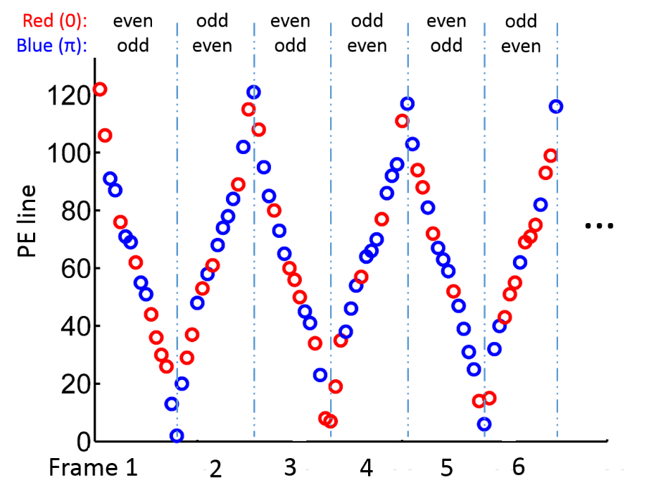
Figure 1: Phase modulation and k-space undersampling
patterns of the proposed ATOMICS technique. For odd frames, even/odd PE lines
have 0 or π phase offset between two simultaneously imaged slices. For even
frames, even/odd PE lines have π or 0 phase offset between slices. Zero and π
phase differences are indicated by red and blue circles respectively.
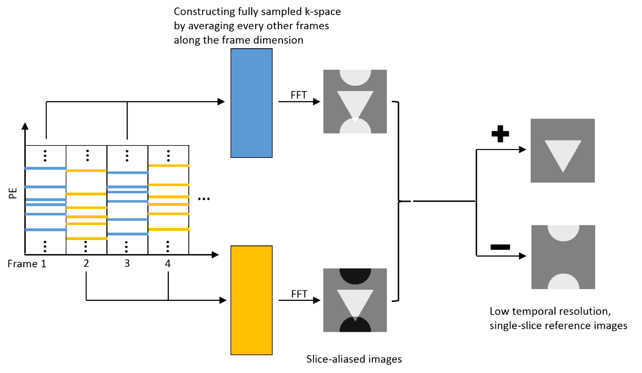
Figure 2: Procedure for extracting single-band reference
images from MB data. First, even and odd frame data are averaged separately.
The reference images are then generated by taking the sum or difference of the
two averages. The non-aliasing reference images are of low temporal resolution
because multiple frames are mixed.
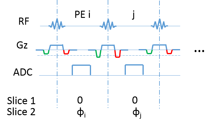
Figure 3: MR signal phase can be modulated by gradients. A
phase difference ϕ can be introduced between slices by a gradient moment Mz=ϕ/γd, where γ is the gyromagnetic ratio and d is
the distance between slices. In multiband bSSFP sequences, the rephrasing (red)
and prephasing (green) gradient lobes are both modified while keeping the total
gradient balanced, i.e., with no net zeroth moment.
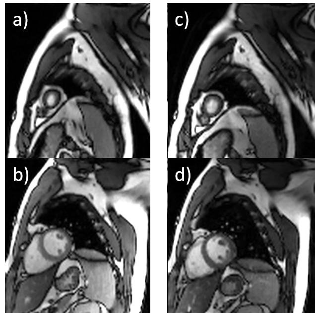
Comparison of CS only and ATOMICS images at the
same cardiac phase. Image a) and b) were two slices acquired separately with CS
cine, while c) and d) were acquired simultaneously with ATOMICS. Heart morphology was well delineated in both groups.

Twelve consecutive cine frames from systole to
diastole cardiac phase. The two slices were acquired simultaneously using ATOMICS,
and the temporal resolution was 2.88 ms × 15 = 43.2 ms. No obvious artifacts
were observed and the wall motion was well preserved.