3461
Quantitative comparison of a brain protocol with and without FRONSAC enhancement
E. H. Bhuiyan1, Y. Rodriguez1, R. Todd Constable1, and G. Galiana1
1Radiology and Biomedical Imaging, Yale University, New Haven, CT, United States
1Radiology and Biomedical Imaging, Yale University, New Haven, CT, United States
Synopsis
FRONSAC has previously been demonstrated as a highly effective approach to using nonlinear gradients to reduce undersampling artifacts, but previous results were limited to proof-of-principle experiments in individual subjects. Here we report quantitative comparisons for a full protocol of standard brain imaging sequences (2D GRE, 2D TSE, 2D T2w-FLAIR, 3D GRE and 3D MP-RAGE) acquired in a cohort of healthy subjects, comparing standard Cartesian to FRONSAC-enhanced acquisitions. Preliminary results confirm that FRONSAC significantly improves undersampling artifacts, measured as RMSE relative to a fully sampled Cartesian reference, while retaining the contrast, SNR, and reliability of standard Cartesian sequences.
Purpose
Fast Rotary Nonlinear Spatial Acquisition (FRONSAC) is a novel approach to accelerated imaging that can significantly accelerate acquisition and consequently reduce the cost of MR imaging. In this study a single FRONSAC waveform is applied to enhance a set of widely used clinical sequences (2D GRE, 3D GRE, 3D MP-RAGE, 2D TSE, and 2D T2w-FLAIR) comparing each to the conventionally acquired image. A cohort of subjects were imaged (n=4 with continuing enrollment) and we report quantitative metrics on both fully sampled and undersampled images. Results confirm that FRONSAC encoding reduces undersampling artifacts and improves RMSE by a factor of 3 while preserving other desirable features of these conventional sequences.Background
FRONSAC has previously been demonstrated as a highly effective approach to using nonlinear gradients to reduce undersampling artifacts, but previous results were limited to proof-of-principle experiments in individual subjects.1-4 Here we present quantitative comparisons for a full protocol of standard brain imaging sequences acquired in a cohort of healthy subjects, comparing standard Cartesian to FRONSAC-enhanced acquisitions. Preliminary results confirm that FRONSAC significantly improves undersampling artifacts while retaining contrast and SNR. Furthermore, results from this larger scan series continue to show no particular sensitivity to realistic experimental conditions (imperfect B1 or off-resonance spins), which further validates that FRONSAC-enhanced images retain the robustness of the underlying Cartesian sequence.Method
Each sequence was tested on four subjects (continuing enrollment) with both Cartesian and FRONSAC encoding for a 128 matrix size and 25cm FOV with transverse multislice geometry. All images were acquired with an 8 channel head coil nested in a 1ch Tx head coil, and bandwidth was 130Hz/pix. 2D sequences were acquired with 4 slices and 3D sequences were acquired with cubic FOV and matrix size. All scans were acquired at full resolution and undersampled retrospectively. All sequences were acquired with the same FRONSAC gradient, using gradient amplitude of 450 mT/m3, 460mT/m3 and 64 mT/m2 with oscillation frequency 10kHz, and waveforms that were triangular analogs of sine/cosine/sine on the C3/S3/Z2 channels, respectively. Fields were mapped as previously described1-3. Because reconstructions employ an empirical measurement of the NLG encoding, concomitant fields and eddy currents simply contribute a small additional encoding which is accounted for in the map and reconstruction. For spin echo sequences, TR/TE=3000ms/21ms and ETL=8, with the center of k-space acquired in the 4th echo. For gradient echo sequences, TR/TE=300/16 in 2D images and 25/10 in 3D images.Results
Figure 1a shows multislice 2D GRE images of a single subject with Cartesian and FRONSAC enhanced sequences at undersampling factors of 2 and 4. Below each are difference images from a fully sampled reference image. Similar data (not shown) was acquired for 2D TSE, 2D T2w-FLAIR, 3D GRE and 3D MP-RAGE. These difference images are quantified as RMSE for each subject and slice, with that data presented as the first panel of Figure 2. Pooled across subjects and slices, the average RMSE at R=4 was 7.6×10⁻⁶ vs 2.8×10⁻⁶ for Cartesian and FRONSAC-enhanced images, respectively. Figure 3 shows a histogram of the pixel by pixel signal ratio in fully sampled Cartesian and FRONSAC GRE images. The mean intensity ratio across pixels is 1.002 with a standard deviation of 0.145. This result, as well as visual inspection of Figure 1, shows that there is no appreciable contrast change from adding FRONSAC encoding to the acquisition. Figure 4 quantifies SNR of Cartesian vs FRONSAC images, again by subject and slice. This reveals comparable SNR between the images, with means of 300 and 330 for Cartesian and FRONSAC R=2 images and 530 and 450 for R=1. Finally, Figure 5 presents data addressing potential spatially varying resolution in Cartesian vs. FRONSAC images. While it is true that nonlinear gradients generally provide spatially varying resolution, the small moments applied in a FRONSAC image correspond only to acquiring on the order of 4 additional lines in k-space which is unlikely to have a visual impact (e.g. resolution corresponding to a 128 matrix at the center and 132 matrix at the edge). This is illustrated by comparing a zoom of a central vs peripheral ROI in Cartesian and FRONSAC images, which shows no detectable difference in the apparent resolution.Discussion
Image contrast was degraded both in Cartesian and FRONSAC due to imperfect B1 profiles, potential subject movement, and imperfect shim. However, these are realistic obstacles in clinical scanning, and FRONSAC-enhanced Cartesian imaging appears to show comparable ability to overcome these imperfections. It is important to note that NLG mapping, once performed on a uniform phantom, does not change with coil loading or sample geometry. Therefore, it is mapped once per coil installation, not for each scan, and there was no evidence that it changes with subject.Conclusion
Data was collected in a cohort of healthy volunteers using a protocol of standard 2D and 3D brain imaging sequences with and without the addition of FRONSAC encoding. Quantitative metrics confirm that FRONSAC encoding shows consistent improvement in the undersampled image quality, while maintaining the contrast, SNR, spatially homogeneous resolution and overall reliability of standard Cartesian imaging.Acknowledgements
We would like to thank Andrew Dewdney (Siemens) and Terry Nixon for supporting the nonlinear gradient hardware.References
- Dispenza NL, Littin S, Zaitsev M, Constable RT, Galiana G. Clinical Potential of a New Approach to MRI Acceleration, Nature Publishing, Scientific Reports 9, Article number: 1912 (2019).
- Luedicke N, Tagare H, Galiana G, Constable RT, editors. Trajectory design of optimized repeating linear and nonlinear gradient encoding using a k-space point spread function metric. ISMRM Annual Meeting; 2016; Singapore.
- Nadine L. Dispenza, Maxim Zaitsev, R. Todd Constable, Gigi Galiana, ``Clinical Imaging Potential of FRONSAC”, invited talk for ISMRM Annual Meeting and Exhibition, Paris, France June 18, 2018.
- E. H. Bhuiyan, Nadine L. Dispenza, R. Todd Constable, and Gigi Galiana, Optimization of a 3-channel gradient waveform for FRONSAC encoding, Proc. Intl. Soc. Mag. Reson. Med. 27 (2019).
Figures
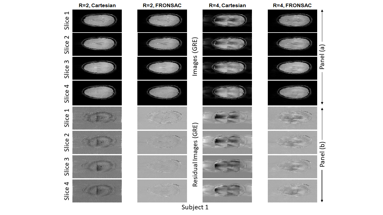
Cartesian vs Cartesian-FRONSAC images, with
residuals. A representative set of 2D
Cartesian and Cartesian-FRONSAC GRE images are shown for a single subject. Residual images, shown below, are obtained by
comparing the R=2 & R=4 images to the normalized Cartesian R=1 images for
the corresponding slices. Similar data
was acquired for 2D TSE, 2D T2w-FLAIR, 3D GRE and 3D MP-RAGE.
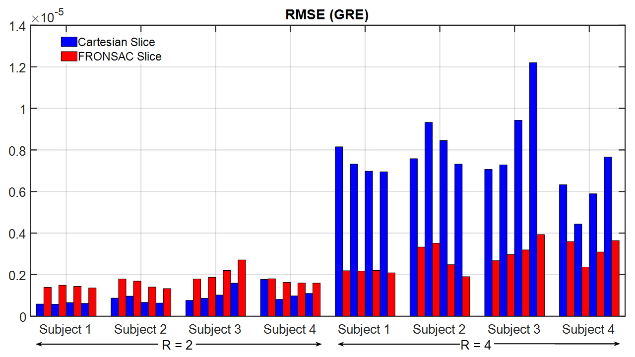
RMSE of Cartesian vs Cartesian-FRONSAC.
This bar graph shows RMSE by slice and subject for Cartesian and
FRONSAC-enhanced GRE relative to the fully sampled Cartesian GRE image. At R=2, both images show minimal undersampling
artifact. Simple Cartesian
reconstructions show slightly lower RMSE, likely due to the fact that the
undersampled images come from the same dataset as the reference image. At higher undersampling factors, the improved
undersampling artifact in Cartesian-FRONSAC leads to a 3 fold improvement in
RMSE, with errors similar to those seen at R=2.
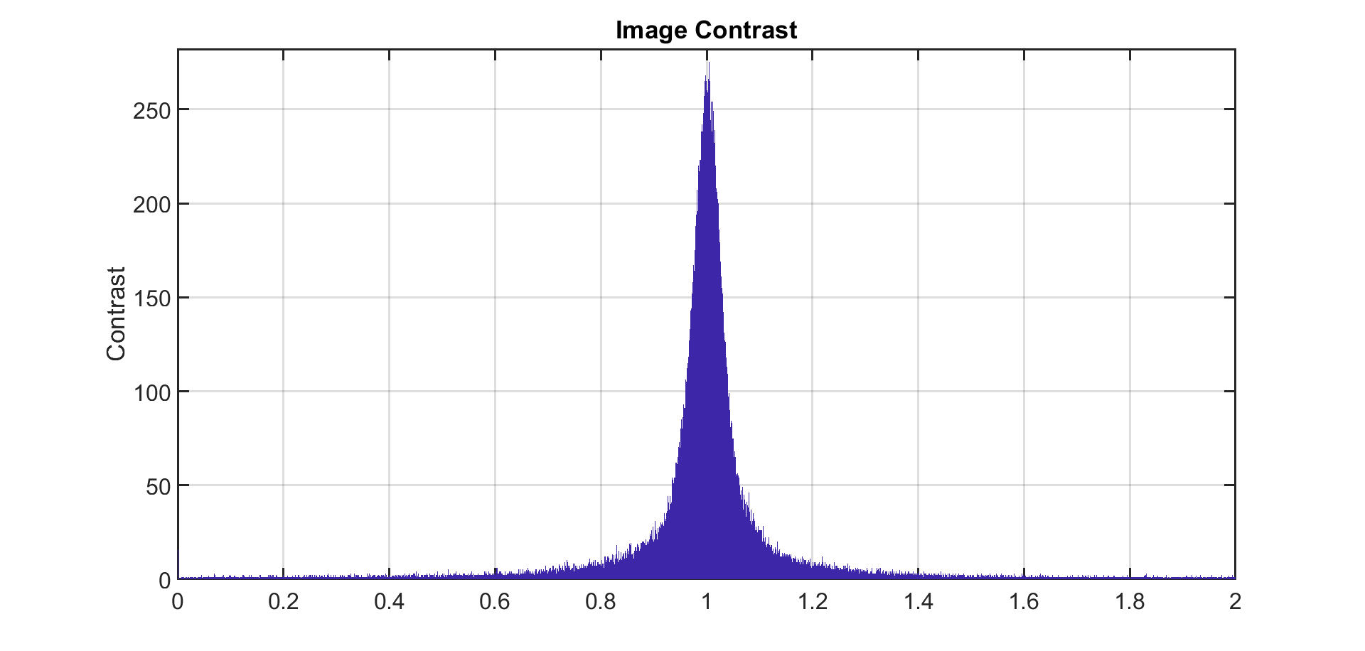
Contrast
of Cartesian vs FRONSAC images. A
pixel-wise calculation of signal in the fully sampled Cartesian images divided
by the signal in fully sampled FRONSAC images demonstrates that contrast is not
altered by FRONSAC encoding. Images were
first masked by SNR to avoid calculating contrast over noisy voxels. Mean
intensity ratio was 1.002 with a standard deviation of 0.145.
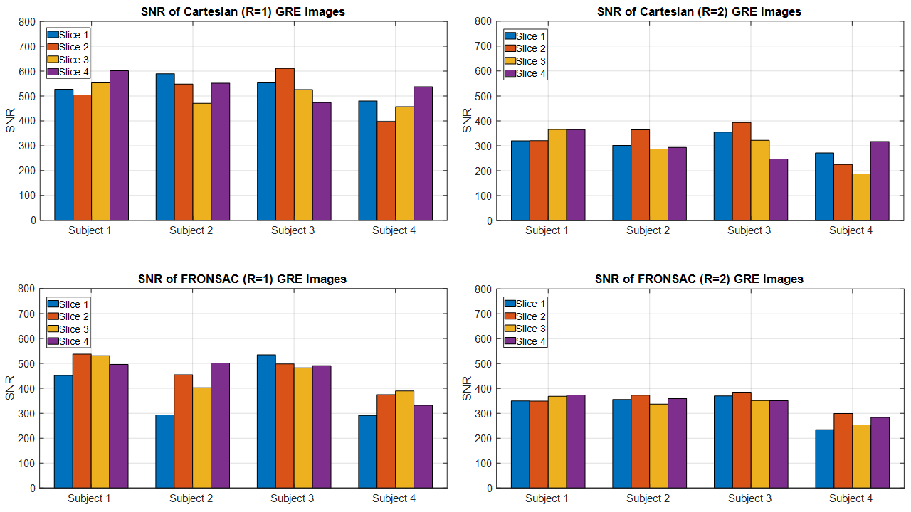
Signal to noise ratio
(SNR) for Cartesian vs Cartesian FRONSAC. Using the same signal and noise ROIs, SNR was calculated across subjects
and slices, showing comparable SNR at equal undersampling.
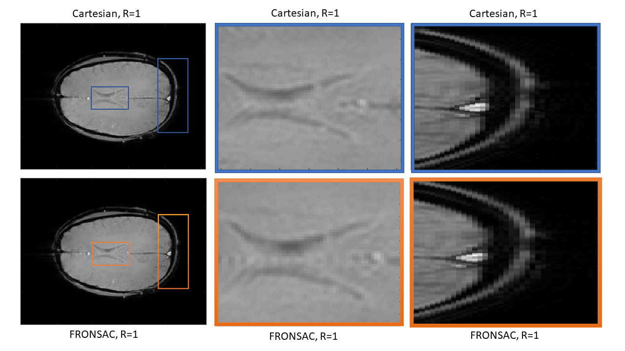
Spatially varying resolution in Cartesian vs.
FRONSAC images for R=1. Because FRONSAC
uses low nonlinear gradient moments, it minimally contributes to spatially
varying resolution. Resolution is
comparable between Cartesian and FRONSAC images at both the center and
periphery of the FOV.