3429
Spin-echo sequence with interleaved spiral acquisition avoids pulsation artifact for sagittal T1-weighted brain imaging at 3.0T
Ke Jiang1, Jiazheng Wang1, and Xiaofang Xu1
1Philips Healthcare Greater China, Beijing, China
1Philips Healthcare Greater China, Beijing, China
Synopsis
T1-weighted imaging is a necessary component of clinical brain MR to provide anatomical information at high spatial resolution. T1w brain image in clinical practice is regularly acquired with two-dimensional turbo-spin-echo sequence, which suffers from the pulsation artifacts in the phase-encode direction from superior sagittal sinus, the straight sinus, and the sigmoid sinus. This study exploits the spiral trajectory for the spin-echo sequence to achieve the T1 weighting and to avoid the pulsation artifacts.
Introduction
T1-weighted imaging is a necessary component of clinical brain MR to provide anatomical information at high spatial resolution. T1w brain image in clinical practice is regularly acquired with two-dimensional turbo-spin-echo sequence, which suffers from the pulsation artifacts in the phase-encode direction from superior sagittal sinus, the straight sinus, and the sigmoid sinus [1]. In contrast, the spiral trajectory, due to its fast imaging nature, is less sensitive to motion, and the absence of a phase-encode direction prohibits the formation of motion artifacts in a certain direction. Despite the intensive research on spiral trajectory for ultra-fast imaging [2], here we demonstrate the capability of spiral acquisition in T1w sagittal brain imaging to avoid pulsation artifacts.Method
The spiral acquisition was implemented in a spin-echo (SE) sequence such that a spiral-out trajectory starts at the echo time, a schematic diagram of the sequence shown in Figure 1. The whole k-space was acquired using multiple interleaves, with an illustration given in Figure 2. The SE-spiral and the traditional TSE images, both with T1 weighting, were collected at 3.0 T (Ingenia Elition, Philips healthcare, Best, the Netherlands) on the brain of healthy volunteer with 32 channel head coil used. Informed consent was obtained from each volunteer and the study was approved by a local IRB. Isotropic FOV and voxel size were acquired for both sequences: 230mm and 0.8mm. Other parameters for SE-Spiral: TR/TE = 600/9.5ms, acquisition window (the duration of a single spiral) = 10ms, interleaves = 33, NSA = 2, scan time = 2.7min. For TSE: TR/TE = 600/19ms, NSA =1, TSE factor = 4, scan time =2.1min. The TR was kept the same for the two sequences to yield image contrasts as similar as possible.Result
Figure 3 shows the T1 weighted brain images obtained from both SE-Spiral and the traditional TSE sequences. Pulsation artifact appeared only on TSE image which was indicated by red arrows. SNR/CNR for SE Spiral and TSE was 42/23.8 and 29/11.2, respectively.Discussion
We demonstrated the feasibility of SE Spiral in avoiding pulsation artifact for human brain imaging at 3T system. Longer acquisition window can lead to less scan time but introduce more blur. In this study acquisition window was set to 10ms and interleaves was set to 33 to achieve the balance between image quality and scan time. The slightly longer TA for SE Spiral compared to TSE was due to NSA (=2) which was implemented to obtain higher SNR.Conclusion
For T1 weighted human brain imaging, SE Spiral acquisition is capable of alleviating pulsation artifact at 3T systemAcknowledgements
No acknowledgement found.References
[1] Katarzyna Krupa, et al. Pol J Radiol. 2015; 80: 93–106.
[2] Curtis L. Johnson, et al. Magnetic Resonance in Medicine. 2013; 70:404–412.
Figures
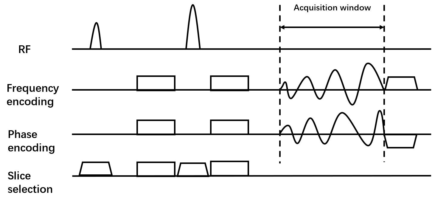
Figure 1. Schematic diagram of SE Spiral pulse sequence.
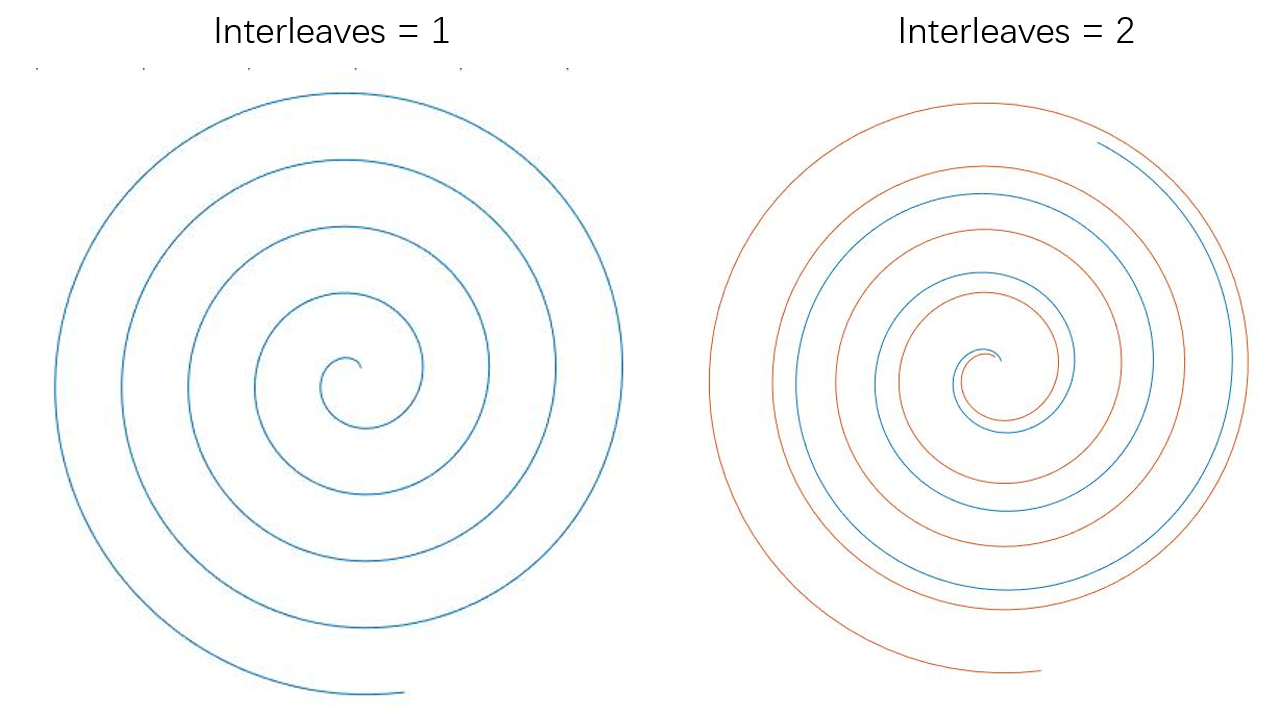
Figure 2. Spiral trajectory with interleaves = 1 (left) and interleaves = 2 (right).
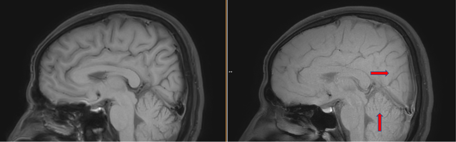
Figure 3. Representative T1 weighted images obtained by SE Spiral (left) and TSE(right). Pulsation artifacts can only be observed on TSE indicated by red arrow.