3421
Susceptibility effects of uHDC material upon static field homogeneity1Radiology, PennState University College of Medicine, Hershey, PA, United States, 2Neurosurgery, PennState University College of Medicine, Hershey, PA, United States
Synopsis
We performed a study to assess the effects of ultra-high dielectric constant material (uHDC) on the static field homogeneity at 3T. We characterized these effects on both a spherical and body phantom with 3D off-resonance field mapping and balanced SSFP imaging. The static field experienced shifts of up to 100 Hz in the presence of uHDC monolithic blocks. Shimming was able to partially compensate for these shifts. Future studies incorporating these materials should take their susceptibility effects into account.
Purpose
Dielectric materials have been utilized to achieve useful gains in SNR and transmit efficiency & homogeneity, at field strengths ranging from 1.5 to 7T. Both aqueous pads filled with slurries1 and monolithic ceramics2-3 have been studied. One concern these materials present that typically goes unnoticed is their effect on static B0 field homogeneity. Potentially, these materials could have a susceptibility value that differs from both air and human tissue. Two prior studies examined the susceptibility effect of a calcium4 or barium5 titanate pad at 7T and determined it to be negligible. To date, no studies have specifically examined ultra-high dielectric constant (uHDC) monolithic ceramic blocks for their effects on static field homogeneity. Here we present such a study, and demonstrate uHDC ceramics can impact the static field.Methods
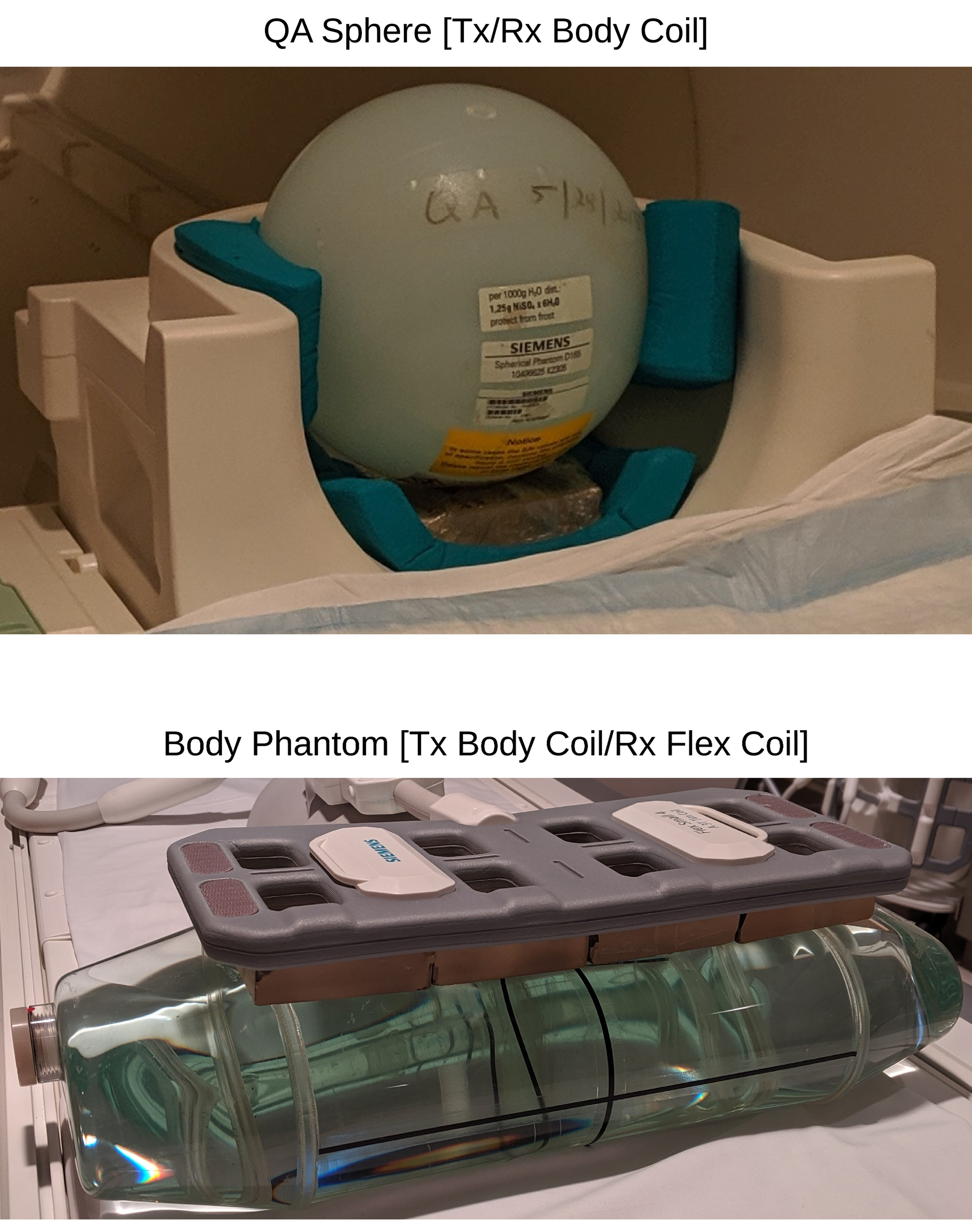
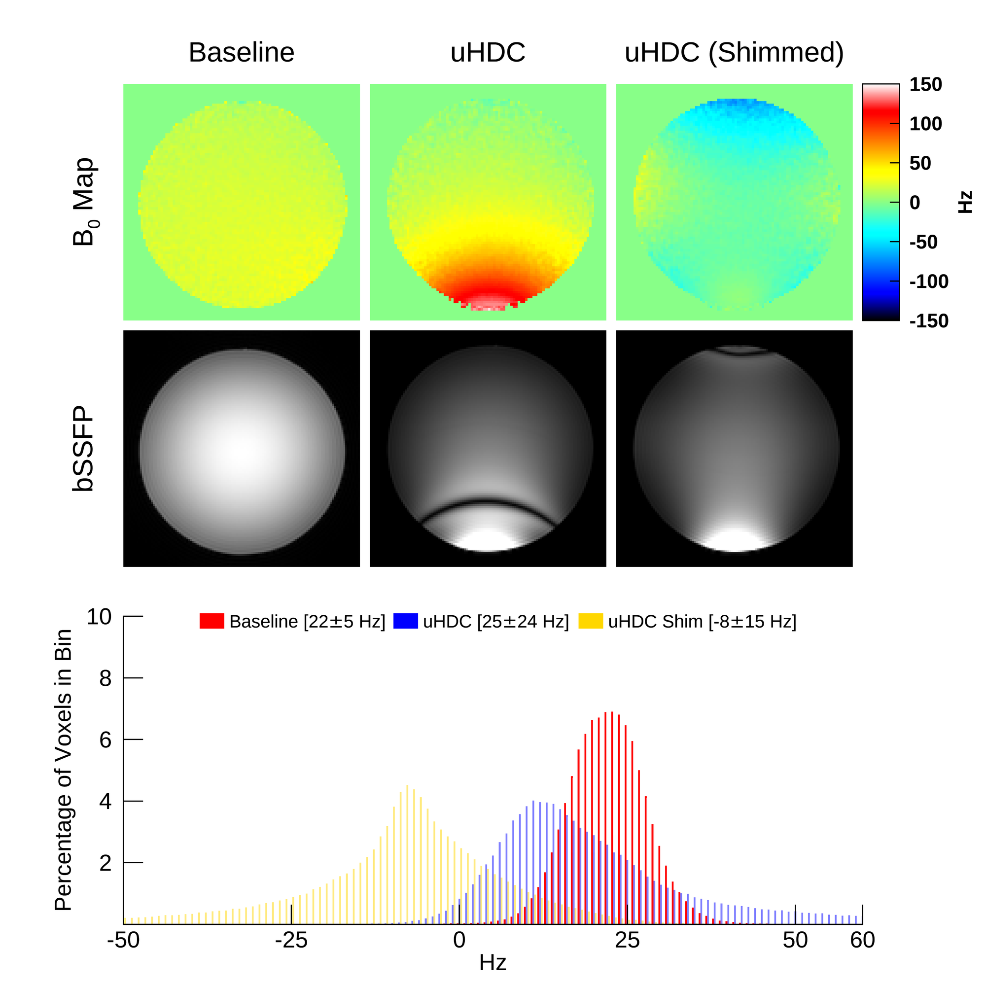
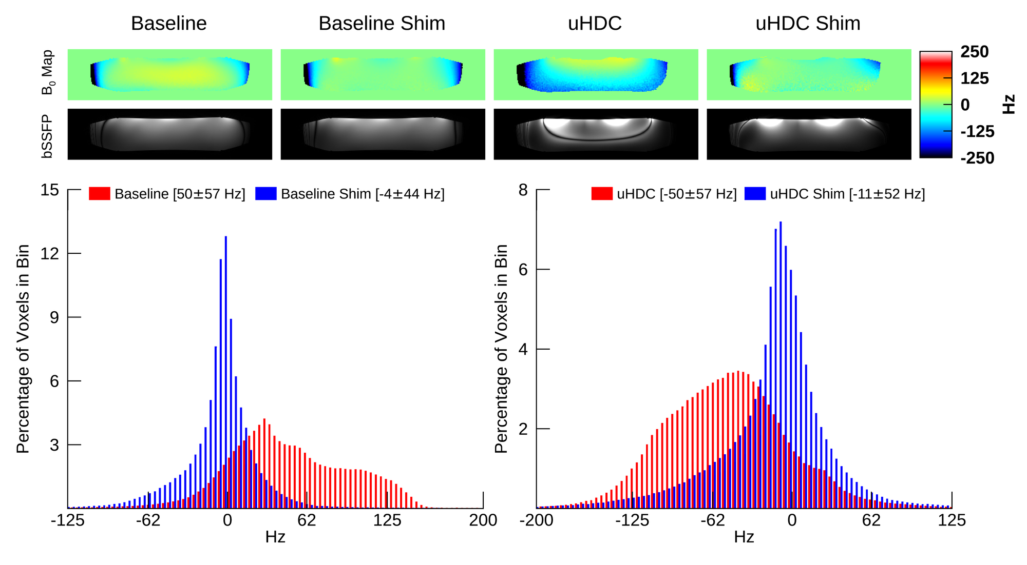
The effect of uHDC material upon static field homogeneity was studied experimentally with two different phantoms, depicted in Figure 1. All data was acquired on a Siemens 3T PrismaFit (Siemens Healthineers, Erlangen, Germany). A 3D off-resonance field map was acquired with two separate spoiled GRE acquisition (TR = 35 ms, TE1/2 = 2 / 2.5 ms, FA = 10 deg, 2 mm3 isotropic). Additionally, a 3D fully balanced SSFP acquisition (TR = 10.28 or 7.51 ms, TE = 5.14 or 3.76, FA = 60 deg, 1.5 x 1.5 x 2 mm) was acquired over the same imaging volume. The imaging volume was set to cover the entire phantom. (1) In the first experiment (top of Fig.1), a spherical Siemens QA phantom (diameter 168 mm, permittivity ~ 80) was scanned with and without a uHDC block present. The scanning was also repeated with the block present and B0 shimming over the entire phantom applied. The block had dimensions of 105 x 80 x 20 mm, and a permittivity of approximately 850 (PZT formulation). This setup was placed within the bottom half of a Siemens 20-Ch head array to provide a support structure. Signal transmission and reception utilized the PrismaFit body coil. (2) In the second experiment (bottom of Fig.1), a Siemens body phantom was scanned with and without 4 uHDC blocks present. The four blocks were identical to the single one utilized in Experiment 1. Signal transmission utilized the PrismaFit body coil, and reception a Siemens 4-Ch flex array. When scanning without the uHDC blocks, they were removed and the flex array placed directly on the top of the phantom. The scanning was also repeated with and without B0 shimming applied over the entire phantom.Results & Discussion
Figure 2 displays the results of the first experiment. A central axial slice of the volume, passing through the middle of the uHDC block, was extracted for display. The off-resonance field map with the uHDC block present experienced a strong shift in the static field, reaching values above 100 Hz. The bSSFP image displayed in the middle row confirms this shift, with a banding artifact visible close to the uHDC block location. B0 shimming (right column) partially mitigates the shift and removes the banding artifact, though another small band appears at the top of the slice. The histogram (bottom) displays the off-resonance distribution throughout the entire phantom, with the mean and standard deviation of each configuration listed in the legend. Shimming reduced the standard deviation with the uHDC block present from 24 Hz to 15 Hz, which was still above the baseline width of 5 Hz. Figure 3 displays the results of the second experiment. In this experiment, shimming was applied both without and with the uHDC block present. The presence of 4 blocks again leads to a strong shift in the field, with a corresponding banding artifact present in the bSSFP image. Both baseline and the uHDC case suffer from a broad distribution of B0 due to the size of the phantom. After shimming, the two cases obtain roughly equivalent B0 distributions.Conclusion
We have presented a study demonstrating the susceptibility effects of uHDC materials upon a spherical and body phantom. Shifts of up to 100 Hz were induced by the uHDC material, and were partially compensated for by static field shimming. While prior studies that examined aqueous barium or calcium titanate pads did not find a susceptibility effect, this may be due to the water in the pad creating a better susceptibility matching condition with human tissue. The actual susceptibility value of the monolithic PZT ceramic has not been characterized. Thus, future studies employing uHDC material should be prepared to take these effects into account.Acknowledgements
This work was supported in part by NIH grants of U01 EB026978References
[1] Luo et al, “Permittivity and performance of dielectric pads with sintered ceramic beads in MRI: early experiments and simulations at 3T”, MRM 2013; 70(1): 269-275
[2] Rupprecht et al, “Improvements of transmit efficiency and receive sensitivity with ultrahigh dielectric constant (uHDC) ceramics at 1.5T and 3T”, MRM 2018; 79(5): 2842-51
[3] Koolstra et al, “Improved Image Quality and Reduced Power Deposition in the Spine at 3 T Using Extremely High Permittivity Materials”, MRM 2018; 79(5): 1192-1199
[4] Teeuwisse et al, “Quantitative Assessment of the Effects of High-Permittivity Pads in 7 Tesla MRI of the Brain”, MRM 2012; 67:1285-1293
[5] Teeuwisse et al, “Simulations of High Permittivity Materials for 7 T Neuroimaging and Evaluation of a New Barium Titanate-Based Dielectric”, MRM 2012; 67:912-918
Figures