3416
Reduction of Static Off-Resonance Artifacts in T2*-Weighted Gradient-Echo MRI1Philips Research, Hamburg, Germany
Synopsis
A reconstruction method is presented to reduce off-resonance induced artifacts in gradient-echo MRI. The method reconstructs the magnetization without the off-resonance induced phase and in this way reduces the effects of intra-voxel dephasing. The method is applied to simulations and volunteer scans, showing reduced signal voids and improved homogeneity in R2*-maps.
Introduction
Gradient echo sequences are susceptible to local variations of the magnetic field (off-resonance). A common gradient echo image artifact caused by off-resonance are signal voids caused by intra-voxel dephasing in regions where the gradient of the off-resonance is large. A number of methods have been proposed in the past to mitigate static off-resonance induced artifacts, for example by correcting the magnitude images by a function based on the local off-resonance gradient [1, 2], or by taking also the neighborhood of any voxel into account [3]. Here we present a new correction algorithm based on demodulating the off-resonance field at a higher spatial resolution, showing a reduction of signal voids and also improving the quantitative accuracy of R2* values.Methods
The algorithm reconstructs the complex magnetization m by iteratively (de)modulating the off-resonance-induced phase at a higher resolution. In this way, signal cancellation from intra-voxel dephasing can be reduced up to the limit where the signal is lost because it is shifted fully outside the acquired region of k-space. The algorithm performs the following steps, where F is the Fourier Transform, a subscript h indicates a quantity at high resolution (upsampling two-fold proved to be sufficient), and r denotes a deringing filter.a) Initialization: m0 = 0, Φh = upsample(exp(-i2π B0 TE))
b) Iteration step i → i+1 (maximum 20 steps):
1. upsample image to high resolution: mh = upsample(mi)
2. modulate upsampled image with off-resonance phase: mh = mh ∙ Φh
3. transform to k-space: sh = F-1(mh)
4. set central part of sh to acquired data s: sh[acq. region] = s
5. transform to image-space: mh = F(sh)
6. demodulate phase: mh = mh / Φh
7. down-sample and dering image: sh = F-1(mh)
mi+1= F(r∙sh[acq.region])
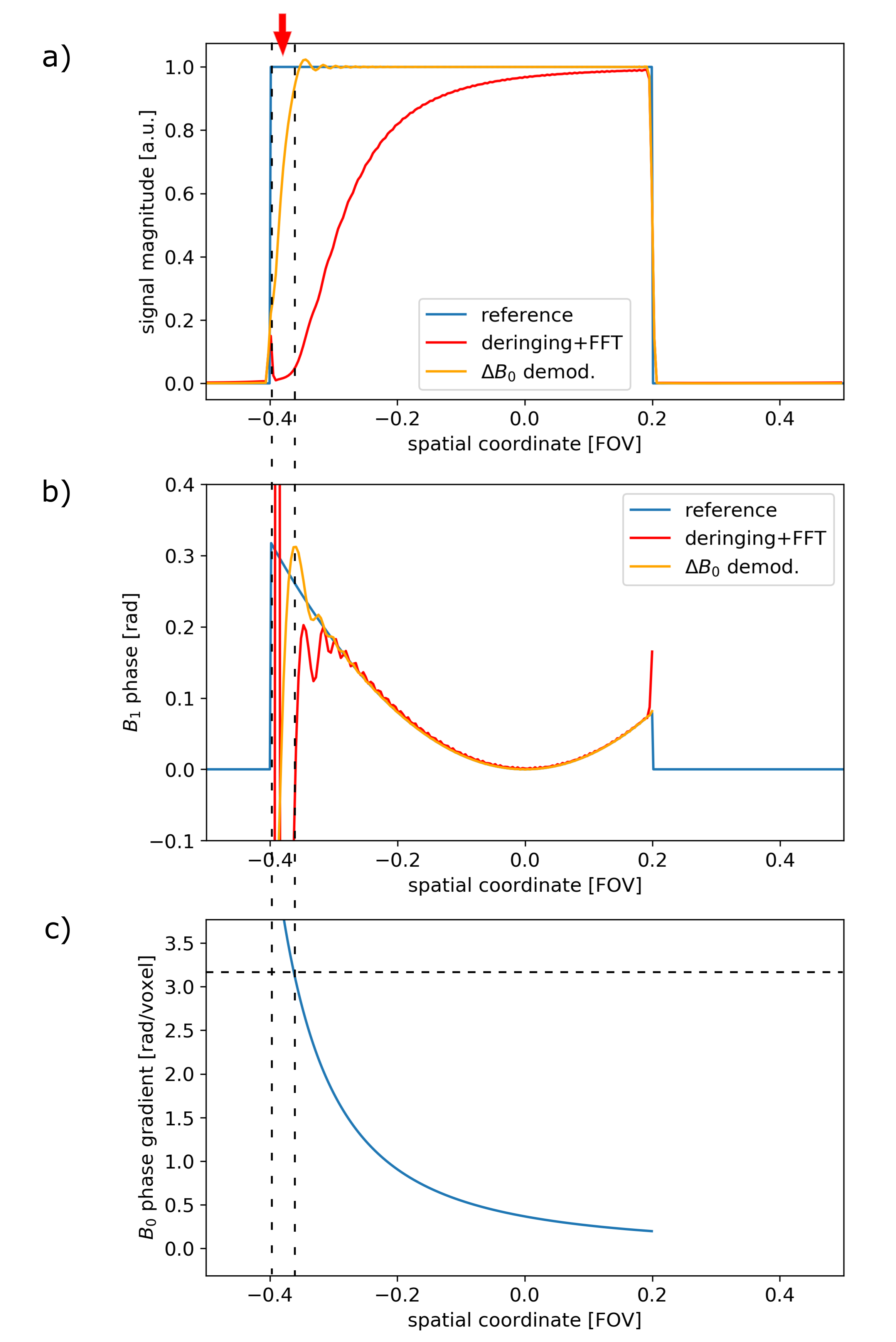
1D simulations are performed to illustrate the performance of the above method, as shown in Figure 1a-b, in blue. Data was simulated on 512 matrix and truncated in Fourier space to the reconstruction matrix of 256 and using a Hamming filter for deringing.
A healthy volunteer's brain was scanned on a 3T scanner (Ingenia, Philips, Best, The Netherlands) with IRB approval using an RF-spoiled gradient-echo sequence (acquisition voxel: 1x1x2mm3, echo-times: (3, 11, 19, 27)ms, TR: 30ms, FA: 14°).
In vivo data was processed offline using Python, the raw k-space data compressed to a single channel and using a Kaiser-Bessel deringing filter. The off-resonance map B0 was estimated from the first two echoes.
Results
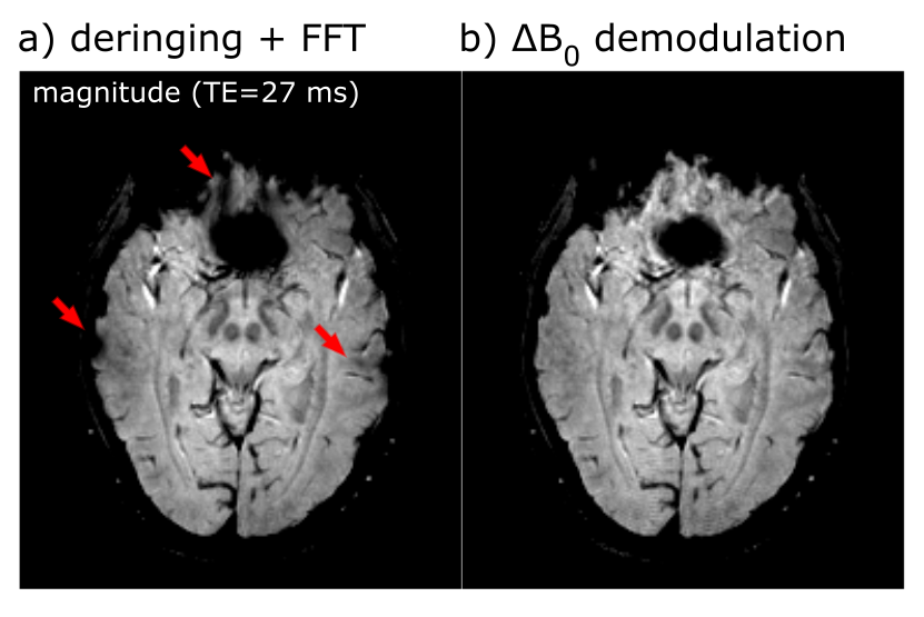
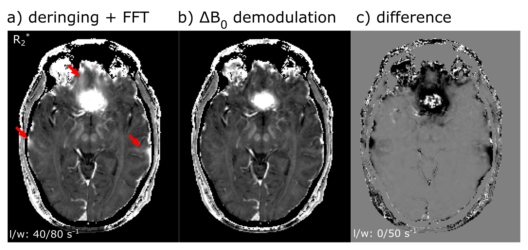
1D simulations: The standard reconstruction (deringing + FFT, Figure 1 a-b, red lines) shows the expected behavior: strong amplitude reduction where the gradient of the B0-induced phase is large. The proposed method (Figure 1 a-b, orange lines) is able to reconstruct the reference magnetization magnitude and phase (blue lines) accurately for most part of the rectangle. However, at the very left edge not all signal can be recovered (See red arrow in top graph). In this region (indicated by the dashed vertical lines) the gradient of the off-resonance-induced phase is larger than $$$\pi$$$ per voxel, which corresponds to shifting the signal from this voxel outside the encoded region in Fourier space.In vivo data: Figure 2a the standard reconstruction (deringing + FFT) with a large signal void above the nasal cavity and weaker signal reductions above the ears. With the proposed method, the signal void is significantly smaller (Figure 2b) and the reduction above the ears has disappeared. This is even more evident in the R2*-maps in Figure 3, particularly in the difference image, Figure 3c. The latter shows a reduction in the estimated R2* values in regions affected by intra-voxel dephasing.
Discussion and Conclusion
We have demonstrated a new method to reduce artifacts caused by strong B0 variations by (de)modulating the off-resonance induced phase at a higher resolution than the acquired data. As shown in the simulation experiments, the complex signal can be faithfully reconstructed up to the expected limit of about $$$\pi$$$/voxel. While a full correction of inhomogeneity-induced artifacts at finite acquisition resolution is not yet achieved, our method fully uses the available information in the raw k-space data and does not simply apply a voxel-by-voxel correction. Possibly, this reduces noise-amplification effects in previously published methods [1-3] which are caused by division by small numbers, although this has not been studied yet. The accuracy of the B0 map is not critical as long as the bulk of the phase-gradients is demodulated.Acknowledgements
No acknowledgement found.References
[1] Fernández‐Seara MA, Wehrli FW, "Postprocessing technique to correct for background gradients in image‐based R2* measurements", Magnetic Resonance in Medicine 44:358-366 (2000)
[2] Hernando
D, Vigen KK, Shimakawa
A, Reeder SB, "R2* mapping in the presence of macroscopic B0 field variations
", Magnetic Resonance in Medicine 68:830-840 (2012)
[3] Yablonskiy D, Sukstanskii A, Luo J, and Wang X, "Voxel Spread Function Method for
Correction of Magnetic Field Inhomogeneity Effects in Quantitative
Gradient-Echo-Based MRI", Magnetic Resonance in Medicine 70:1283–1292
(2013)
Figures