3407
BMART-Enabled Field-Map Combination of Phase-Cycled Projection-Reconstruction Cardiac Cine for Banding-Free Balanced SSFP1Electrical Engineering, Stanford University, Stanford, CA, United States, 2Medical Biophysics, Western University, London, ON, Canada
Synopsis
For banding-free cardiac cine bSSFP imaging, we present a non-Cartesian accelerated frequency-modulated sequence that acquires three phase-cycles within a short breath-hold. A projection-reconstruction trajectory is used and data is also acquired on the rewinds, enabling generation of a B0 field map using BMART. This facilitates field-map combination of the phase-cycle images, which results in more homogeneous blood pool and reduced signal contributions from out-of-slice spins than root-sum-of-squares.
Introduction
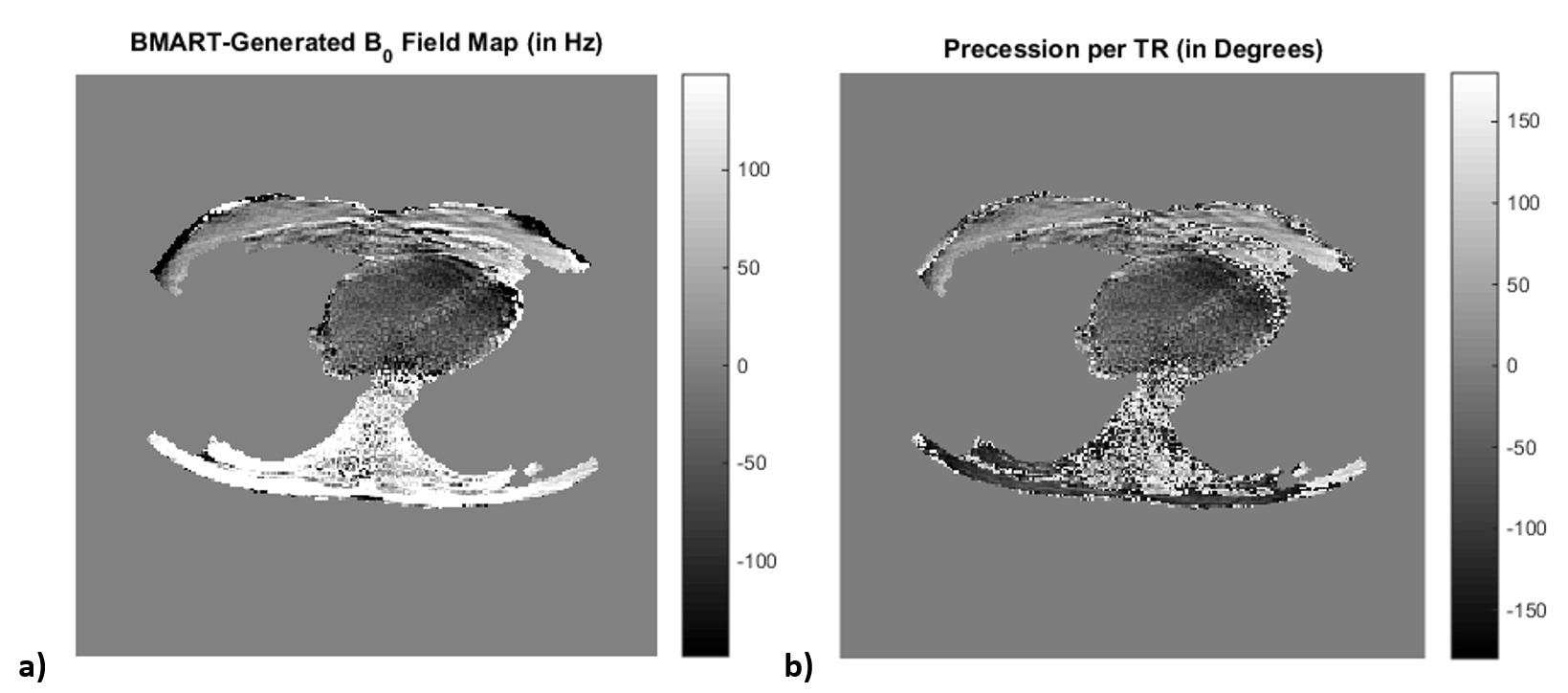
For banding artifact reduction in cardiac cine balanced SSFP imaging, a highly accelerated frequency-modulated sequence can be used to acquire three phase-cycles within a short breath-hold.1 However, due to the complexities of flow artifacts near banding,2,3 the images still have significant artifactual intensity variations after root-sum-of-squares (SOS) combination. In addition, through-plane flow near a phase-cycle’s stopband may cause that component image to include significant contributions from out-of-slice spins,2 and this signal persists in the SOS-combined images. Acquiring a B0 field map enables inclusion of only passband signal in the final combined image and exclusion of stopband and bright-band signal, facilitating a flatter spectral profile after phase-cycle combination and reducing the contributions from out-of-slice spins in the final image.4 However, the method in [4] requires an additional scan and introduces the potential of misregistration between the field map and the cine data. For non-Cartesian balanced SSFP acquisitions where the rewinds fully sample k-space, B0 Mapping using Rewinding Trajectories (BMART) can be used to acquire a field map during the imaging scan itself with minimal increase to the TR.5 Here, we illustrate the feasibility of using a projection-reconstruction (PR) trajectory for an accelerated frequency-modulated cine sequence in conjunction with BMART to acquire three phase-cycles and a field map within a short breath-hold, enabling use of field-map combination when reconstructing the final cine images.Methods
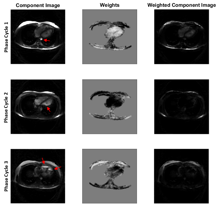
In the PR trajectory, the spokes span all 360o so that the rewinds cover k-space. An undersampled segmented acquisition with a reduction factor of 7.1 is used, but the k-space sampling pattern is shifted every cardiac phase and between phase-cycles. Therefore, averaging the cardiac phases and phase-cycles generates fully sampled data for parallel-imaging calibration (similar to earlier Cartesian work1) and field-map generation. The sequence was implemented at 1.5T for use with an eight-channel cardiac coil. It acquires a cine loop of an axial slice with 36 cm field of view, 1.7 mm resolution, 1.3 ms TE, 3.4 ms TR, and 40 ms temporal resolution. This TR is 0.18 ms longer than that of a sequence that does not acquire data on the rewinds. The scan time is 13 heartbeats, including the first heartbeat of discarded acquisitions for signal stabilization. The frequency modulation scheme of [1] is applied, obviating the need for any other stabilization heartbeats. A fully balanced SSFP sequence (i.e., no partial dephasing) is used. The shim was offset prior to imaging to create challenging off-resonance conditions.The cine data is reconstructed using ESPIRiT with $$$\ell1$$$ finite-difference regularization. For the calculation of the B0 map using BMART, the data up to half the k-space extent is sorted into 25 bins based on the average delay between the normal and rewind acquisition at that k-space location. The precession per TR $$$\phi$$$ is calculated from the B0 map, and a triangular weighting function $$$w(\phi) = \textrm{tri}((\phi-180)/120)$$$ is used to smoothly merge the passbands of the three phase-cycles for each cardiac phase. Note that phase-cycling shifts over the cardiac cycle due to the frequency modulation scheme, so the weighting of the three images at any given location also changes with cardiac phase.
Results
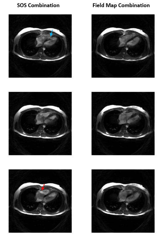
In a volunteer, the weights derived from the B0 map (Figure 1a) mask out the banding and near-band flow artifacts in and around the heart in the component images (Figure 2). As a result, for all cardiac phases, the field-map combinations have more homogeneous blood pools and substantially reduced hyperintense regions than the SOS combinations (selected cardiac phases are shown in Figure 3).Discussion and Conclusion
We provide a proof-of-concept that a PR trajectory can be used for a frequency-modulated cine sequence, enabling reconstruction of three phase cycles and a field map from data acquired within a short breath-hold. Acquiring data on the rewinds to enable generation of a field map using BMART requires only a 5% lengthening of the TR but facilitates field-map combination of the phase-cycle images. The resulting combined images show more homogeneous blood pool and reduced signal contributions from out-of-slice spins.The correspondence of the weights with the banding in and near the heart suggests that the field map generated using BMART is accurate in the region of interest. Although, in this work, data from all cardiac phases were averaged before calculating the field map, it may be possible to reconstruct field maps for each cardiac phase to capture changes in the field over the cardiac cycle, or a sliding window may possibly be used to balance the SNR and temporal resolution of the field map. The field map here may have phase-wrap errors in the subcutaneous fat. Since this application only uses $$$\phi$$$ and not B0 itself, use of a TR that is an integer multiple of the time difference between the normal and rewind acquisitions at the center of k-space may warrant investigation. In addition, measurement of the gradient waveforms and more accurate characterization of the gradient delays may improve the accuracy of the field map.
The basic parallel imaging reconstruction with finite-difference regularization used here to reconstruct the individual phase-cycles images seemed to result in good quality, but incorporation of the phase-cycle redundancy regularization in [1] to better support higher acceleration should be investigated.
Acknowledgements
We would like to thank GE Healthcare and NIH HL127039 for supporting this project.References
1. Datta, A, Nishimura, DG, Baron, CA. Banding‐free balanced SSFP cardiac cine using frequency modulation and phase cycle redundancy. Magn Reson Med. 2019; 82: 1604– 1616. https://doi.org/10.1002/mrm.27815
2. Markl M, Pelc NJ. On flow effects in balanced steady-state free precession imaging: Pictorial description, parameter dependence, and clinical implications. J Magn Reson 2004; 20:697–705.
3. Storey P, Li W, Chen Q, Edelman RR. Flow artifacts in steady-state free precession cine imaging. Magn Reson Med 2004; 51:115–122.
4. Datta A, Nishimura DG. Field map combination method for multiple-acquisition bSSFP. In Proceedings of the 25th Annual Meeting of ISMRM, Honolulu, May 2017. p.454.
5. Baron CA, Nishimura DG. B0 mapping using rewinding trajectories. Magn Reson Med 2016. doi: 10.1002/mrm.26391.
Figures