3385
Characterization and elimination of X-nuclear eddy current artifacts on clinical MR systems
Mary A McLean1,2, Scott Hinks3, Joshua Kaggie1, Ramona Woitek1, Frank Riemer4, Martin Graves1, Dominick McIntyre2, Ferdia Gallagher1, and Rolf Schulte5
1Dept of Radiology, University of Cambridge, Cambridge, United Kingdom, 2Cancer Research UK Cambridge Institute, University of Cambridge, Cambridge, United Kingdom, 3GE Healthcare, Waukesha, WI, United States, 4MMIV, Dept of Radiology, Haukeland University Hospital, Bergen, Norway, 5GE Healthcare, Munich, Germany
1Dept of Radiology, University of Cambridge, Cambridge, United Kingdom, 2Cancer Research UK Cambridge Institute, University of Cambridge, Cambridge, United Kingdom, 3GE Healthcare, Waukesha, WI, United States, 4MMIV, Dept of Radiology, Haukeland University Hospital, Bergen, Norway, 5GE Healthcare, Munich, Germany
Synopsis
Using pulse-acquire spectroscopy, line-shape distortions characteristic of eddy currents were demonstrated for X-nuclei, which were not seen for 1H, on systems from multiple vendors. The severity of these appeared correlated with the amplitude of the f0 eddy current frequency compensation term applied by the system along the axis of the applied spoiler gradient. A proposed correction to eddy current compensation taking account of the X-nuclear gyromagnetic ratio was shown to dramatically reduce these distortions on a GE system. The same correction was also shown to improve the quality of non-Cartesian imaging (spirals and cones).
Introduction
Visual inspection of X-nuclear spectra and images sometimes suggests the presence of eddy currents not seen on 1H-MRS, which is very sensitive to eddy current effects in general1. We hypothesized that this was because the software-defined frequency pre-emphasis applied to correct for the spatially independent but time-varying eddy current field Δf0(t) needs to be adjusted for the gyromagnetic ratio (γ) of the nucleus being observed. Here we present a method to characterize the severity of eddy current artifacts on different nuclei, axes, and scanners, and we demonstrate a solution on a GE scanner which greatly improves quality of X-nuclear images and spectra.Methods
Experiments were performed on 3 T clinical systems at three sites: System A (MR750, GE Healthcare, Waukesha WI); System B (Skyra-fit, Siemens Healthcare, Erlangen, Germany); and System C (Achieva, Philips Healthcare, Best, Holland). All sites used similar quadrature birdcage head coils dually resonant for 1H and either 23Na, 13C or 31P (Rapid, Rimpar, Germany). Spectral distortions were assessed using spherical phantoms, and a resolution phantom filled with 80 mM saline was used for 3D 23Na imaging. Eddy current distortion is maximized by minimizing the delay between the large spoiler gradient pulse at the end of one TR period and the acquisition in the next. Therefore, pulse-acquire spectra with matched parameters were collected for 1H and X-nuclei (23Na, 31P, and/or 13C) using the minimum allowed TR. Data were collected with the spoiler pulse applied along each of the three orthogonal axes in turn. Modifications to eddy current compensation system calibration files on System A optimized for each X-nucleus were developed and applied, by multiplying the correction terms applied for Δf0(t) by the ratio of the gyromagnetic constants γX-nucleus /γ1H. Data were compared with and without these corrections using slice-selective MRS, 2D spirals2, and 3D cones3.Results and Discussion
On System A (GE), small distortions due to eddy currents were seen in water 1H-MRS in axial and coronal orientations (Fig 1d and g), but not sagittal (Fig 1a). In 23Na-MRS, there was still no evidence of eddy currents in the sagittal orientation, but the distortion was greater than seen for 1H in coronal orientations, and very much greater than 1H in axial orientations (Fig 1b, e ,h). This mirrored the relative amplitudes of coefficients for ‘very long time constant’ f0 correction terms within the eddy current correction calibration file, which were 0, 0.6 and 4.4 respectively in the sagittal, coronal and axial orientations. Spectra were re-acquired following rescaling the eddy current f0 correction terms for sodium as described above (Fig 1c, f, i), resulting in distortion free 23Na spectra.With the default eddy current compensation, low frequency, non-Cartesian X-nuclear coronal images were consistently displaced superiorly relative to the 1H image. When the rescaled f0 correction terms were used for compensation, no displacement was observed (Fig 2). Similar displacement effects were observed on System A when imaging 23Na using 3D non-Cartesian sequences such as cones. Additionally, when using a short TR there was considerable blurring and distortion: rescaling the eddy current f0 correction terms for sodium resulted in markedly improved image quality (Fig 3).
A similar effect was seen on System B (Siemens), where the 1H MRS appeared clear of eddy current distortion but there was marked evidence of eddy currents on the X-nucleus (Fig 4). On System C (Philips), there were some distortions, but they were similar between nuclei, whereas Systems A and B showed much worse effects on X-nuclei than 1H. It seems likely that Philips either follow a different method or do not correct for eddy-current induced frequency errors at all.
Within sites, the severity of distortion was similar between axes on System B (Siemens), and differed markedly between axes on System A (GE). The presence of residual minor 1H eddy currents on System A in the coronal and axial orientations suggests that a recalibration of the correction terms is required. However, if these residual eddy currents were the source of the X-nuclear distortions, they would be expected to be of similar magnitude along the coronal and axial directions as seen for 1H, instead of the large differences between these axes which were seen for 23Na (Fig 1). These differences between axes must therefore be related not to residual eddy currents but to the applied correction itself.
Rescaling the eddy current frequency pre-emphasis was shown to improve the quality of not only spectroscopy but also fast imaging of X-nuclei using spiral and 3D cones sequences. These techniques are widely used in clinical research, and overcompensation of eddy current B0 terms may have been having quite widespread effects for many years.
Conclusion
Spectroscopy using a spoiler gradient and the minimum allowed TR presents a simple, quick and effective method to evaluate the relative performance of eddy current compensation for different nuclei, axes and manufacturers. We demonstrated greater distortions due to eddy currents on X-nuclei than on 1H, and presented a modification to markedly improve X-nuclear image quality.Acknowledgements
Collection of the data presented in this study was funded by the Cancer Research UK Cambridge Centre, National Institute of Health Research-Cambridge Biomedical Research Centre, and Addenbrooke’s Charitable Trust. Thanks to Damian McHugh and Sha Zhao (University of Manchester) and Adrian Carpenter (Cambridge) for data collection.References
[1] Klose U. In vivo proton spectroscopy in presence of eddy currents. Magn Reson Med 1990; 14:26-30. [2] Wiesinger F, Weidl E, Menzel MI, Janich MA, Khegai O, Glaser SJ, Haase A, Schwaiger M, Schulte RF. IDEAL spiral CSI for dynamic metabolic MR imaging of hyperpolarized [1-(13)C]pyruvate. Magn Reson Med 2012; 68:8-16. [3] Riemer F, Solanky BS, Stehning C, Clemence M, Wheeler-Kingshott CA, Golay X. Sodium (23Na) ultra-short echo time imaging in the human brain using a 3D-Cones trajectory. MAGMA 2014; 27:35-46.Figures
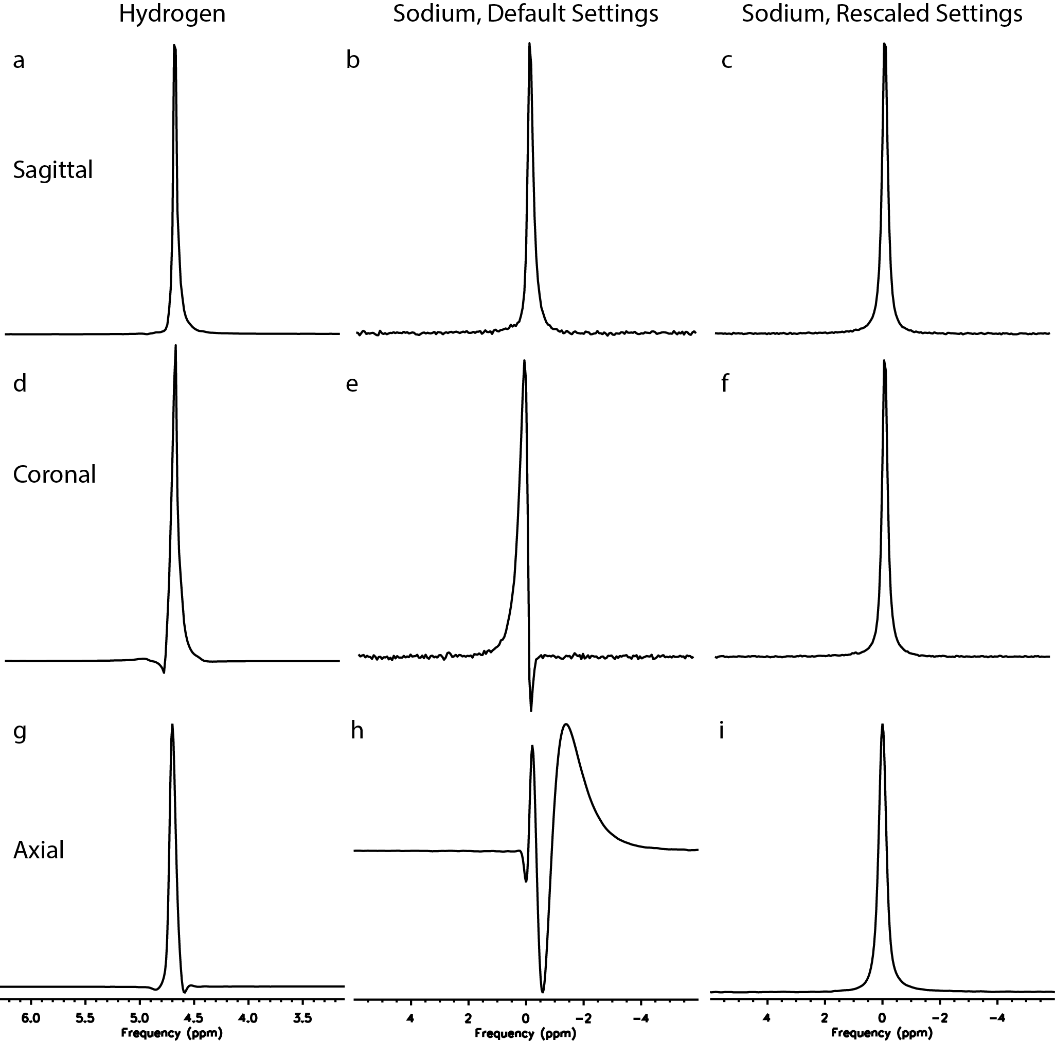
Figure 1: Eddy current distortions in spectra from a spherical phantom acquired on
System A using (left) 1H-MRS,
(middle) 23Na-MRS with
default eddy current correction applied, and (right) 23Na-MRS with a tailored eddy current
correction.
Figure 2: Spatial mis-registration of spiral X-nuclear images. With the default eddy current
correction settings (left), a coronal spiral image of a spherical phantom containing 1M
13C-bicarbonate (overlaid in colour) is seen to be offset in the SI
direction relative to the corresponding 1H MRI image. With corrected
eddy current f0 terms (right), this offset is reduced.
Figure 3: Distortion of
X-nuclear cones images. With the
default eddy current correction settings (top), a series of 23Na
axial 3D cones images of a phantom containing 80mM saline acquired at short TR (46ms)
is seen to be blurred and distorted. This is particularly evident in the
coronal and sagittal projections inset to the right. With corrected eddy current f0
terms (bottom), the blurring and distortion are greatly reduced.
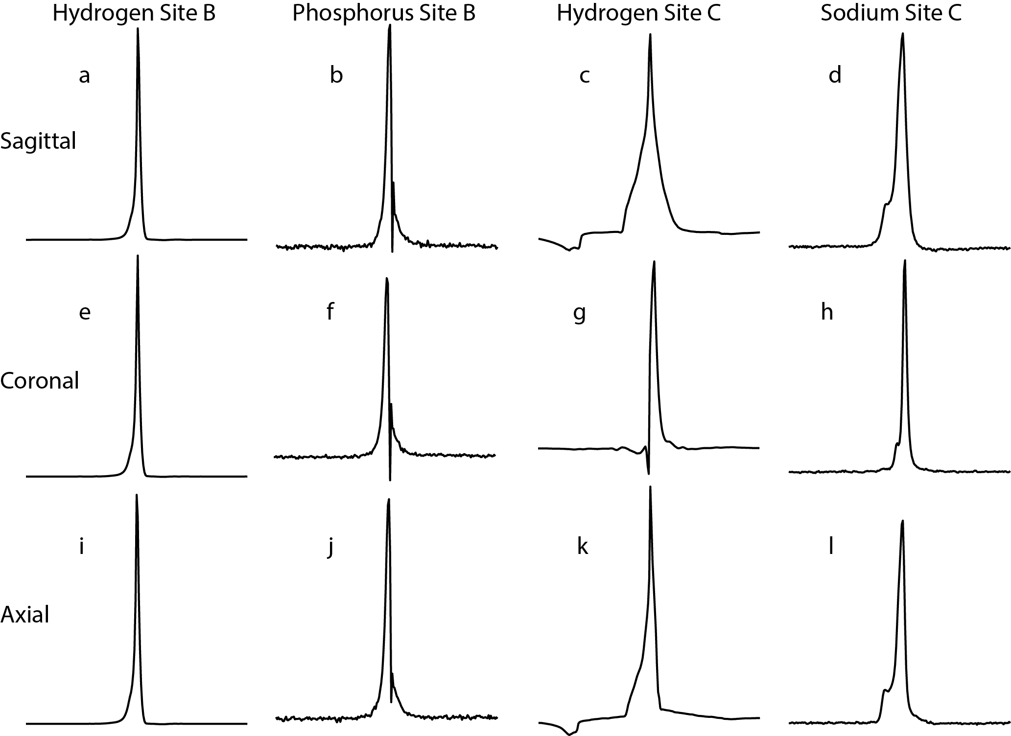
Figure 4: 1H and X-nuclear
eddy currents on Systems B and C. Similar experiments to Figure 1 were
performed on systems from different manufacturers. On System B (Siemens Skyra),
31P spectra acquired along all three axes showed clear signs of eddy
currents which were absent on 1H. On System C (Philips Achieva), the
lineshape distortions were similar between 1H and the X-nucleus (23Na).