3376
Methods and challenges for body fat composition and hepatic fat fraction measurement in a multi-site study using MRI
SHILPY CHOWDHURY1, SPENCER LOONG1, JOAN SABATE1, and SAMUEL BARNES1
1LOMA LINDA UNIVERSITY, LOMA LINDA, CA, United States
1LOMA LINDA UNIVERSITY, LOMA LINDA, CA, United States
Synopsis
Running a large longitudinal multi-center study to measure visceral fat and hepatic fat fraction present unique challenges. We scanned fat/water and ex vivo liver phantoms across all sites and over time to show consistency. SliceOmatic and ImageJ were used for quantification of visceral fat and LCModel for hepatic fat fraction. Visceral fat showed a good correlation with hepatic fat and demographics. The use of phantoms and careful design of the protocol can help address challenges of a longitudinal study and help maintain consistency across sites.
INTRODUCTION
MRI is commonly used to measure visceral fat and hepatic fat fraction (HFF).1 However, performing these measurements in a multi-site longitudinal study presents unique challenges. In particular, maintaining consistent image and spectroscopy quality and consistent post processing on all participants and over time can be difficult. This study presents how these challenges were addressed in a longitudinal multi-center study.METHODS
This is an ongoing longitudinal multi-site clinical trial involving four universities. Here we present baseline cross-sectional data from the first 200 participants. All participants underwent a non-contrast abdominal MRI on a 3T Siemens MR scanner. The study was approved by the Institutional Review Board and written informed consent was obtained from all participants. MRI scanners at all sites scanned two phantoms: two ex-vivo preserved human livers, and a fat/water phantom. The phantoms were scanned with the human imaging protocol. The ex-vivo livers were formalin fixed livers, one chosen from an individual with no history of liver pathology, and the other with a history of non-alcoholic fatty liver disease. The livers were used to verify consistent hepatic fat fraction measurements across all sites and over time. The fat/water phantom was constructed to verify consistent fat volume measurements across sites and over time. The phantom was an approximately 4 liter container that contained about 3.2 liters of 5% agarose gel with 0.03% sodium azide, and 0.8 liters of lard. The lard was contained in 20 ml sealed plastic tubes and embedded in the agarose gel. A DIXON axial vibe two-echo sequence was used for measuring visceral fat. Slice coverage was enough to cover 4 cm above the dome of the liver to 7 cm below the top of the iliac crest. Acquisition was acquired in a single breath hold. Parameters for this sequence were: slices 96, FOV 400 mm, slice thickness 3.5 mm, TR 5 ms, TE (1.23, 2.46 ms), flip angle 9 deg. Image processing was performed using sliceOmatic (for segmentation) with a watershed algorithm and ImageJ was used for thresholding using intermodes. Statistical analysis was performed using SPSS. A Pearson correlation was run to show correlation of visceral fat with HFF and with demographics like age, weight, waist circumference and body mass index. P value of <0.05 was considered statistically significant.RESULTS
All sites demonstrated consistent results, the phantom scans showed similar values across all sites and over time (figure 2 and figure 3). The spectroscopy fat fraction measurements showed a standard deviation of 1.05% and 0.41% on the liver with the higher and lower fat percentage, respectively. The fat volume measurements showed a standard deviation of 14.1ml. Visceral adipose tissue showed a good correlation with hepatic fat fraction, and also with age, weight, waist circumference and body mass index (figure 1 and figure 4).DISCUSSION
Segmenting the images to separate subcutaneous from visceral fat is robust and consistent using a watershed algorithm in SliceOMatic. The internodes thresholding algorithm2 was used to threshold the images. It was found to give the most consistent and accurate results on the phantom data and was adaptable enough to adjust to the changes in signal intensity across slices.Ensuring the acquisition of high-quality spectra is a particular challenge in a large study. Spectral quality is harder to evaluate at the time of the exam compared to standard MR images, so the quality of the data can have a lot of variation. To improve consistency the MR spectroscopy was performed twice at the same position on all participants. One acquisition is performed at the beginning of the exam and another at the end of the exam. Automatic shimming is forced to run before each MR spectroscopy acquisition. The best spectrum is selected based on the quality of the spectra and the fitting. This increased the consistency of the data and decreased the number of subject call-backs to <1%.
For cross-sectional data we used iliac crest as the landmark and made visceral fat measurements for the 50 slices above the iliac crest. However, for longitudinal measurements simple alignment based on the bony anatomy is insufficient due to changing internal position of the visceral fat. The position of the internal organs and visceral fat varies significantly with diaphragm position and inspiration amount. This can cause multi-centimeter shifts in organ/visceral fat position from one breath hold to the next. A manual review of the data and selecting of slices that cover the same internal anatomy with a similar appearance of the visceral fat is required.
CONCLUSION
Body composition and hepatic fat fraction measurements can be consistent across centers and longitudinally with careful selection of acquisition and processing parameters.Acknowledgements
This study was part of the Habitual Diet and Avocado Trial and received funding from the Hass Avocado Board.References
- Peter M Graffy and Perry J Pickhardt. Quantification of hepatic and visceral fat by CT and MR imaging: relevance to the obesity epidemic, metabolic syndrome and NAFLD. Br J Radiol. 2016; 89 (1062): 20151024.
- Judith M.S. Prewitt and Mortimer L. Mendelsohn. The analysis of cell images. Annals of the New York Academy of Sciences. 1966; 128:1035-1053.
Figures
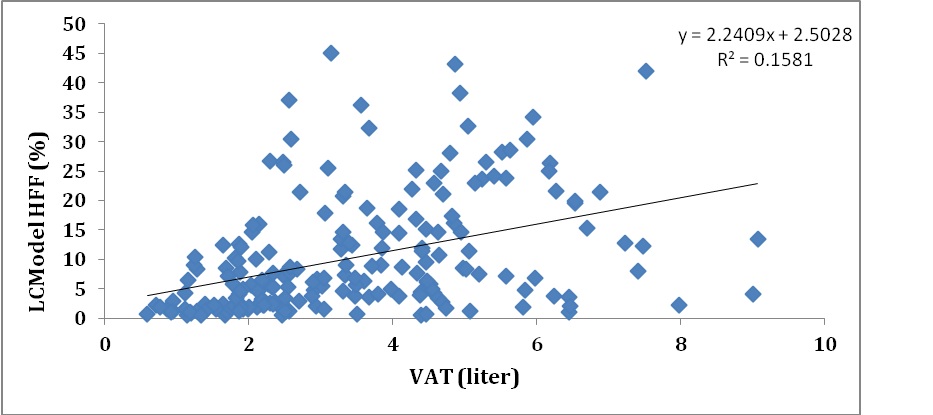
Correlation
between Visceral Adipose Tissue (VAT) and HFF using LCModel

Boxplot
showing fat/water phantom measurements across sites and over time
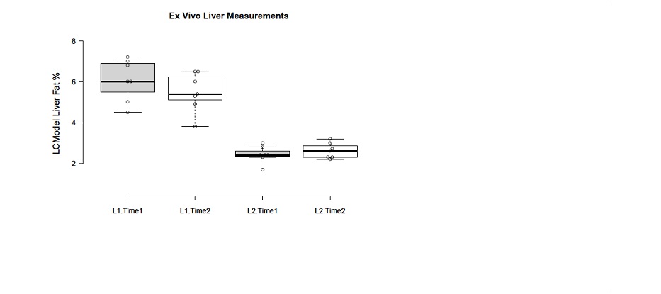
Boxplot
showing Ex vivo Liver measurements across sites and over time
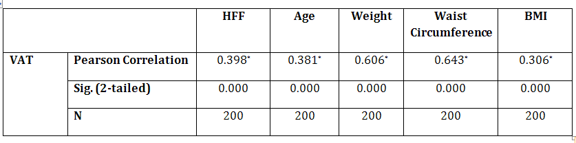
Correlation
between VAT and HFF, age, weight, waist circumference and BMI. *Correlation
is significant with p<0.0001 (2-tailed).