3374
Noise Reduction and Ghosting Alleviation in ASL Perfusion Measurement using Multi-dimensional Integration (MDI)1UIH America, Inc., Houston, TX, United States, 2United Imaging Healthcare, Shanghai, China
Synopsis
3D GRASE (GRAdient and Spin Echo) pulse sequence has been widely employed as the readout in arterial spin labeling (ASL) applications, given its efficient acquisition and relatively long lasting signal intensities. However, the inherent weakness of GRASE, such as vulnerability to motions, can induce ghosting artifacts to perfusion imaging and quantitative cerebral blood perfusion (CBF) maps. Herein, we propose applying the Multi-Dimensional Integration (MDI) algorithm to processing the perfusion and CBF maps, by which method noticeable alleviation of motion ghosting can be obtained, and the imaging noise is reduced as well.
Introduction
ASL is a useful technique mainly applied in brain imaging for CBF measurement without using any exogenous contrast agent.1 Using 3D GRASE as the readout of ASL, ghosting along phase encoding direction may be caused by motion during the acquisition time. Specifically, multiple shots in phase encoding direction are usually utilized in GRASE to acquire a 3D k-space volume. This usually leads to inconsistent phases among shots due to motion, and consequentially ghosting artifacts in imaging. The ghosting can be more undesirable in producing perfusion weighted images and CBF maps, since both of them are generally in relatively low SNR due to the nature of low ASL signals. The conventional method of magnitude subtraction requires control and labeling images being generated with a multi-channel combination algorithm (e.g. SOS), so that the resulted image quality may not be optimal in the sense that the noise in such low SNR images may vary spatially. In this work, we propose that these issues can be overcome by using a convenient yet comprehensive strategy, namely Multi-Dimensional Integration (MDI),2 in which the complex signal ratios of raw images are extracted without a direct signal combination of channels and repetitions. Moreover, because the ratios are independent of the control-labeling repetitions, the data inconsistency between consecutive repetitions arising from motion becomes lessened, leading to alleviation of ghosting artifacts.Methods
Considering the calculation for perfusion weighted images, instead of directly subtracting channel combined magnitude images, the complex ratio of control and labeling pairs for each channel and repetition is calculated. Then the calculated complex ratios are summed up over all channels and repetitions, and the magnitude of which gives an overall ratio (Rctrl/label). Consequently, unity is subtracted from it, followed by multiplication by the magnitude of labeling image. This rearranged subtraction protocol is expressed as equation 1 below:$$\left[\mid\sum_{ch=1}^M\sum_{rep=1}^N\left(\frac{S_{ctrl}\left(ch,rep\right)}{S_{label}\left(ch,rep\right)}\right)\mid-1\right]\cdot M_{label}\tag{1}$$
where Sctrl and Slabel are the complex images from control and labeling acquisitions respectively; Mlabel is the magnitude of labeling image; ch and rep denote the channel index and repetition index, respectively; and M and N represent total numbers of channels of head coil and repetitions, respectively. Similarly, CBF is calculated as follows in equation 2:
$$\left[\mid\sum_{ch=1}^M\sum_{rep=1}^N\left(\frac{S_{ctrl}\left(ch,rep\right)}{S_{label}\left(ch,rep\right)}\right)\mid-1\right]\cdot \mid\sum_{ch=1}^M\left(\frac{\sum_{rep=1}^NS_{label}\left(ch,rep\right)}{S_{M_{0}}\left(ch\right)}\right)\mid\cdot\lambda\tag{2}$$
where $$$S_{M_{0}}$$$ is the complex proton density weighted image (M0) and λ signifies the coefficient containing all required parameters for CBF calculation suggested by the ASL consensus.1 $$$S_{M_{0}}$$$ is independent of repetition number as it is acquired only once in an ASL scan protocol. In this equation, two complex ratios are calculated and no magnitude images are required.
For demonstration, ASL experiment was performed with a healthy volunteer at 3.0 T using a uMR 780 scanner (United Imaging Healthcare, Shanghai, China) equipped with a 24-channel head coil. A 3D variable-flip-angle based GRASE sequence was employed for data acquisition. TR/TE = 4643/13.8 ms. An acquisition matrix of 64×64 was applied. 32 slices with thickness of 4 mm were collected with 10% slice oversampling. The ASL labeling duration was set to 1800 ms followed by 1800 ms post-labeling delay.
Results
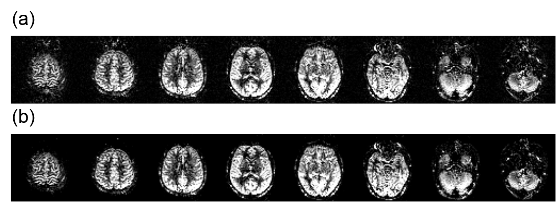
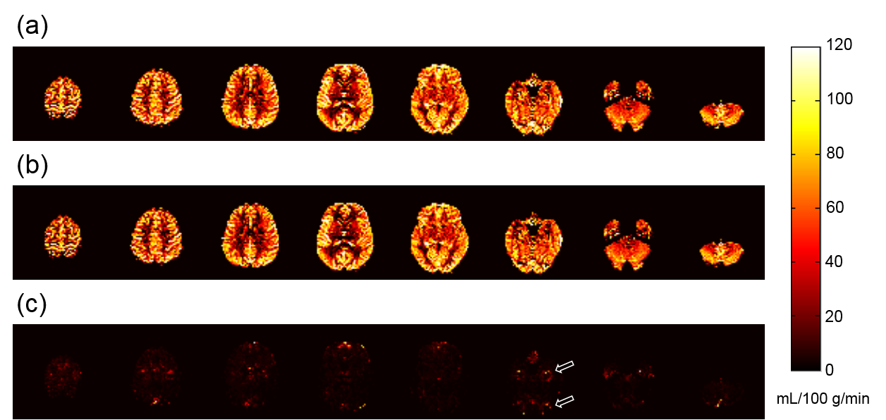
Figure 1 shows the perfusion images based on (a) SOS magnitude subtraction and (b) MDI for 8 slices from the ASL experiment. It is evident that the overall noise level of images by MDI method in Fig. 1(b) is suppressed compared with images based on SOS algorithm. Meanwhile, the promiscuous signal outside of brain tissue in Fig. 1(a), which can be partially attributed to motion ghosting, is largely reduced with MDI as exhibited in Fig. 1(b).Figure 2 (a, b) demonstrates the CBF maps with (a) the SOS method in comparison with using (b) MDI from the same experiment. Because the CBF maps in Fig 2(a) and (b) are masked based on image registration algorithm, the background noise and ghosting are not considered for comparison. The difference between the two methods are shown in Figure 2(c). The most evident difference indicated by the hollow arrows, is the ghosting artifacts associated with eye ball movements. Besides, the difference images do not generally exhibit a pattern corresponding to brain anatomy or perfusion contrast, thus should bear a large contribution of noise. Similarly to the perfusion images in Fig. 1, this noise in CBF images shown in Fig. 2(b) is reduced with MDI.
Discussion and Conclusion
The pivot of MDI is that the irrelevant dimensions with any interested quantity are operated individually in the quantity mapping. Without directly combining these dimensions, noise distribution is not modified and no mapping bias is introduced, especially for maps with low signal to noise ratio. The signal ratios are usually employed for removal of the irrelevant dependence. As for ASL, the channel and repetition are irrelevant dimensions for perfusion mapping. Taking advantage of MDI, the relevance is thus eliminated while calculating the complex ratios, through which optimal noise distribution can be achieved. The additional benefit with this strategy is that the data inconsistency due to motion is mitigated. Both the perfusion and CBF maps show reduction of noise and ghosting artifacts in the axial slices. Taken as a whole, this MDI scheme applied in ASL presents a reliable practice and gives insights into obtaining perfusion mapping with greater accuracy in clinical examinations and studies.Acknowledgements
No acknowledgement found.References
1. Alsop, David C., et al. "Recommended implementation of arterial spin‐labeled perfusion MRI for clinical applications: A consensus of the ISMRM perfusion study group and the European consortium for ASL in dementia." Magnetic resonance in medicine 73.1 (2015): 102-116.
2. Ye, Yongquan, et al. "MR Relaxivity Mapping using multi-dimensional integrated (MDI) complex signal ratio." ISMRM 27th Annual Meeting & Exhibition, Montreal, QC, Canada; 2019, p 4392.
Figures