3369
The geometric solution to banding mitigates motion artifact in bSSFP imaging1Radiology, University of Washington, Seattle, WA, United States, 2Radiology, University of Washington, SEATTLE, WA, United States, 3Radiology, University of British Columbia, Vancouver, BC, Canada
Synopsis
Widespread use of phase-cycled bSSFP imaging is hampered by problematic sensitivity to artifacts caused by susceptibility and motion. The geometric solution (GS), which can eliminate susceptibility-related banding and signal modulation, has previously show relative insensitivity to motion. Here, GS-bSSFP is evaluated using a phantom and in humans, imaging the skull base. The GS mitigates motion artifact in both paradigms and is particularly resilient when one of its four phase cycles is corrupted by motion.
Introduction
Balanced steady state free precession (bSSFP) imaging boasts high SNR efficiency but is plagued by dark artifacts. While susceptibility-induced banding and signal modulation are primary concerns, other darkening artifacts including those caused by motion can be problematic. The geometric solution1 (GS) can demodulate bSSFP of banding, and recent simulations and in vivo work have shown an insensitivity to motion2,3. Here we evaluate the GS performance using a water-flow phantom and in vivo while imaging the human skull base (including the internal auditory canal (IAC) and orbits, where aqueous/vitreous humor flow, ocular motion, cerebrospinal fluid flow, and cisternal vessel pulsation can occur) to demonstrate that the GS can mitigate motion artifacts.Methods
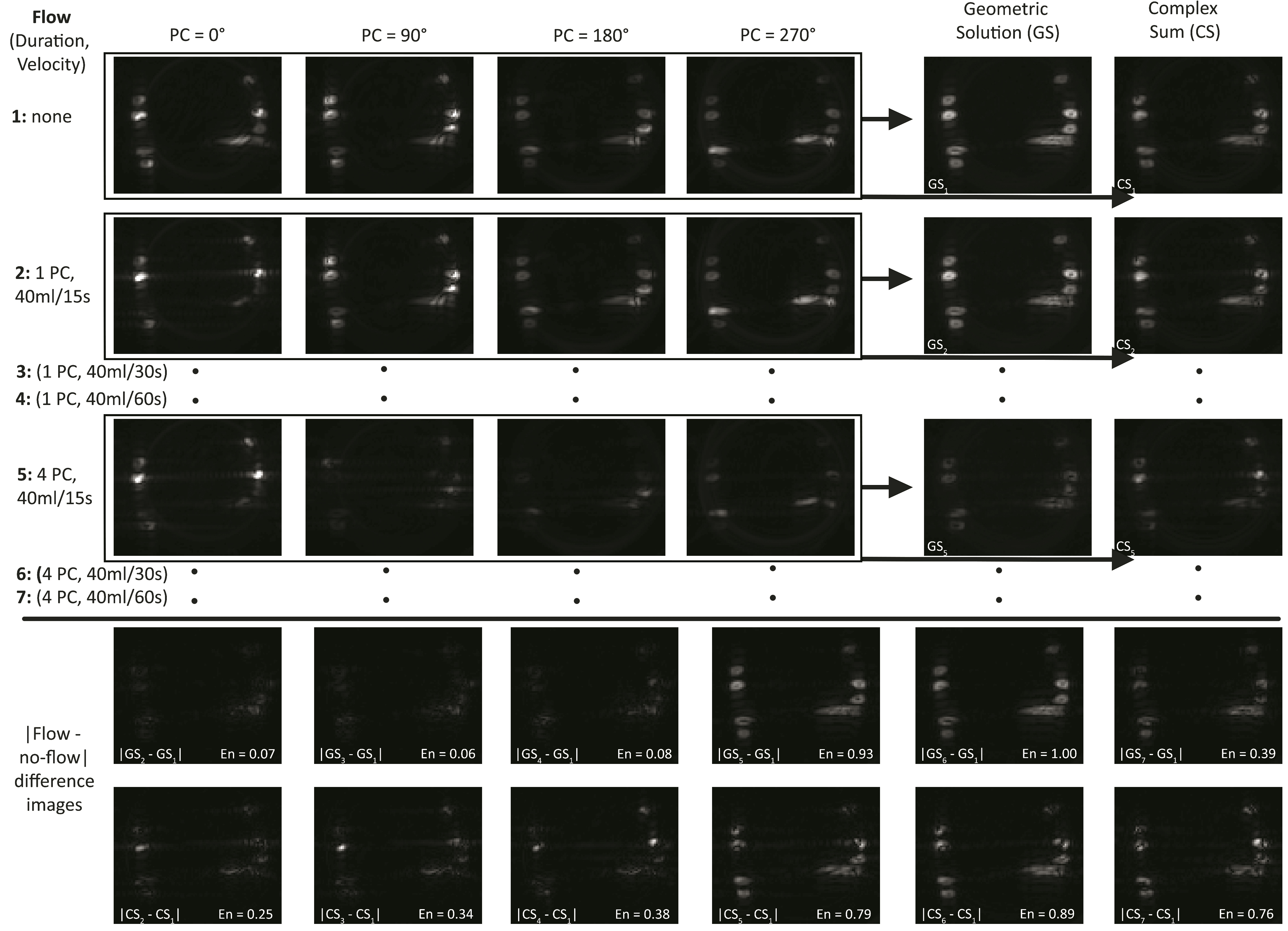
A plastic bottle was wrapped with ¼” inner diameter plastic tubing and placed in the MRI scanner. Water was pumped through the tube at controlled velocity and duration. Figure 1 details the 0°, 90°, 180°, and 270° phase-cycled (PC) bSSFP images that were acquired 1: without flow, during 1PC (0°) at velocities 2: 40ml/15s, 3: 40ml/30s, and 4: 40ml/60s, and during all 4PC at velocities 5: 40ml/15s, 6: 40ml/30s, and 7: 40ml/60s. The Complex Sum (CS) and GS were computed in each of the seven scenarios as previously discussed1,4.In order to gauge flow artifact in flow scenario X, residual flow artifact was computed by the absolute value of its complex difference from the 1: non-flow map, say |GSx – GS1|, as at the bottom of Fig. 1. A quantitative estimate of the total flow artifact may then be made via the total energy of each difference map:
$$En=\sum_{all pixels}(GS_X-GS_1)^2$$
En energy was computed for each flow/reconstruction scenario, normalized to the largest value, and listed on its corresponding difference map.
An institutional review board approved 21 patients undergoing a clinically indicated temporal bone (IAC protocol) MRI of the skull base for evaluation of treated vestibular schwannoma to received added bSSFP sequences acquired with PC = 0°, 90°, 180°, 270° in both sagittal and axial orientations on Philips Ingenia 3T scanners. Scan parameters were FA/TR/TE = 30°-45°/4.8-5.5ms/1.9-2.2ms, 316/314-316/42-100 matrix size and 0.57/0.57/1mm voxel size along frequency/phase/slice directions, respectively. The GS and CS were computed, and signal-normalized to avoid bias. Three fellowship-trained neuroradiologists blinded to sequence type, imaging parameters, clinical presentation, and patient disposition evaluated standard bSSFP, GS-bSSFP, and CS-bSSFP for the degree of dark artifact in the IAC and orbits of each patient using a 5-point Likert scale (1 = significant to 5 = none). Prevalence of dark artifact was compared between techniques using the Wilcoxon rank-sum test or the Sign test, clustered by subject to account for multiple readers.
Results
The bottom of Figure 1 illustrates that water velocities have minimal effect on energy En, however, 4PC flow durations showed significant increase in En relative to 1PC durations for both the GS and CS. The GS showed remarkably low energy for 1PC flow, consistently less than 30% of the corresponding 1PC CS energy. Neither technique performed well given flow during all phase cycles.Figure 2 depicts a patient’s orbits in standard bSSFP, GS-bSSFP, and CS-bSSFP axial images. The GS exhibits relatively minimal vitreous darkening when compared with the CS and standard bSSFP, with sharp margins of orbital structures.
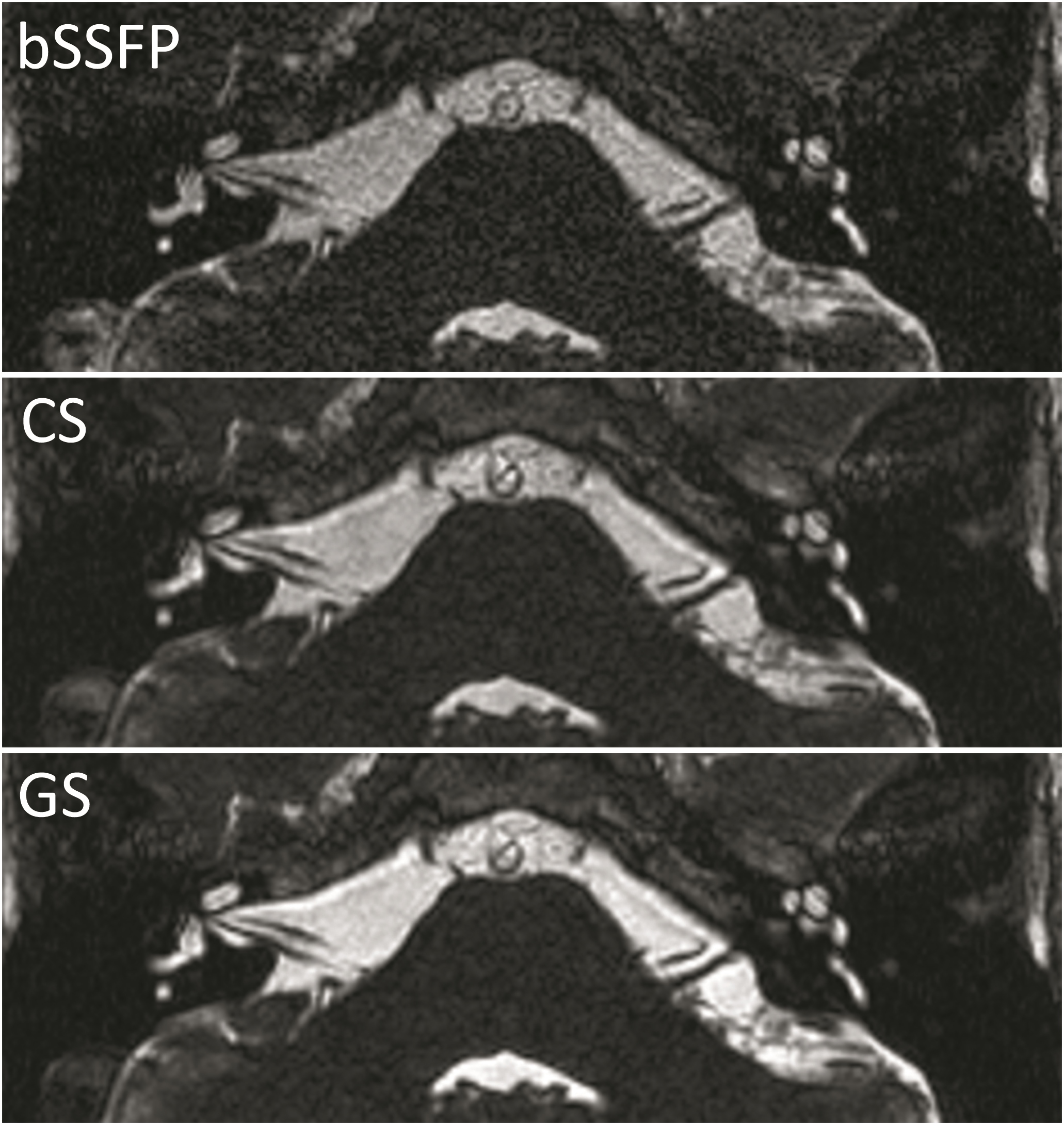
Figure 3 depicts a patient’s IAC in a standard bSSFP image, and GS-bSSFP and CS-bSSFP reconstructions. The GS displays good contrast, cranial nerve demarcation and slightly better CSF homogeneity when compared to the other techniques.
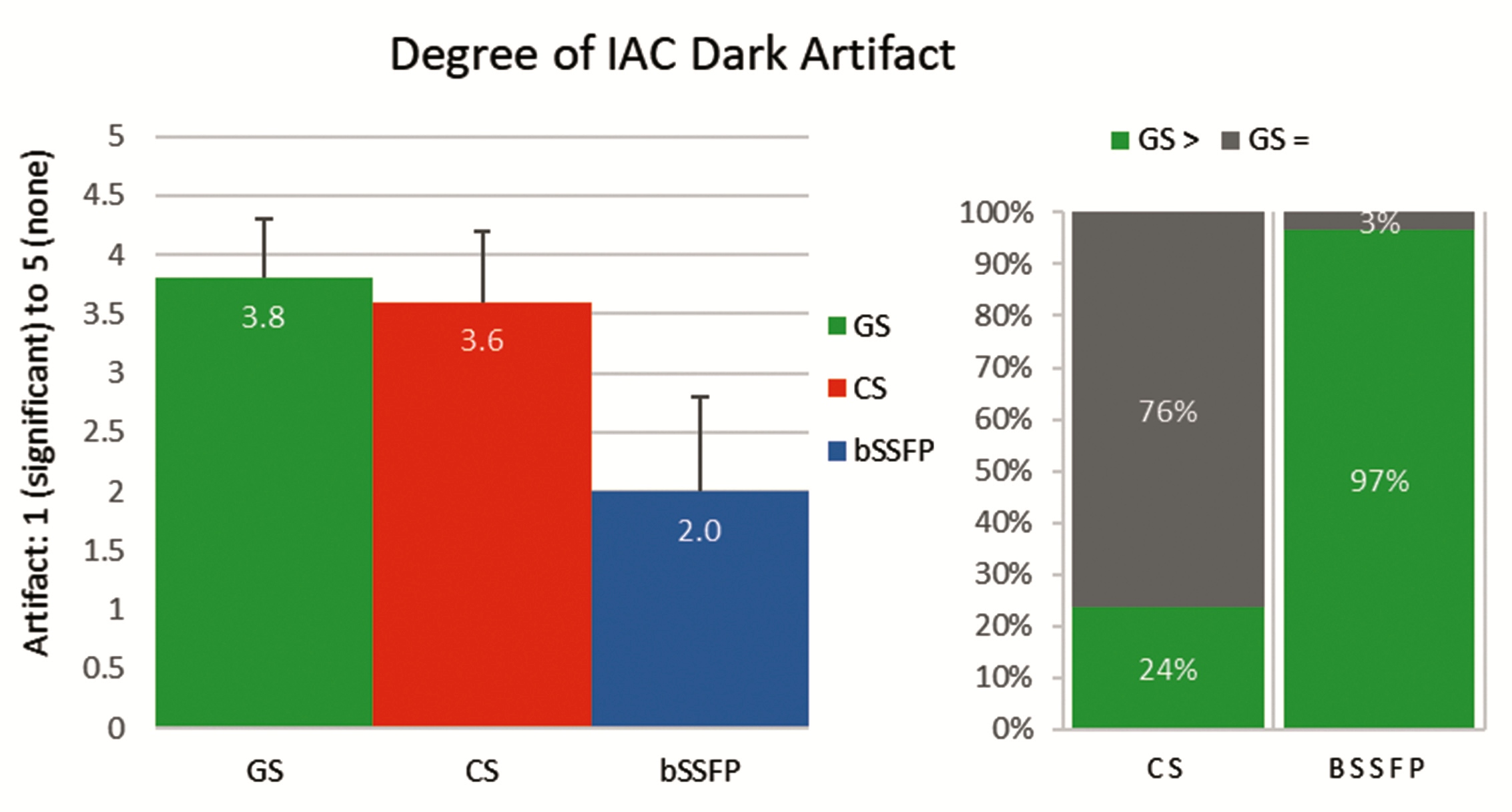
Figure 4 shows that the GS was assessed to have slightly less dark artifact in the IAC than the CS and much less than standard bSSFP, with p-values 0.001 and <0.001 respectively. The GS had less artifact than the CS in 24% and the same in 76% of reads, and less artifact than bSSFP in 97% and the same in 3% of reads; the GS was never rated to have more dark artifact in the IAC than the CS or standard bSSFP.
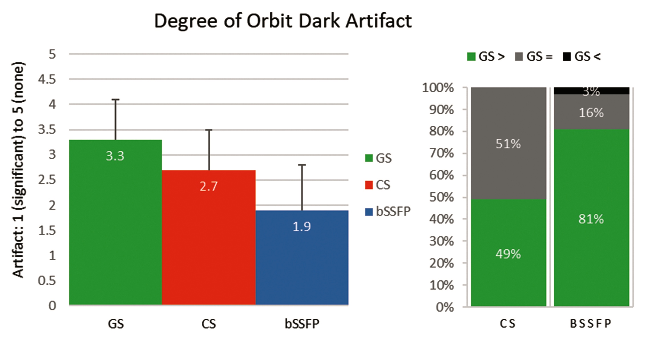
Figure 5 depicts that the GS was assessed to have significantly less dark artifact in the orbits than the CS and standard bSSFP (p<0.001 for both). The GS had less artifact than the CS in 49% and the same in 51% of reads, and less artifact than bSSFP in 81%, the same in 16%, and more in 3% of reads (2/63); the GS was never rated to have more dark artifact in the orbits than the CS.
Discussion
Phantom and in vivo results indicate that the GS shows capacity for flow artifact mitigation when motion occurs during only one phase cycle, while motion during all phase cycles reduces the GS performance considerably. This may be explained by the recent discovery that the GS is insensitive to motion’s noise-like effects when those effects are oriented in a radial direction in the complex plane with respect to a spoke in the GS cross-solution3. This logic breaks down when all four PCs are corrupts by motion, although further testing of variable flow duration is necessary.Conclusion
The GS is a robust MRI technique that not only yields high SNR efficiency and signal demodulation, but also demonstrates relative insensitivity to motion artifacts in a phantom and in vivo.Acknowledgements
NoneReferences
[1] Xiang QS, Hoff MN. Banding artifact removal for bSSFP imaging with an elliptical signal model. Magn Reson Med. 2014;71:927–933.
[2] Hoff MN, Wilson GJ, Xiang XS, Andre JB. Artifact Correction in Temporal Bone Imaging with GS-bSSFP. Proc ISMRM 2014; 22:1627.
[3] Hoff MN, Andre JB, Xiang QS. On the Resilience of GS-bSSFP to Motion and other Noise-like Artifacts, Proc ISMRM 2015; 23:818.
[4] Hoff MN, Andre JB, Xiang QS. Combined Geometric and Algebraic Solutions for Removal of bSSFP Banding Artifacts with Performance Comparisons, Magn Reson Med. 2017;77:644–654.
Figures