3363
Using NMR field probes to flag significant head movement at 7T1Physics, Sir Peter Mansfield Imaging Centre, University of Nottingham, Nottingham, United Kingdom
Synopsis
A fixed array of NMR field probes has proved to be a valid tool for monitoring the effects of a wide range of head movements in a 7T scanner. Here, we use this set up to detect significant field changes in order to give early feedback on head motion. This information could be used in deciding to stop a scan or to reacquire all or part of the k-space data in order to avoid acquisition of motion corrupted image data. This method can be implemented without requiring image sequence modification nor rigid coupling of a motion marker to the head.
Introduction
Head movements during MRI cause artefacts in the resulting images. A variety of Motion Correction (MoCo) techniques have been developed for correcting these artefacts prospectively or retrospectively [1]. However, if the level of movement is larger than the effectiveness range of the particular MoCo technique, the image results will still be corrupted. Under these circumstances, or when no MoCo is applied, it would be useful to have a system which could indicate when head movement that is large enough to cause image artefact has occurred. This would potentially allow a choice to be made to reacquire all, or part of, the k-space data needed for image production. In this work, the possibility of using NMR field probes to flag up significant motion has been explored using measurements of extra-cranial magnetic field perturbations which were concurrently acquired with optical monitoring of head pose. The proposed approach allows the detection of significant head movement without requiring image sequence modification [2] nor rigid coupling of a motion marker to the volunteer’s head.Methods
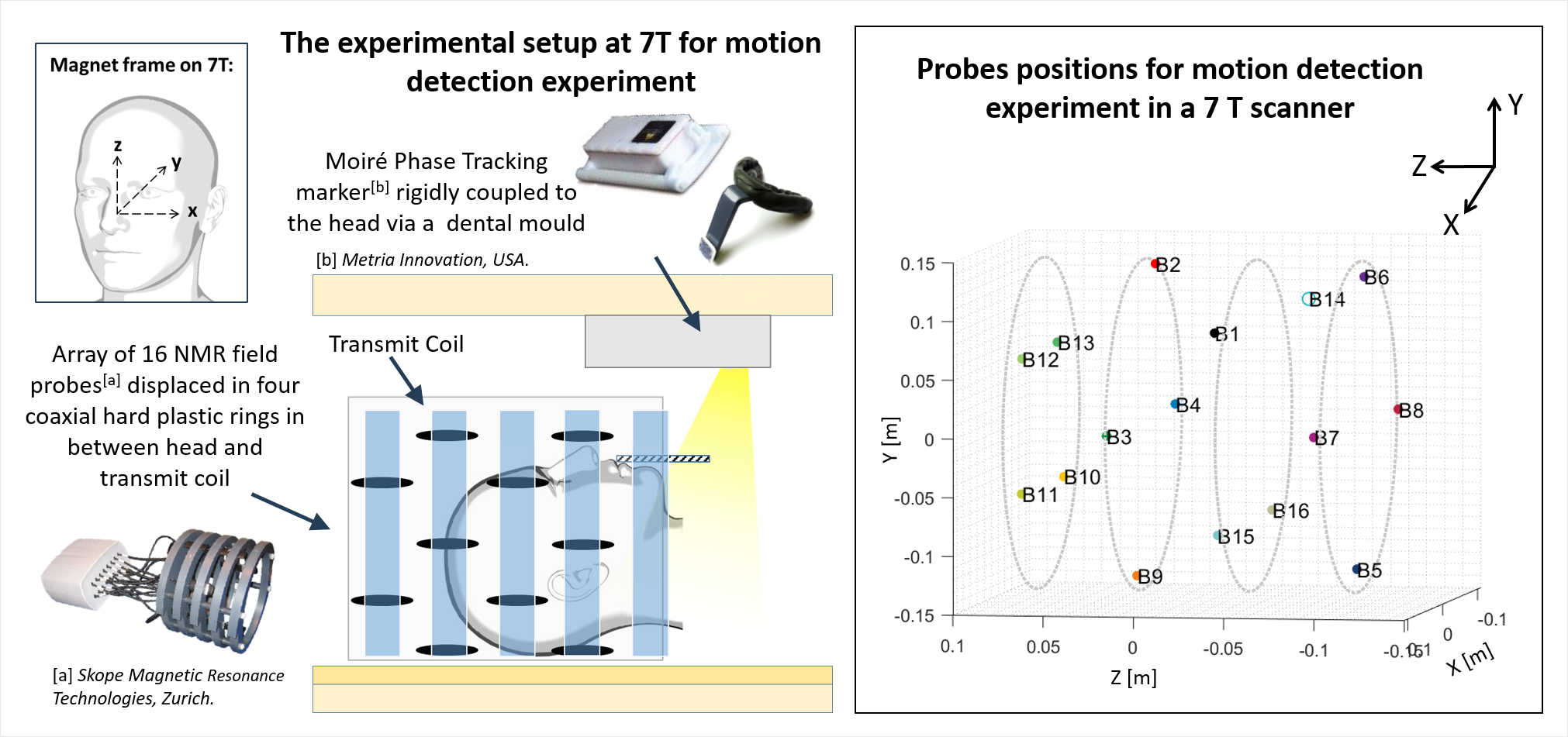
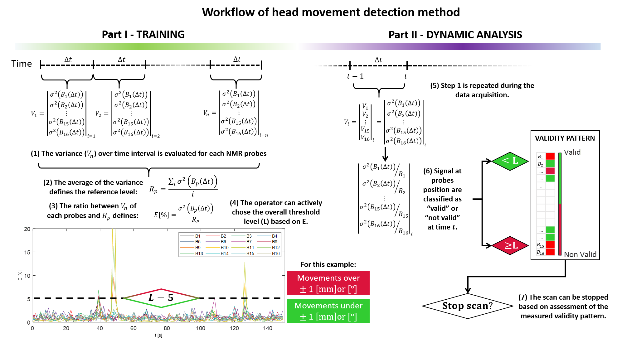
A set of 16 NMR field probes (Skope) was positioned around the head in a customised cylindrical holder [4] formed from four coaxial rings (Figure 1). Extracranial magnetic field changes produced by a range of different head movements executed by four subjects were measured using these field probes at a sampling rate of 6.67 s-1, whilst head pose was simultaneously monitored using a Moire’ Phase Tracking (MPT) system, comprising an optical camera and an MPT marker attached to a dental mould. The detection of significant motion is based on a comparison of the variance of the field measurements during a short time interval to the average value of the variance measured with the subject at rest in the scanner. The aim is to identify a reference level (L) for the field variance at each probe (p) that indicates an unacceptable level of head movement. Here, the level (L) is based on the ratio (E) of the measured field variance at each time step ( σ2(Bp(Δt)) ) to the average value of the variance measured in the rest condition (R). Any probe for which the level is exceeded is classified as indicating an invalid state. The complete workflow of the method is shown in Figure 3. For this analysis, Δt was set equal to ten time steps (corresponding to 1.5 s). Figure 4 shows a forecast of which probes are most likely to detect head movements during the different conditions (“Rest”, “Feet wiggling”, “Head Shake” and “Head Nod”) based on applying PCA to the variance of the data.Results
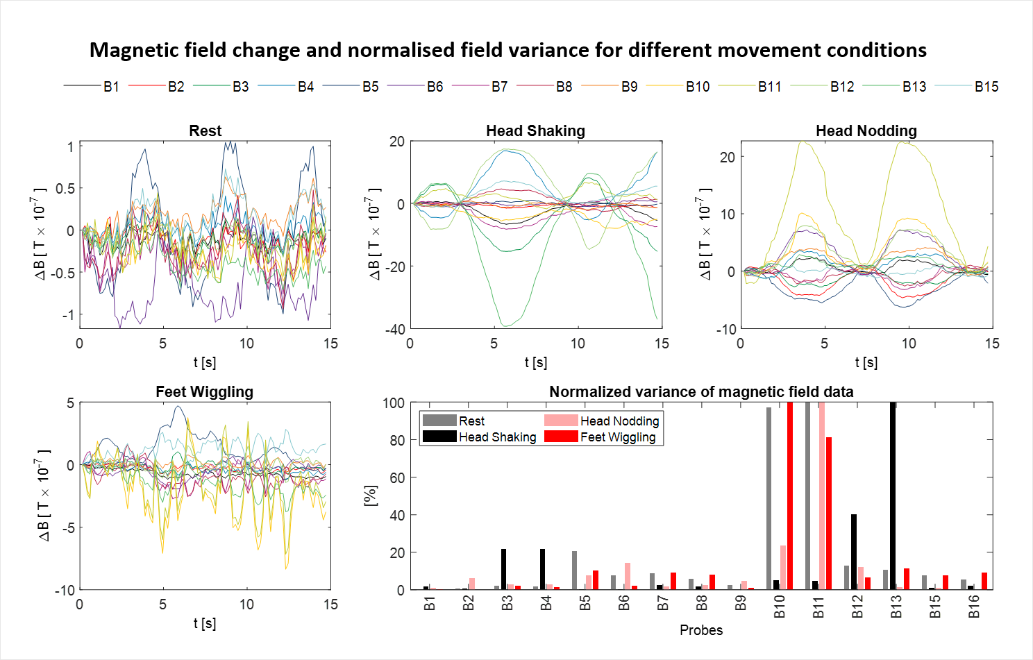
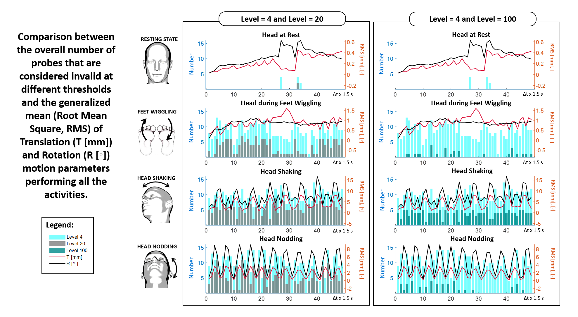
The plots in Figure 2 show the extra-cranial changes in magnetic field for different head movements for one subject. Larger changes are measured for larger head movements, as expected. Analysis of the pattern of variation of field variances over the different probes shows that the range of field variation depends on the probe position and type of movement. The variation of the number of probes classified as invalid with time during the different movement conditions is shown in Figure 5 for different values of the threshold level. The concurrent variation of the RMS displacement and rotation relative to the starting position (measured using the MPT system) is also shown. These results indicate that by varying the threshold level it is possible to adjust the level of movement above which a significant number of probes are classified as invalid. This would allow the level of motion at which a problem is flagged to be adjusted by the operator.Discussion and Conclusion
This work has demonstrated that it is possible to develop a motion detection tool for a 7T MRI scanner based on the use of NMR field probes. This tool does not require image sequence modification [2] or use of markers that are directly coupled to the head [4]. For these experiments the field probes were sited in a bespoke holder, which took the place of the standard receiver coil array, but in future the probes could be placed between the receiver array and volume transmit coil. Finding the correct criteria to use in deciding when to stop a scan automatically or to rescan some k-space data so as to avoid significant motion artefacts will require further work. Possible criteria are based on adjusting the level L and evaluating the number of probes that are invalid over a given period of time.Acknowledgements
No acknowledgement found.References
[1] McLaren review J. McLaren Prospective motion correction in brain imaging: A review 2013 March; 69, 621-63
[2] Aranovitch A. et al, “Prospective motion correction with NMR markers using only native sequence elements”, Magn Reson Med. 2018; 79:2046–2056
[3] Bortolotti L. et al, “Non-contact measurement of head movements inside a 7 T Scanner using a 16-channel field camera”, ISMRM2019, Abstract number: 4432.
[4] Bortolotti L. et al, “Simulation of external magnetic field changes due to head motion during 7 Tesla MRI scan”, ISMRM2019; Abstract number: 4431.
[5] Bortolotti L. et al, “Using a field camera to monitor head movement in 7T scanner”, ESMRMB2019, A-1346
[6] Andre, Jalal B., et al. "Toward quantifying the prevalence, severity, and cost associated with patient motion during clinical MR examinations."Journal of the American College of Radiology” 12.7 (2015): 689-695.
[7] Simon Pascal Gross, Diss. ETH No. 23739, Zurich, 2016.
Figures