3333
High-resolution multifrequency MR Elastography of liver tumors using a compact tabletop MRI Scanner1Department of Radiology, Berlin, Germany, 2Institute of Medical Informatics, Berlin, Germany
Synopsis
MR elastography is a clinical imaging technique for the mapping of viscoelastic tissue properties on the millimeter scale. However, basic studies in tissue samples require a compact MRE setup which provides sub-millimeter resolution. We here introduce compact 0.5-Tesla tabletop multifrequency MRE for high-resolution mapping of viscoelasticity and water diffusion in VX2-liver tumors and surrounding liver tissue. Ex-vivo tumors were stiffer, more viscous and showed higher water diffusivity than adjacent tissue. In tumors, viscosity was inversely correlated with water diffusion while in liver tissue, stiffness was inversely correlated with diffusion.
Introduction:
Liver cancer is the third most common cause of cancer deaths worldwide with an increasing incident1. Malignant hepatic mass formation in patients is associated with abnormal stiffness of liver and tumors, as well as high tumor viscosity2-4 . These parameter changes can be mapped for diagnostic purposes by clinical MR elastography (MRE) on the millimeter scale. However, basic studies in tissue samples require a compact MRE setup which provides sub-millimeter resolution. We here introduce compact 0.5-Tesla tabletop multifrequency MRE for high-resolution mapping of viscoelasticity and water diffusion in VX2-liver tumors and surrounding liver tissue.Method:
VX2 tumor cells were implanted into the liver of nine healthy New Zealeand white rabbits. The animals were sacrificed approximately 4 weeks after tumor injection, when the lesion grew to approximately 1 cm³ size. Tumors with surrounding liver tissue were harvested post mortem and transferred into a rigid glass tube (Fig 1A-B).MRE and diffusion weighted imaging (DWI) were performed in a 0.5-Tesla compact tabletop MRI scanner (Pure Devices GmbH, Würzburg, Germany) (Fig 1C). The MRE technique is explained in detail in 5. In brief, mechanical frequencies of 800, 1000, 1200, 1400, 1600, 1800 and 2000Hz were induced with a piezoelectric driver which was mounted on the tissue containing glass tube. The shear wave fields were acquired using a spin-echo imaging sequence. MRE data were processed by the tomoelastography pipeline which yields maps of shear wave speed (c in m/s) and the phase angle of the complex shear modulus (φ in rad also termed 'fluidity') with a resolution of 150×150×3000 µm6,7. Apparent diffusion coefficient (ADC) was mapped by DWI using a spin-echo sequence with one pair of split diffusion gradients to minimize eddy-currents while generating high b-values of 50, 175, 300, 550, 675 and 800 s/mm². A region of interest was manually drawn based on the MRE and DWI magnitude images.
Results:
Figure 2 shows representative maps of two cases of tumor and liver tissue. The shown tumors differ in size and mechanical heterogeneity: while the majority of the tumor shows increased shear wave speed c and phase angle of the complex modulus φ, regions with low values within the tumor are also observed. Overall, VX2 tumors appear to have a higher shear wave speed c than the surrounding liver (VX2: 4.14±0.9 m/s vs. liver: 2.60±0.49 m/s, P<0.001). Additionally, tumorous tissue showed increased φ- and ADC-values compared with surrounding liver (VX2: 0.84±0.16 rad vs. 0.69±0.12 rad and 8.60E-10±2.47E-10mm²/s vs. 6.76E-10±2.82E-10 mm²/s, respectively, both P<0.05). MRE and DWI values are given in figure 2 and table 1. φ-values of tumorous tissue were negatively correlated with tumoral ADC (P<0.05, R=-0.79), whereas c-values of liver tissue were negatively correlated with ADC-values of the liver (P<0.05, R=-0.72).Discussion and Conclusion:
We demonstrated that MRE can be performed in a compact tabletop MRI scanner at low field strength for investigations of tissue samples with sub-millimeter resolution. The compact MRE device is portable and facilitates investigations of fresh tissue samples in the operating theater. Similar to in-vivo liver tumors in patients, we observed abnormally high stiffness and fluidity (φ) values in VX2- tumor samples. Intriguingly, we observed a negative correlation of tumor fluidity and water diffusion. This negative correlation might be explained by the restricted extracellular space within the tumor due to cell proliferation8. Cell accumulation leads to friction due to hydrophobic interactions between the lipophile cell membrane and the components of extracellular matrix when shear forces are applied as in MRE. Additionally, the decreased amount of extracellular space might cause restriction of free water diffusion. However, these tentative interpretations require further investigations by histology and more cases.The presented MRE technique facilitates mechanical parameter mapping in ex-vivo tissue samples and is therefore well suited for studies in tumors. Further investigations and correlation analyses with histopathological examinations have to be performed in order to understand the microscopic mechanisms that potentially influence global mechanical properties and water diffusion of liver tumors.
Acknowledgements
No acknowledgement found.References
1. Bray, F., et al. Global cancer statistics 2018: GLOBOCAN estimates of incidence and mortality worldwide for 36 cancers in 185 countries. CA Cancer J Clin 68, 394-424 (2018).
2. Venkatesh, S.K., et al. MR elastography of liver tumors: preliminary results. AJR Am J Roentgenol 190, 1534-1540 (2008).
3. Garteiser, P., et al. MR elastography of liver tumours: value of viscoelastic properties for tumour characterisation. Eur Radiol 22, 2169-2177 (2012).
4. Shahryari, M., et al. Tomoelastography distinguishes non-invasively between benign and malignant liver lesions. Cancer Research, canres.2150.2019 (2019).
5. Braun, J., et al. A compact 0.5T MR elastography device and its application for studying viscoelasticity changes in biological tissues during progressive formalin fixation. Magnetic Resonance in Medicine 79, 470-478 (2018).
6. Tzschatzsch, H., et al. Tomoelastography by multifrequency wave number recovery from time-harmonic propagating shear waves. Med Image Anal 30, 1-10 (2016).
7. Hirsch, S., et al. MR Elastography of the Liver and the Spleen Using a Piezoelectric Driver, Single-Shot Wave-Field Acquisition, and Multifrequency Dual Parameter Reconstruction. Magn Reson Med 71, 267-277 (2014).
8. Svolos, P., et al. The role of diffusion and perfusion weighted imaging in the differential diagnosis of cerebral tumors: a review and future perspectives. Cancer Imaging 14, 20 (2014).
Figures
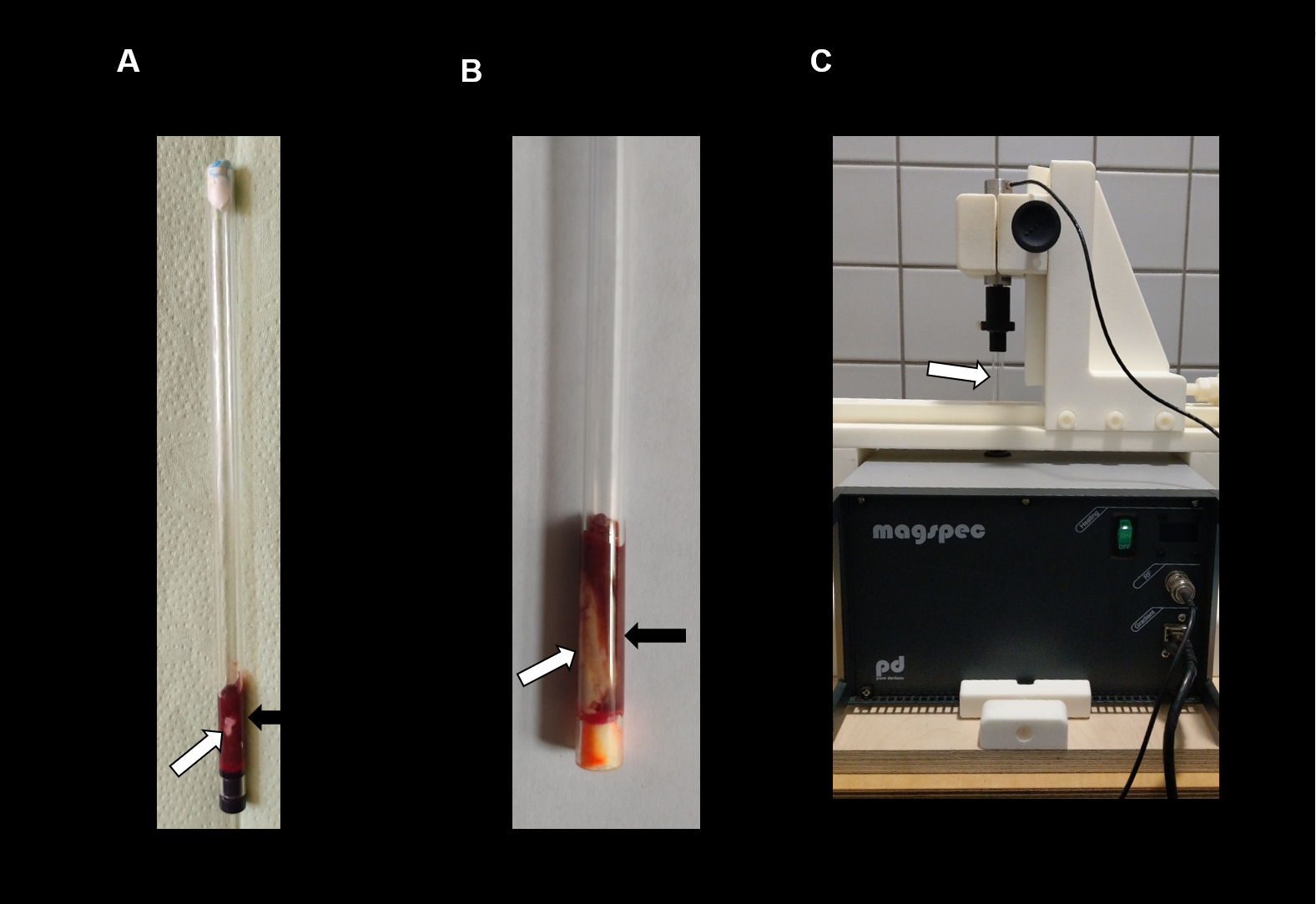
Tabletop MRI and MRE setup: (A) and (B) show two different tissue samples of VX2 tumor (white arrow) and surrounding liver tissue (black arrow). Tissue is placed within a rigid glass tube of 7 mm diameter. (C) Tabletop MRE setup: the glass tube (white arrow) is placed into the tabletop MRI device and mounted on a 3D printed scaffold for the piezoelectric actuator. Harmonic mechanical frequencies are induced along the tube direction.
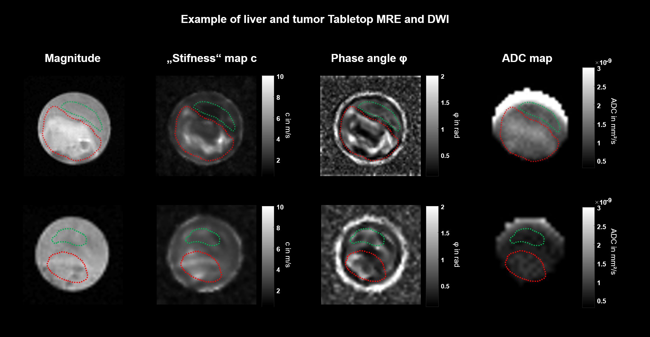
High-resolution stiffness and fluidity mapping of VX2 liver tumors. Magnitude of MRE, stiffness maps (shear wave speed c in m/s), phase angle of the complex shear modulus φ (fluidity) and apparent diffusion coefficient (ADC) are shown for two cases. VX2 (red region of interest) is stiffer and more fluid than surrounding liver tissue (green region of interest). ADC is elevated compared to the surrounding liver tissue.

Mean values of mechanical and apparent diffusion coefficient (ADC) parameters of VX2 tumor and surrounding liver tissue. * P< 0.05, **P < 0.01, ***P < 0.001.

Correlation analysis of mechanical parameters shear wave speed c (stiffness) and phase angle φ (fluidity) and the apparent diffusion coefficient (ADC) for tumor and liver tissue. Tumor fluidity shows a negative correlation with tumor ADC. A similar correlation is observed for liver stiffness versus liver ADC. R, Pearson's correlation coefficient.
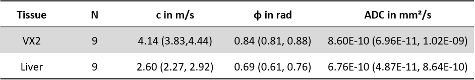
Table 1: Mechanical properties and ADC of VX2 and liver
VX2 tumors show increased mechanical values as well as increased ADC compared to the surrounding liver. c, shear wave speed; φ, phase angle of the complex shear modulus; ADC, apparent diffusion coefficient.